This website uses cookies to ensure you get the best experience on our website.


- Table of Contents
Plan HSPB2 paraffin-section IHC around cytoplasmic staining in heart and skeletal muscle (HPA tissue IHC). This guide covers fixation consistency, tissue controls and chromogenic scoring while accounting for the reported mismatch between antibody staining and RNA expression (HPA tissue IHC).

Expected localisation, validated protocols, controls and antibodies — the at-a-glance facts below, then the full design guide.
| Expected localisation | Cytoplasmic tissue staining (HPA tissue IHC); nuclear foci annotated (UniProt) | |
| Staining pattern | Cytoplasmic cardiomyocytes and skeletal myocytes (HPA tissue IHC) | |
| Antigen retrieval | EDTA pH 8.0 HIER, heat-mediated (datasheet PA1802) | |
| Positive control | Heart muscle+3 more · see all | |
| Negative control | Appendix+4 more · see all |
| Fixation | Keep fixation consistent across sections. (standard IHC practice; not target-specific) | |
| Caveat | Antibody staining has low agreement with RNA expression (HPA tissue IHC) | |
| Regulation | Preferential expression in skeletal and heart muscle (UniProt) | |
| Isoform / epitope | No annotated isoforms; one 1–182 chain (UniProt) |
The catalog antibody protocol uses EDTA pH 8.0 retrieval (datasheet: PA1802). One published gastric tissue microarray protocol uses gastric enzyme retrieval (PMC12162319).
| Sample | Paraffin-embedded human heart tissue; fixative not specified (datasheet PA1802) |
| Fixation | Image fixative and duration unreported (datasheet PA1802); verify before use. |
| Sectioning | 4–5 µm sections on charged slides (standard) |
| Deparaffinisation | Xylene, graded ethanol series to water (standard) |
| Antigen retrieval | Heat retrieval: EDTA pH 8.0 (datasheet PA1802); 20 min, 95–100 °C (standard) |
| Peroxidase block | 3% H2O2, 10 min, room temperature (standard) |
| Blocking | 10% goat serum (datasheet PA1802) |
| Primary antibody | Rabbit anti-HSPB2, 2 μg/ml (datasheet PA1802) |
| Primary incubation | Overnight at 4 °C (datasheet PA1802) |
| Detection | HRP-conjugated secondary, DAB chromogen (datasheet PA1802) |
| Counterstain | Hematoxylin, blue, dehydrate and mount (standard) |
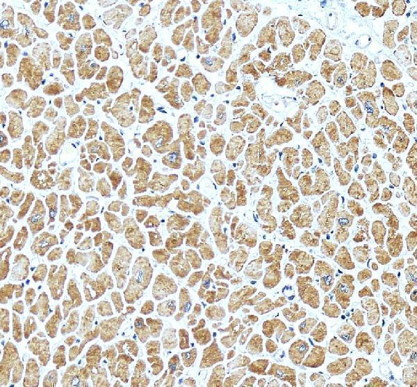
| Expected result | HSPB2-positive staining in cardiomyocytes of heart muscle (HPA tissue IHC: Medium). HPA tissue profile: Cytoplasmic expression in several tissues, including heart and skeletal muscle. No signal in the no-primary control. |
HSPB2 is reported in the cytoplasm and nucleus, including nuclear foci; it has no transmembrane segment (UniProt Q16082). In tissue IHC, expect cytoplasmic staining in cardiomyocytes and skeletal myocytes, with staining also reported in kidney tubule and placental decidual cells (HPA tissue IHC). HPA rates its tissue staining Approved but reports low consistency with RNA expression, so interpret the pattern alongside controls (HPA tissue IHC).
| Cytoplasmic staining in cardiomyocytes or skeletal myocytes, around the reported medium level (HPA tissue IHC). | This fits the reported tissue pattern and UniProt's cytoplasmic location (HPA tissue IHC; UniProt Q16082). Compare the cells and compartment with the control section; a brown deposit elsewhere does not establish HSPB2 expression (general IHC practice). |
| Predominantly cell-surface, extracellular or broadly nonspecific nuclear staining, with little cytoplasmic signal. | Recheck compartment assignment and controls: HSPB2 has no transmembrane segment, while UniProt reports cytoplasm and nuclear foci (UniProt Q16082). Nuclear staining alone is not automatically false; HPA also reports nucleoplasm in ICC-IF (HPA subcellular). |
| Prominent staining in appendix glandular cells or breast adipocytes, where HPA reports no detection (HPA tissue IHC). | Treat this as discordant with those cell-specific observations, not proof that the entire tissue is negative (HPA tissue IHC). Cross-reactivity, endogenous detection activity or misidentified cells are possibilities to test with controls (general IHC practice). |
| Brown precipitate spreads across stroma, lumen and multiple cell types without clear cellular boundaries. | A diffuse deposit cannot be confidently scored as cytoplasmic HSPB2 staining (general IHC practice; HPA tissue IHC). Check whether the same pattern appears when the primary antibody is omitted; that comparison can reveal detection-system background (general IHC practice). |
| No signal in cardiomyocytes or skeletal myocytes on a slide intended as a positive control (HPA tissue IHC). | First assess tissue preservation, antibody and detection controls, and whether the expected cells are present (general IHC practice). HPA reports medium staining rather than universal staining and notes low agreement with RNA, so one blank section does not settle expression (HPA tissue IHC). |
| Which cells provide a tissue benchmark? | Heart cardiomyocytes and skeletal myocytes have medium HPA staining; kidney tubule and placental decidual cells are additional reported medium-positive cell populations (HPA tissue IHC). These are cell-specific observations, not guarantees for every section (HPA tissue IHC). |
| How strong is the tissue evidence? | HPA labels tissue IHC Approved and lists antibody HPA079414 as IHC Approved, while explicitly noting low consistency between staining and RNA expression (HPA tissue IHC; HPA antibodies). Read intensity with that limitation in mind (HPA tissue IHC). |
| Does protein structure predict a membrane pattern? | UniProt lists the 182-residue chain without a signal peptide, propeptide or transmembrane segment, and assigns cytoplasm and nucleus (UniProt Q16082). Those annotations support checking compartment, but do not establish antigen-retrieval or fixation sensitivity (UniProt Q16082). |
| Which sampled cells make useful low-signal comparisons? | HPA reports no detection in appendix glandular cells and breast adipocytes, and low staining in several other named cell populations (HPA tissue IHC). Match the actual cell type before using any tissue as a comparison (HPA tissue IHC; general IHC practice). |
| IF/ICC Q&A: Should Golgi signal be expected on this IHC slide? | HPA reports mainly Golgi localization, with additional nucleoplasm, in ICC-IF images; UniProt also reports cytoplasm and nuclear foci (HPA subcellular; UniProt Q16082). For paraffin IHC, judge against HPA's tissue-level cytoplasmic pattern; ICC-IF localization is a separate observation (HPA tissue IHC; HPA subcellular). |
| Situation | Likely cause | Next action |
|---|---|---|
| The positive-control muscle section is blank. | The expected cells may be absent or the staining run may have failed; HPA reports medium staining in cardiomyocytes and skeletal myocytes (HPA tissue IHC; general IHC practice). | Confirm cell identity and section integrity, then review retrieval, antibody application and detection against the run controls (general IHC practice). Do not infer target-specific retrieval sensitivity from the HPA pattern (HPA tissue IHC). |
| Most cells and extracellular areas stain brown. | Nonspecific binding or detection background may obscure a cellular pattern (general IHC practice). | Compare a primary-omission control, review blocking and washes, and score only staining with identifiable cell boundaries and compartment (general IHC practice). |
| An expected low-signal cell population appears strongly positive. | Cell misidentification, cross-reactivity or endogenous enzyme activity may explain the discordance (general IHC practice; HPA tissue IHC). | Verify the cell type and compare primary-omission and detection controls; keep HPA's cell-specific no-detection calls and low RNA concordance in view (HPA tissue IHC; general IHC practice). |
| Signal seems confined to cell surfaces. | That distribution conflicts with UniProt's cytoplasm/nucleus annotation and absence of a transmembrane segment (UniProt Q16082). | Inspect morphology and counterstain, then compare the IHC-validated antibody's control pattern before assigning HSPB2 positivity (general IHC practice; HPA antibodies). |
| Only nuclei stain. | Nuclear localization is possible, but UniProt specifies nuclear foci and HPA's tissue summary emphasizes cytoplasm (UniProt Q16082; HPA tissue IHC). | Check whether staining is focal within identified cells, review background controls, and avoid calling uniform nuclear deposit a confirmed tissue pattern on that evidence alone (general IHC practice; HPA tissue IHC). |
| Reported medium-positive cells stain unevenly across a section. | Technical variation or differences among sampled cells may affect interpretation; the HPA entry supplies a reported level, not a per-cell acceptance threshold (HPA tissue IHC; general IHC practice). | Compare intact regions and run controls, record the affected cell population and distribution, and avoid upgrading patchy signal solely to match the HPA label (general IHC practice; HPA tissue IHC). |



![HPA ICC-IF BJ [Human fibroblast] — HSPB2 (HPA079414)](https://images.proteinatlas.org/79414/2095_B4_2_blue_red_green.jpg)
![HPA ICC-IF BJ [Human fibroblast] — HSPB2 (HPA079414)](https://images.proteinatlas.org/79414/2095_B4_5_blue_red_green.jpg)
Comprehensive Human Protein Atlas IHC scoring per tissue (reliability: Approved — Low consistency between antibody staining and RNA expression data.). Rows are taken directly from the HPA tissue chart — click any row's HPA link to view the source.
| Tissue | Cell type | Level | Evidence | Source |
|---|---|---|---|---|
| Heart muscle | Cardiomyocytes | Medium | Protein (IHC) | HPA → |
| Kidney | Cells in tubules | Medium | Protein (IHC) | HPA → |
| Placenta | Decidual cells | Medium | Protein (IHC) | HPA → |
| Skeletal muscle | Myocytes | Medium | Protein (IHC) | HPA → |
| Tissue | Cell type | Level | Evidence | Source |
|---|---|---|---|---|
| Appendix | Glandular cells | Not detected | Protein (IHC) | HPA → |
| Breast | Adipocytes | Not detected | Protein (IHC) | HPA → |
| Caudate | Neuronal cells | Not detected | Protein (IHC) | HPA → |
| Cerebellum | Cells in granular layer | Not detected | Protein (IHC) | HPA → |
| Cervix | Glandular cells | Not detected | Protein (IHC) | HPA → |
Use the paraffin-section IHC result as a starting point, then check cell type, compartment and staining controls before interpreting HSPB2 signal.
PA1802 has IHC images from human paraffin-embedded heart and rat frozen cardiac muscle, plus a HeLa ICC image; listed reactivity covers human, mouse and rat (catalog: image captions, reactivity).
PA1802 will render with its human heart paraffin-section IHC figure (PA1802 IHC caption). The catalog also documents rat frozen cardiac muscle IHC and HeLa ICC; it lists human, mouse and rat reactivity (catalog: image captions, reactivity).
Which to pick: For tissue IHC, choose PA1802: its own figure documents paraffin-embedded human heart with EDTA retrieval at pH 8.0; the fixative is unreported (PA1802 IHC caption). For IF/ICC, PA1802 lists ICC and shows a HeLa ICC image, but its application list does not include IF; its host is rabbit and clonality is unreported (catalog: applications, ICC caption, host, clone). For cross-species work, PA1802 lists human, mouse and rat reactivity, with IHC images from human and rat tissue (catalog: reactivity, IHC captions).