HSPB8 / Heat shock protein beta-8 · IHC design guide

Design Immunohistochemistry for HSPB8

Plan HSPB8 paraffin IHC using the cytoplasmic tissue pattern and reported positive and undetected cell populations as controls (HPA tissue IHC). The catalog antibody’s tissue IHC example uses 1 µg/mL primary antibody with chromogenic detection (datasheet A02492-2).

Evidence assembled Oct 2026 · For research use; verify linked source records and product datasheet before use
Immunohistochemistry protocol sheet for HSPB8 (IHC for HSPB8): expected localisation Cytoplasmic staining in several tissues (HPA tissue IHC), antibody A02492-2, validated IHC image, and IHC protocol steps
Printable HSPB8 IHC protocol sheet — expected localisation Cytoplasmic staining in several tissues (HPA tissue IHC), antibody A02492-2, controls and protocol steps. Open the full HSPB8 IHC guide →

HSPB8 Immunohistochemistry Experimental Design Guide

Expected localisation, validated protocols, controls and antibodies — the at-a-glance facts below, then the full design guide.

Must know before staining
Expected localisation Cytoplasmic staining in several tissues (HPA tissue IHC)
Staining pattern Cytoplasmic staining across several tissue cell types (HPA tissue IHC)
Antigen retrieval Citrate pH 6 HIER, heat-mediated (datasheet A02492-2)
Positive control ⓘ Cerebral cortex+4 more · see all
Negative control ⓘ Adipose tissue+3 more · see all
Important caveats
Reasons your staining may differ from the expected pattern.
Fixation Matched tissue-IHC evidence does not establish the fixation claim. Validate the specimen-specific method before use. (selected-SKU IHC image A02492-2)
Caveat Antibody staining and RNA show low consistency (HPA tissue IHC)
Regulation Skeletal muscle RNA is tissue enhanced (HPA tissue RNA)
Isoform / epitope No annotated isoforms; one 1–196 chain (UniProt)
Section 1

Recommended HSPB8 IHC & IF Protocols

The catalog antibody’s IHC-P protocol is paired with four published HSPB8 staining protocols covering muscle, nerve, bladder and spinal cord tissue (datasheet A02492-2; PMC articles below).

Recommended immunohistochemistry (IHC-P) protocol parameters
SampleParaffin-embedded mouse pancreas tissues; fixative not specified (datasheet A02492-2)
FixationImage fixative and duration unreported (datasheet A02492-2); verify before use.
Sectioning4–5 µm sections on charged slides (standard)
DeparaffinisationXylene, graded ethanol series to water (standard)
Antigen retrievalHeat retrieval: Citrate pH 6, 20 min (datasheet A02492-2)
Peroxidase block3% H2O2, 10 min, room temperature (standard)
Blocking10% goat serum (datasheet A02492-2)
Primary antibodyRabbit anti-HSPB8, 0.5-1μg/ml (datasheet A02492-2)
Primary incubationOvernight at 4 °C (datasheet A02492-2)
DetectionStreptavidin-biotin complex (SABC), DAB chromogen (datasheet A02492-2)
CounterstainHematoxylin, blue, dehydrate and mount (standard)
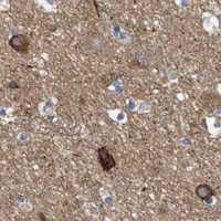
Expected resultHSPB8-positive staining in neuronal cells of cerebral cortex (HPA tissue IHC: High). HPA tissue profile: Cytoplasmic expression in several different tissue types. No signal in the no-primary control.
💡Decision noteStart with heat-mediated citrate retrieval at pH 6 for the catalog antibody (datasheet A02492-2). Published studies also used EDTA retrieval (PMC10777032; PMC6194336).
Section 2

What Is the Expected HSPB8 Staining Pattern?

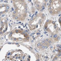
HSPB8 should show predominantly cytoplasmic staining in several tissue types (HPA: tissue IHC). Cytosolic localisation is supported by ICC-IF, while nuclear localisation is also reported; HSPB8 has no transmembrane segment (HPA: subcellular; UniProt Q9UJY1 topology). HPA reports high staining in specific neuronal, epithelial, alveolar, glandular and trophoblastic cells, but rates its tissue IHC profile Approved with low consistency between antibody staining and RNA expression (HPA: tissue IHC).

What am I looking at on my slide?
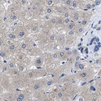
Cytoplasmic signal in cerebral cortex neurons or lung alveolar cells.This fits the reported compartment and cell types (HPA: tissue IHC, High in neuronal and alveolar cells). Judge staining in the identified cells rather than treating every cell in the section as an expected positive (HPA: tissue IHC).
Strong membrane-rim or extracellular staining dominates, with little intracellular signal.That distribution is unexpected for a protein reported in the cytoplasm and nucleus, with no transmembrane segment (UniProt Q9UJY1 localisation and topology). Check morphology and detection background before interpreting it as HSPB8 (general IHC practice).
Strong staining appears in adipocytes or bone-marrow hematopoietic cells.HPA reports these cells as Not detected (HPA: tissue IHC). Investigate antibody cross-reactivity or endogenous detection activity; a discordant cell pattern alone does not establish which cause applies (general IHC practice).
Color spreads across tissue, empty spaces or section edges without a clear cell pattern.Diffuse deposition does not reproduce HPA's cell-associated cytoplasmic profile (HPA: tissue IHC). Review blocking, washes and detection controls for nonspecific background (general IHC practice).
No staining appears in a well-preserved, HPA-reported High cell population.Absence in a reported positive population warrants a run check, but HPA notes low consistency between antibody staining and RNA expression (HPA: tissue IHC). Confirm cell identity, then review controls and IHC conditions before calling the sample negative (general IHC practice).
💡Expected HSPB8 appearanceA convincing positive is cell-associated, mainly cytoplasmic staining in an HPA-reported High population, such as skin keratinocytes; broad extracellular color or dominant membrane-rim staining is suspect (HPA: tissue IHC, High in keratinocytes; UniProt Q9UJY1 topology).
How each factor affects the staining
Compartment and cell identityInterpret cytoplasmic staining in the named positive cells first (HPA: tissue IHC). Nuclear staining is possible, so it is not automatically an artefact; UniProt reports movement to nuclear foci during heat shock (HPA: subcellular; UniProt Q9UJY1 localisation).
Choice of comparison tissueHPA reports High staining in cervix and esophagus squamous cells, pancreatic exocrine cells, placental trophoblasts, and stomach glandular cells; it reports Not detected in ovarian stroma and splenic red-pulp cells (HPA: tissue IHC). These are cell-specific reference patterns, not guarantees for every specimen.
Evidence and antibody scopeThe tissue profile is Approved despite low agreement with RNA expression, and antibody HPA015876 is Approved for IHC (HPA: tissue IHC; HPA: antibody validation). Interpret unexpected patterns cautiously; this record does not provide an IHC Enhanced designation (HPA: antibody validation).
Topology and processingUniProt lists a 1–196 chain, no signal peptide or propeptide, and no transmembrane segment (UniProt Q9UJY1 processing and topology). These features support checking intracellular localisation; they do not identify a retrieval condition or establish fixation sensitivity.
IF/ICC Q&A: should a nuclear signal be rejected?No. HPA reports mainly cytosolic localisation with additional supported nucleoplasmic localisation in ICC-IF, and UniProt reports nuclear foci during heat shock (HPA: subcellular; UniProt Q9UJY1 localisation). Those observations do not set the expected nuclear intensity in paraffin IHC.
Why is my staining missing, weak or wrong?
SituationLikely causeNext action
Positive reference cells show no chromogenic signal.The IHC run may have failed, or the sampled section may lack the expected cells; HPA's tissue profile also has low RNA agreement (HPA: tissue IHC).Verify the named cell population and inspect a concurrently stained reference and detection controls; then review retrieval and primary-antibody conditions as general IHC practice.
Signal is mainly at cell borders or outside cells.The pattern conflicts with reported cytoplasmic and nuclear localisation and the lack of a transmembrane segment (UniProt Q9UJY1 localisation and topology).Check section morphology and compare a no-primary control for detection background; repeat staining if the compartment remains unclear (general IHC practice).
Adipocytes stain strongly while expected positive cells do not.HPA reports adipocytes as Not detected, making this a discordant cell pattern (HPA: tissue IHC). Cross-reactivity or endogenous detection activity is possible (general IHC practice).Confirm cell identity and compare no-primary and detection controls; interpret the result cautiously until the source of signal is resolved (general IHC practice).
Background obscures cell boundaries throughout the section.Diffuse color can arise from nonspecific antibody or detection signal (general IHC practice). It cannot be scored against HPA's cell-specific profile (HPA: tissue IHC).Review blocking, washing and detection controls, then adjust the general IHC workflow according to the observed background source (general IHC practice).
Nuclear staining accompanies cytoplasmic staining.Additional nuclear localisation is supported by ICC-IF, and nuclear foci are reported during heat shock (HPA: subcellular; UniProt Q9UJY1 localisation).Check whether nuclear color is confined to identifiable cells and compare background controls; do not reject it solely because it is nuclear (general IHC practice).
A weak glandular signal seems inconsistent with a High reference.HPA reports Low staining in rectum, prostate and breast glandular cells, but High staining in stomach glandular cells (HPA: tissue IHC). Glandular identity alone does not predict intensity.Confirm the organ and named cell type before comparing intensity; score the observed cellular compartment and reference pattern separately (HPA: tissue IHC; general IHC practice).

Sample controls for HSPB8 IHC & IF

🧪Run pancreas first: exocrine glandular cells should stain (HPA: High in pancreatic exocrine glandular cells; selected-SKU caption: mouse pancreas IHC). Use adipose tissue as the negative tissue; adipocytes should show no specific stain, while any cells on the pancreas slide treated as internal negatives should show counterstain without DAB signal only after their negative status is independently verified (HPA: Not detected in adipocytes; standard IHC practice).
Positive control tissue: Cerebral cortex (Neuronal cells, HPA High)
Negative control tissue: Adipose tissue (HPA Not detected)
ICC-IF cell lines (HPA subcellular resource): HPA ICC-IF images show HSPB8 in MCF-7, U-251MG, U2OS, with annotated localisation: Cytosol (supported) (HPA subcellular).
Technical controls: Include a no-primary, secondary-only control and a concentration-matched rabbit IgG isotype control matched to the primary’s clonality; confirm specificity with knockout tissue or a validated immunizing-peptide block (selected-SKU caption: rabbit primary; standard IHC practice). Block endogenous peroxidase and check for endogenous biotin background in pancreas when using the caption’s biotin-based, DAB detection (selected-SKU caption: biotinylated secondary, streptavidin-biotin complex and DAB; standard IHC practice).
⚠️Feasibility: The selected-SKU paraffin-section caption reports citrate retrieval at pH 6 for 20 minutes and overnight primary incubation at 4°C, but its fixative is unreported; no target-specific fixation window or fixation effect is supplied (selected-SKU caption). Retrieval is part of the demonstrated IHC procedure, though its necessity has not been established by a comparison without retrieval (selected-SKU caption). The supplied evidence does not establish that frozen sections or IF are easier; HPA supports cytosolic and additional nucleoplasmic localization for IF/ICC interpretation, while the biotin-based pancreas IHC workflow requires a background check (HPA: subcellular summary; selected-SKU caption; standard IHC practice).

HPA tissue IHC evidence for HSPB8

Comprehensive Human Protein Atlas IHC scoring per tissue (reliability: Approved — Low consistency between antibody staining and RNA expression data.). Rows are taken directly from the HPA tissue chart — click any row's HPA link to view the source.

Positive expression · recommended positive controls

TissueCell typeLevelEvidenceSource
Cerebral cortex Neuronal cells High Protein (IHC) HPA →
Cervix Squamous epithelial cells High Protein (IHC) HPA →
Esophagus Squamous epithelial cells High Protein (IHC) HPA →
Lung Alveolar cells High Protein (IHC) HPA →
Pancreas Exocrine glandular cells High Protein (IHC) HPA →

Undetected expression · recommended negative controls

TissueCell typeLevelEvidenceSource
Adipose tissue Adipocytes Not detected Protein (IHC) HPA →
Bone marrow Hematopoietic cells Not detected Protein (IHC) HPA →
Ovary Ovarian stroma cells Not detected Protein (IHC) HPA →
Spleen Cells in red pulp Not detected Protein (IHC) HPA →
Section 3

Advanced HSPB8 IHC Tips

Troubleshoot HSPB8 staining in paraffin sections by checking retrieval, cell type, compartment and controls before interpreting chromogenic signal.

How should I retrieve HSPB8 in paraffin sections when staining is weak?
Start with heat-mediated retrieval in citrate buffer at pH 6 for 20 minutes (datasheet A02492-2). In the documented mouse pancreas section, the catalog antibody was then applied at 1 μg/mL overnight at 4°C, followed by biotinylated secondary antibody and DAB detection (datasheet A02492-2). If staining remains weak, check deparaffinisation, heating consistency and whether the tissue lifted or dried before changing retrieval conditions; process a reference pancreas section alongside the test sections (datasheet A02492-2: mouse pancreas IHC). Compare changes using the same antibody concentration and DAB development time, since simultaneous changes make the cause of improvement difficult to identify.
Can I attribute weak HSPB8 staining to overfixation?
Target-specific fixation sensitivity is unknown: the selected paraffin-section caption does not state a fixative or fixation duration (datasheet A02492-2). Record each specimen’s fixative, time in fixative and processing history, then compare sections prepared under matched conditions before assigning weak signal to fixation. Begin troubleshooting with the documented citrate pH 6, 20-minute heat retrieval and 1 μg/mL primary incubation overnight at 4°C (datasheet A02492-2). Include a concurrently stained reference pancreas section, because documented staining there provides a run control while leaving the effect of fixation on HSPB8 unresolved (datasheet A02492-2: mouse pancreas IHC).
Should nuclear DAB signal count as HSPB8 staining?
Score cytoplasmic staining as the expected tissue pattern, since the tissue profile describes cytoplasmic expression across several tissue types (HPA: tissue IHC profile). Nuclear localisation is biologically possible: HSPB8 is listed in the cytoplasm and nucleus, with translocation to nuclear foci during heat shock (UniProt Q9UJY1: subcellular location). Assess nuclear signal separately from cytoplasmic intensity and require a distinct intracellular pattern rather than diffuse colour over nuclei and surrounding tissue. Compare the pattern with a no-primary control and with matched sections; a nuclear result needs independent support before it is interpreted as stress-related redistribution rather than staining artefact.
Could an HSPB8 variant or masked epitope explain discrepant staining?
The supplied record lists 0 isoforms, a 196-aa chain and an sHSP domain spanning residues 74–185 (UniProt Q9UJY1: isoforms, processing and domains). It also lists phosphorylation and arginine methylation sites, including serines 24 and 57 and arginines 71 and 78 (UniProt Q9UJY1: modified residues). These annotations do not establish where the catalog antibody binds or whether a modification alters binding, so request epitope information before attributing an IHC difference to either mechanism. Compare matched sections with consistent citrate pH 6 retrieval and antibody incubation, and treat any epitope explanation as a hypothesis until experimentally tested (datasheet A02492-2: IHC method).
How can I check HSPB8 localisation by multiplex IF?
Use the separate IF/ICC workflow to pair HSPB8 with a marker for the expected cell type; for pancreas, exocrine glandular cells are reported as high in tissue IHC (HPA: pancreas tissue IHC). Select spectrally separated fluorophores and assign the brighter channel after checking unstained tissue for autofluorescence, then collect single-stain controls to assess bleed-through. Permeabilise for an intracellular epitope because HSPB8 has no transmembrane segment and is reported in the cytoplasm and nucleus (UniProt Q9UJY1: topology and subcellular location). Inspect cytosolic and nuclear channels separately, since cytosol is the main supported location and nucleoplasm an additional supported location in ICC/IF images (HPA: subcellular localisation).
What should I check when HSPB8 DAB staining is diffuse?
Run a no-primary control and inspect whether colour follows cells, tissue edges or the entire section before changing antibody concentration. The documented assay used 10% goat serum block, 1 μg/mL primary antibody, a biotinylated secondary antibody and streptavidin–biotin detection with DAB (datasheet A02492-2). Check the peroxidase block as a general chromogenic IHC step, and assess endogenous biotin as a possible contributor when using this detection system. Rinse consistently, keep sections wet and compare DAB development times across matched sections; interpret diffuse signal cautiously where the no-primary control also develops colour.
How should I quantify HSPB8 staining across tissue sections? ⚠ ANSWER MARKED FOR VERIFICATION
Define the tissue compartment and cell population before scoring, then record the percentage of positive cells and staining intensity to calculate an H-score using a consistent scoring scale. For pancreas, specify whether the analysis covers exocrine glandular cells, which are listed as high in tissue IHC (HPA: pancreas tissue IHC). Normalise positive-cell counts to the number of evaluable cells, or stained area to measured tissue area in mm², and exclude folds, edges and necrotic regions consistently. Keep retrieval, exposure to primary antibody and DAB development matched, and report cytoplasmic and nuclear measurements separately because both locations are annotated for HSPB8 (UniProt Q9UJY1: subcellular location).
How can I distinguish genuine HSPB8 signal from an artefact?
Look first for intracellular cytoplasmic staining in an expected cell population; exocrine glandular cells in pancreas are reported as high, whereas adipocytes are reported as not detected in the HPA tissue set (HPA: pancreas and adipose tissue IHC). A nuclear pattern requires separate evaluation because nuclear localisation and heat-shock-associated nuclear foci are annotated for HSPB8 (UniProt Q9UJY1: subcellular location). Reject staining concentrated at cut edges or necrotic regions, and check no-primary sections for endogenous enzyme or detection-system colour. Treat apparent agreement with tissue expression cautiously, since the HPA tissue IHC entry is approved but reports low consistency between antibody staining and RNA expression (HPA: reliability description).
Boster reagents

Best HSPB8 / Heat shock protein beta-8 IHC Antibodies

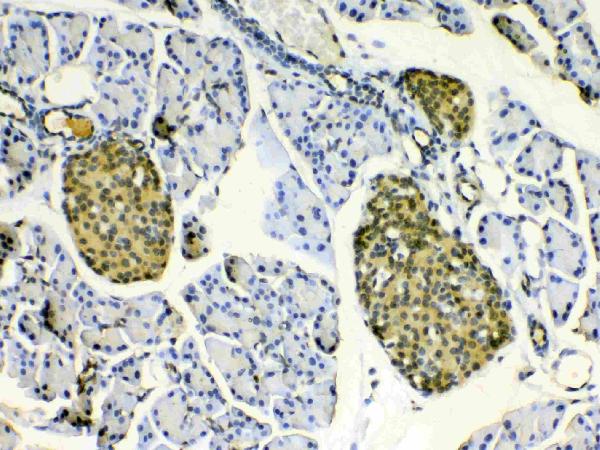
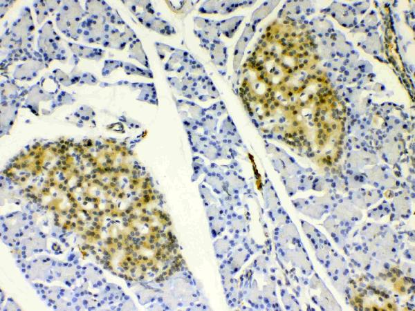
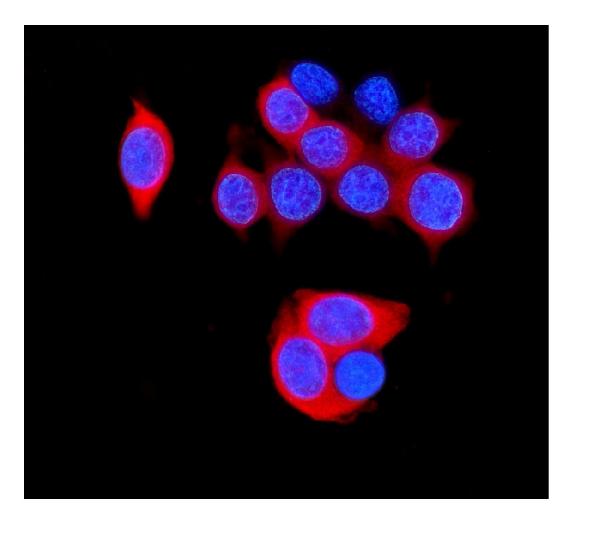
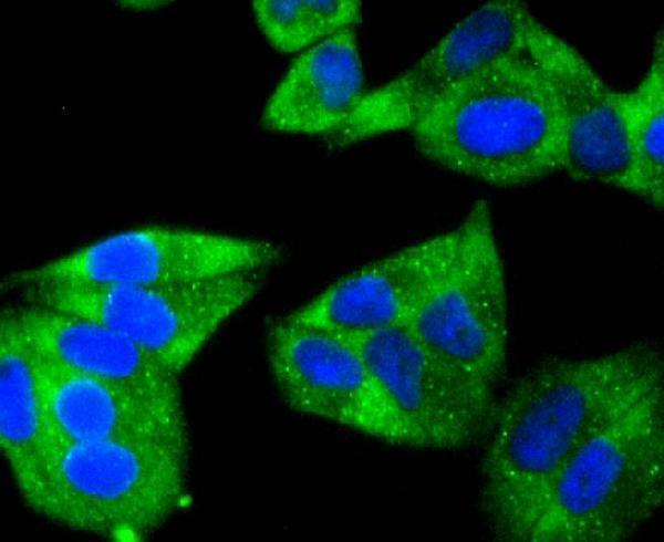
HSPB8 examples cover paraffin-section IHC in mouse pancreas (A02492-2 image caption), ICC in HeLa cells (A02492 image caption), and IF in A431 cells (M02492-2 image caption).

Real IHC data IHC analysis of HSPB8/Hsp22 using anti-HSPB8/Hsp22 antibody (A02492-2). HSPB8/Hsp22 was detected in paraffin-embedded section of mouse pancreas tissues. Heat mediated antigen retrieval was performed in citrate buffer (pH6, epitope retrieval solution) for 20 mins. The tissue section was blocked with 10% goat serum. The tissue section was then incubated with 1μg/ml rabbit anti-HSPB8/Hsp22 Antibody (A02492-2) overnight at 4°C. Biotinylated goat anti-rabbit IgG was used as secondary antibody and incubated for 30 minutes at 37°C. The tissue section was developed using Strepavidin-Biotin-Complex (SABC)(Catalog # SA1022) with DAB as the chromogen.
Anti-HSPB8/Hsp22 Antibody ®
Cat # A02492-2
Real IF data ICC staining Hsp22 in Hela cells (green). The nuclear counter stain is DAPI (blue). Cells were fixed in paraformaldehyde, permeabilised with 0.25% Triton X100/PBS.
Anti-HSP22 HSPB8 Antibody
Cat # A02492
Real IF data IF analysis of HSPB8/Hsp22 using anti-HSPB8/Hsp22 antibody (M02492-2). HSPB8/Hsp22 was detected in immunocytochemical section of A431 cells. Enzyme antigen retrieval was performed using IHC enzyme antigen retrieval reagent (AR0022) for 15 mins. The cells were blocked with 10% goat serum. And then incubated with 5μg/mL mouse anti-HSPB8/Hsp22 Antibody (M02492-2) overnight at 4°C. DyLight®488 Conjugated Goat Anti-Mouse IgG (BA1126) was used as secondary antibody at 1:100 dilution and incubated for 30 minutes at 37°C. The section was counterstained with DAPI. Visualize using a fluorescence microscope and filter sets appropriate for the label used.
Anti-HSPB8/Hsp22 Antibody ® (monoclonal, 7D8)
Cat # M02492-2

A02492-2 shows IHC in paraffin-embedded mouse pancreas (A02492-2 image caption), while A02492 shows ICC in HeLa cells (A02492 image caption). M02492-2 shows IF in A431 cells (M02492-2 image caption).

Which to pick: For tissue IHC, choose A02492-2: its mouse pancreas caption documents paraffin sections, citrate retrieval at pH 6 for 20 minutes, and DAB detection; the fixative is unreported (A02492-2 IHC image caption). For IF/ICC, A02492 has a HeLa ICC image, while monoclonal M02492-2 has an A431 IF image (A02492 and M02492-2 image captions; catalog: M02492-2 clone 7D8). For cross-species IHC, A02492-2 lists human, mouse, and rat reactivity and has paraffin-section IHC images for all three (catalog: A02492-2; A02492-2 IHC image captions).

Each figure is that product's own IHC / IF validation image from its datasheet.

References

  1. UniProt Consortium. UniProt entry Q9UJY1 (HSPB8_HUMAN, Heat shock protein beta-8).
  2. Human Protein Atlas. HSPB8 tissue IHC expression (reliability: Approved).
  3. Human Protein Atlas. HSPB8 subcellular location (ICC-IF): Mainly localized to the cytosol. In addition localized to the nucleoplasm..
  4. Human Protein Atlas. HSPB8 antibody validation summary (1 antibodies).
  5. A knock-in/knock-out mouse model of HSPB8-associated distal hereditary motor neuropathy and myopathy reveals toxic gain-of-function of mutant Hspb8. Acta neuropathologica 2018 — PMC5756276.
  6. HSPB8 Facilitates the Oncogenesis and Advancement of Bladder Cancer via Activation of HSP27. Journal of Cancer 2024 — PMC10777032.
  7. Small heat shock proteins are induced during multiple sclerosis lesion development in white but not grey matter. Acta neuropathologica communications 2015 — PMC4688967.
  8. Heat shock proteins are differentially expressed in brain and spinal cord: implications for multiple sclerosis. Clinical and experimental immunology 2018 — PMC6194336.
  9. PubMed PMID:11085516 — UniProt-cited evidence.
  10. PubMed PMID:11342557 — UniProt-cited evidence.
  11. PubMed PMID:10833516 — UniProt-cited evidence.