This website uses cookies to ensure you get the best experience on our website.


- Table of Contents
Plan paraffin IHC for HSPG2 using its observed cytoplasmic and extracellular matrix staining (HPA tissue IHC). Start with the catalog antibody at 0.5–1 μg/mL (datasheet: 0.5–1 μg/mL) and assess staining against appropriate controls.

Expected localisation, validated protocols, controls and antibodies — the at-a-glance facts below, then the full design guide.
| Expected localisation | Extracellular matrix and cytoplasm, including endothelium (HPA tissue IHC) | |
| Staining pattern | Cytoplasmic staining in several tissues; matrix positivity (HPA tissue IHC) | |
| Antigen retrieval | Citrate pH 6.0 HIER, 95–98 °C, 20 min (rule: cytoplasmic / membrane antigen) | |
| Positive control | Heart muscle | |
| Negative control | Adrenal gland+4 more · see all |
| Fixation | Keep fixation consistent across samples. (standard IHC practice; not target-specific) | |
| Caveat | Secretion can complicate RNA–protein agreement in tissue (HPA tissue IHC) | |
| Regulation | Stimulus-dependent regulation not specified (UniProt) | |
| Isoform / epitope | No isoforms listed; map epitopes against endorepellin/LG3 (UniProt) |
The catalog antibody’s IHC-P protocol is accompanied by published HSPG2 methods for plaque, brain tissue array, and kidney sections (PMC7508285; PMC2904485; PMC12747916).
| Sample | Tissue sections; selected-image fixative not specified (standard IHC workflow) |
| Fixation | Image fixative and duration unreported (datasheet PB9277); verify before use. |
| Sectioning | 4–5 µm sections on charged slides (standard) |
| Deparaffinisation | Xylene, graded ethanol series to water (standard) |
| Antigen retrieval | Heat-induced epitope retrieval in citrate buffer, pH 6.0, 20 min at 95–98 °C (standard rule: cytoplasmic / membrane antigen) |
| Peroxidase block | 3% H2O2, 10 min, room temperature (standard) |
| Blocking | 10% normal serum of the secondary host, 30 min, room temperature (standard) |
| Primary antibody | Rabbit anti-HSPG2, 0.5-1μg/ml (datasheet PB9277) |
| Primary incubation | Overnight at 4 °C (standard) |
| Detection | HRP-polymer secondary, DAB chromogen 5–10 min (standard) |
| Counterstain | Hematoxylin, blue, dehydrate and mount (standard) |
| Expected result | HSPG2-positive staining in cardiomyocytes of heart muscle (HPA tissue IHC: Medium). HPA tissue profile: Cytoplasmic expression in several tissue types, including endothelial cells. Positivity in extracellular matrix. No signal in the no-primary control. |
HSPG2 is secreted into extracellular matrix and basement membranes and has no transmembrane segment (UniProt P98160). HPA reports extracellular matrix positivity and cytoplasmic staining in several tissues, including endothelial cells; cardiomyocyte staining is medium (HPA tissue IHC: Approved).
| ECM positivity and medium cardiomyocyte staining (HPA tissue IHC). | Matches the reported tissue pattern (HPA tissue IHC; UniProt P98160 localisation). |
| Nuclear-only staining. | Suspect artefact; HSPG2 is secreted to ECM and basement membranes (UniProt P98160 localisation). |
| Adrenal glandular cell staining (HPA: Not detected). | Consider cross-reactivity or endogenous detection activity (HPA tissue IHC; general IHC practice). |
| Diffuse staining across the section. | Consider nonspecific binding or detection background (general IHC practice). |
| No cardiomyocyte signal in heart muscle (HPA: Medium). | Check assay performance against this reported positive cell type (HPA tissue IHC). |
| Protein processing (UniProt P98160). | Endorepellin spans 3687–4391 and LG3 spans 4197–4391; check antibody epitope (UniProt P98160). |
| IHC antibody validation (HPA antibodies). | Four listed antibodies are IHC Approved; no ICC status is listed (HPA antibodies). |
| IF/ICC expected pattern? | Secreted protein; HPA provides no ICC-IF images or main location (UniProt P98160; HPA subcellular). |
| Situation | Likely cause | Next action |
|---|---|---|
| No heart cardiomyocyte signal (HPA: Medium). | Low assay sensitivity is possible (general IHC practice). | Check a positive control; titrate primary and optimize retrieval (general IHC practice). |
| Diffuse section background. | Nonspecific binding is possible (general IHC practice). | Titrate primary and review blocking and washes (general IHC practice). |
| Nuclear-only staining. | Off-target staining is possible (UniProt P98160 localisation; general IHC practice). | Run a no-primary control and reassess antibody specificity (general IHC practice). |
| Adrenal glandular staining (HPA: Not detected). | Cross-reactivity or endogenous activity is possible (general IHC practice). | Run a no-primary control; check endogenous enzyme blocking (general IHC practice). |
| Signal in a no-primary control. | Detection reagent binding or endogenous activity is possible (general IHC practice). | Review secondary detection and endogenous enzyme blocking (general IHC practice). |
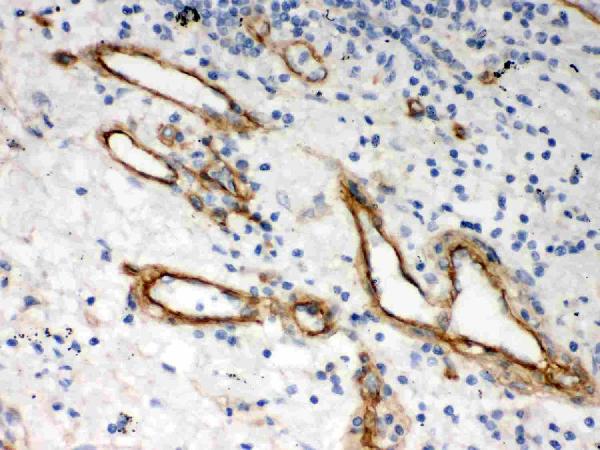
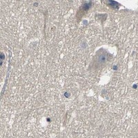


Comprehensive Human Protein Atlas IHC scoring per tissue (reliability: Approved — At least one protein variant secreted, tissue location of RNA and protein might differ and correlation is complex.). Rows are taken directly from the HPA tissue chart — click any row's HPA link to view the source.
| Tissue | Cell type | Level | Evidence | Source |
|---|---|---|---|---|
| Heart muscle | Cardiomyocytes | Medium | Protein (IHC) | HPA → |
| Tissue | Cell type | Level | Evidence | Source |
|---|---|---|---|---|
| Adrenal gland | Glandular cells | Not detected | Protein (IHC) | HPA → |
| Caudate | Glial cells | Not detected | Protein (IHC) | HPA → |
| Cerebellum | Cells in granular layer | Not detected | Protein (IHC) | HPA → |
| Cerebral cortex | Glial cells | Not detected | Protein (IHC) | HPA → |
| Duodenum | Glandular cells | Not detected | Protein (IHC) | HPA → |
Troubleshoot HSPG2 paraffin section IHC by checking retrieval, matrix localization, and compartment aware controls; use IF to clarify ambiguous spatial patterns (UniProt P98160; HPA tissue IHC).
PB9277 has real IHC data from human lung cancer tissue in paraffin sections (catalog: PB9277 figure tag and IHC image caption). No IF data are supplied (catalog: PB9277 IF images).
PB9277 is listed for human IHC, with an IHC(P) image from human lung cancer tissue (catalog: PB9277 applications, reactivity, and IHC image caption). Its listed IHC dilution is 0.5–1 μg/ml (catalog: PB9277 IHC dilution).
Which to pick: Choose PB9277 for human tissue IHC: it is a rabbit antibody listed for IHC, and its image documents IHC(P) on human lung cancer tissue (catalog: PB9277 host, applications, reactivity, and IHC image caption). The caption supports paraffin sections; the fixative and clonality are unreported (catalog: PB9277 IHC image caption and clone). No IF/ICC application or nonhuman reactivity is listed, so the payload offers no validated IF/ICC or cross-species choice (catalog: PB9277 applications and reactivity).