This website uses cookies to ensure you get the best experience on our website.


- Table of Contents
Plan HTRA2 staining in paraffin sections using its observed cytoplasmic tissue pattern (HPA tissue IHC) and expected mitochondrial location (UniProt). Interpret results with the tissue staining reliability caveat in mind (HPA tissue IHC).

Expected localisation, validated protocols, controls and antibodies — the at-a-glance facts below, then the full design guide.
| Expected localisation | Cytoplasmic in tissue (HPA tissue IHC); mitochondrial intermembrane space expected (UniProt) | |
| Staining pattern | Cytoplasmic staining in most tissues (HPA tissue IHC) | |
| Antigen retrieval | EDTA pH 8.0 HIER, heat-mediated (datasheet A01941-3) | |
| Positive control | Adrenal gland+4 more · see all | |
| Negative control | Heart muscle+4 more · see all |
| Fixation | Keep fixation conditions consistent across sections. (standard IHC practice; not target-specific) | |
| Caveat | Antibody staining has low consistency with RNA expression (HPA tissue IHC) | |
| Regulation | Apoptotic stimuli release HTRA2 into cytosol (UniProt) | |
| Isoform / epitope | Four isoforms; maturation removes residues 32–133, so check epitope position (UniProt) |
The catalog antibody protocol uses EDTA retrieval (datasheet A01941-3). Two published HTRA2 IHC protocols provide liver and frontal cortex examples (PMC6829542; PMC6657433).
| Sample | Paraffin-embedded human liver cancer tissue; fixative not specified (datasheet A01941-3) |
| Fixation | Image fixative and duration unreported (datasheet A01941-3); verify before use. |
| Sectioning | 4–5 µm sections on charged slides (standard) |
| Deparaffinisation | Xylene, graded ethanol series to water (standard) |
| Antigen retrieval | Heat retrieval: EDTA pH 8.0 (datasheet A01941-3); 20 min, 95–100 °C (standard) |
| Peroxidase block | 3% H2O2, 10 min, room temperature (standard) |
| Blocking | 10% goat serum (datasheet A01941-3) |
| Primary antibody | Rabbit anti-HTRA2, 2-5 μg/ml (datasheet A01941-3) |
| Primary incubation | Overnight at 4 °C (datasheet A01941-3) |
| Detection | HRP-conjugated secondary, DAB chromogen (datasheet A01941-3) |
| Counterstain | Hematoxylin, blue, dehydrate and mount (standard) |
| Expected result | HTRA2-positive staining in glandular cells of adrenal gland (HPA tissue IHC: Medium). HPA tissue profile: Cytoplasmic expression in most tissues. No signal in the no-primary control. |
HTRA2 is predominantly in the mitochondrial intermembrane space, with a membrane segment at residues 105–125; apoptotic stimuli can release it into the cytosol (UniProt O43464 localization and topology). In tissue IHC, expect cytoplasmic staining in many cell types, including glandular, neuronal, respiratory epithelial, endothelial and hematopoietic cells reported at medium intensity (HPA tissue IHC). Interpret this pattern cautiously: HPA rates its tissue IHC reliability Uncertain because antibody staining and RNA expression have low consistency (HPA tissue IHC).
| Cytoplasmic, granular staining in adrenal glandular cells or cerebral cortex neurons. | This fits the mitochondrial location and HPA's Medium staining in those cells (UniProt O43464 localization; HPA tissue IHC). Granularity is an expected visual interpretation, not proof of mitochondrial identity on a chromogenic section (general IHC practice). |
| Predominantly nuclear staining in otherwise intact cells. | A predominantly nuclear pattern lacks support from the reported mitochondrial location and cytoplasmic tissue profile (UniProt O43464 localization; HPA tissue IHC). Treat it as suspect and compare with controls; cytosolic release after apoptosis does not establish nuclear localization (UniProt O43464 localization; general IHC practice). |
| Strong signal in cardiomyocytes or skeletal myocytes. | HPA reports HTRA2 as Not detected in these cells, so strong staining needs scrutiny (HPA tissue IHC). That category is an observation, not proof of biological absence, especially given HPA's Uncertain IHC reliability (HPA tissue IHC). Check for cross-reactivity or endogenous chromogen activity (general IHC practice). |
| Even color across cells, stroma and empty areas. | Diffuse staining without a cellular pattern is more consistent with background than the reported cytoplasmic distribution (HPA tissue IHC; general IHC practice). Compare a no-primary control, then inspect blocking, washes and chromogen development before assigning an HTRA2 score (general IHC practice). |
| No stain in a tissue reported at medium intensity. | Absent signal in adrenal glandular cells or cerebral cortex neurons conflicts with HPA's observations but does not establish HTRA2 absence (HPA tissue IHC). Review section integrity, detection controls and the antibody's IHC validation before interpreting the specimen (general IHC practice; HPA antibody validation). |
| Tissue and cell choice | HPA reports Medium staining in adrenal glandular cells, marrow hematopoietic cells and cerebral cortex neurons, but Not detected in cardiomyocytes and skeletal myocytes (HPA tissue IHC). Use the former as observed comparators, while retaining the Uncertain tissue IHC reliability caveat (HPA tissue IHC). |
| Processing and epitope position | The precursor contains a propeptide at residues 32–133 and a mature chain at 134–458 (UniProt O43464 processing). If an antibody recognizes the removed region, its staining may differ from an antibody recognizing the mature chain; the supplied antibody records give no epitope positions, so this remains conditional (UniProt O43464 processing; HPA antibody records). |
| Apoptotic state | HTRA2 can leave mitochondria for the cytosol after apoptotic stimuli (UniProt O43464 localization). A less punctate cytoplasmic pattern may therefore merit review alongside cell morphology; the tissue IHC profile alone does not identify apoptosis or validate such a shift (UniProt O43464 localization; HPA tissue IHC). |
| Antibody evidence | The two listed IHC antibodies, HPA027366 and CAB004004, each have Uncertain IHC validation, while HPA006602 has Supported ICC validation (HPA antibody records). Neither ICC support nor tissue expression by itself upgrades a particular IHC result (HPA antibody records; HPA tissue IHC). |
| IF/ICC Q: What pattern should I expect? | A: Mitochondrial localization is Supported in HPA ICC-IF, with images listed for HEK293, Rh30 and U2OS (HPA subcellular; HPA antibody records). That evidence informs the compartment call; this IHC section supplies no IF/ICC protocol (HPA subcellular; general guide scope). |
| Situation | Likely cause | Next action |
|---|---|---|
| Weak or absent signal in an HPA medium-staining comparator. | The stain may have failed, or the chosen section may not contain the reported cell population (HPA tissue IHC; general IHC practice). | Confirm the target cells on the counterstain, then check the IHC-validated antibody and a working detection control; optimize retrieval and antibody concentration within the selected IHC workflow (general IHC practice). |
| Signal appears mainly nuclear. | This does not match the reported mitochondrial and cytoplasmic pattern (UniProt O43464 localization; HPA tissue IHC). | Compare no-primary and positive controls, inspect morphology and repeat with an independently validated antibody if available before scoring it as HTRA2 (general IHC practice; HPA antibody validation). |
| Strong staining appears in HPA 'Not detected' cells. | Cross-reactivity or endogenous detection activity is possible; HPA's negative observation is not absolute ground truth (HPA tissue IHC; general IHC practice). | Run a no-primary control and an appropriate endogenous-activity block for the chromogenic system; compare the suspect cells with HPA-reported medium-staining cells (general IHC practice; HPA tissue IHC). |
| Diffuse brown haze obscures cellular boundaries. | Background from detection chemistry, insufficient blocking or overdevelopment can obscure a cytoplasmic pattern (general IHC practice; HPA tissue IHC). | Inspect a no-primary section, adjust blocking and washes, and shorten chromogen development as indicated by control staining (general IHC practice). |
| Cytoplasmic staining looks diffuse rather than granular. | HTRA2 can enter the cytosol after apoptotic stimuli, but a diffuse chromogenic pattern can also be background (UniProt O43464 localization; general IHC practice). | Review cell morphology and a no-primary control; report a localization shift only with supporting context, and retain HPA's Uncertain IHC caveat (general IHC practice; HPA tissue IHC). |
| Results disagree between two antibodies. | The listed IHC antibodies both have Uncertain validation, and their epitope positions are unspecified (HPA antibody records). | Compare each antibody's compartment pattern and controls in the same cell population; do not infer which recognizes the precursor or mature chain without epitope evidence (UniProt O43464 processing; HPA antibody records; general IHC practice). |
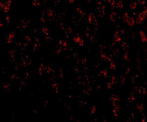
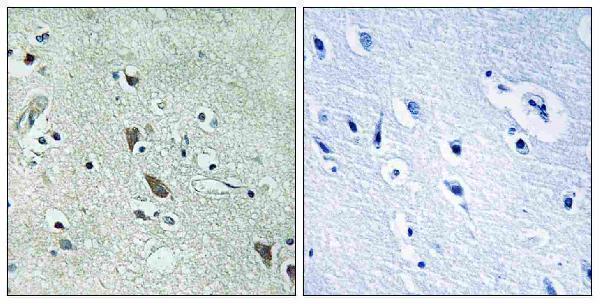
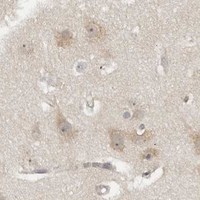


Comprehensive Human Protein Atlas IHC scoring per tissue (reliability: Uncertain — Low consistency between antibody staining and RNA expression data. Pending external verification.). Rows are taken directly from the HPA tissue chart — click any row's HPA link to view the source.
| Tissue | Cell type | Level | Evidence | Source |
|---|---|---|---|---|
| Adrenal gland | Glandular cells | Medium | Protein (IHC) | HPA → |
| Appendix | Glandular cells | Medium | Protein (IHC) | HPA → |
| Bone marrow | Hematopoietic cells | Medium | Protein (IHC) | HPA → |
| Breast | Glandular cells | Medium | Protein (IHC) | HPA → |
| Bronchus | Respiratory epithelial cells | Medium | Protein (IHC) | HPA → |
| Tissue | Cell type | Level | Evidence | Source |
|---|---|---|---|---|
| Heart muscle | Cardiomyocytes | Not detected | Protein (IHC) | HPA → |
| Ovary | Ovarian stroma cells | Not detected | Protein (IHC) | HPA → |
| Skeletal muscle | Myocytes | Not detected | Protein (IHC) | HPA → |
| Soft tissue | Chondrocytes | Not detected | Protein (IHC) | HPA → |
| Spleen | Cells in red pulp | Not detected | Protein (IHC) | HPA → |
Use the catalog antibody’s paraffin-section conditions as a starting point, then interpret staining against HTRA2 localisation and the reported uncertainty in tissue staining (datasheet A01941-3; UniProt O43464; HPA tissue IHC).
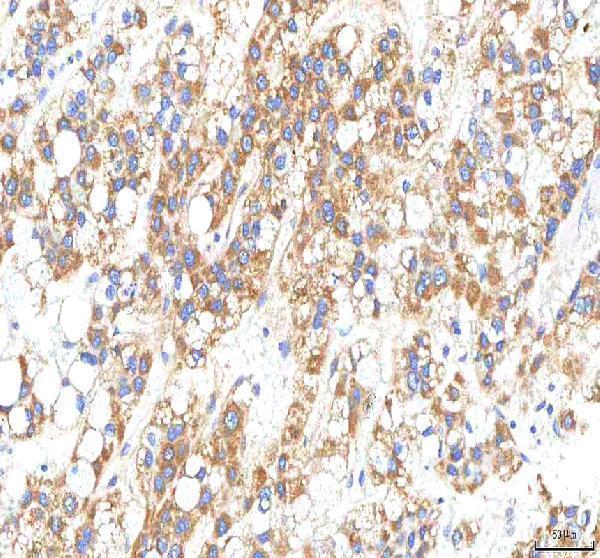
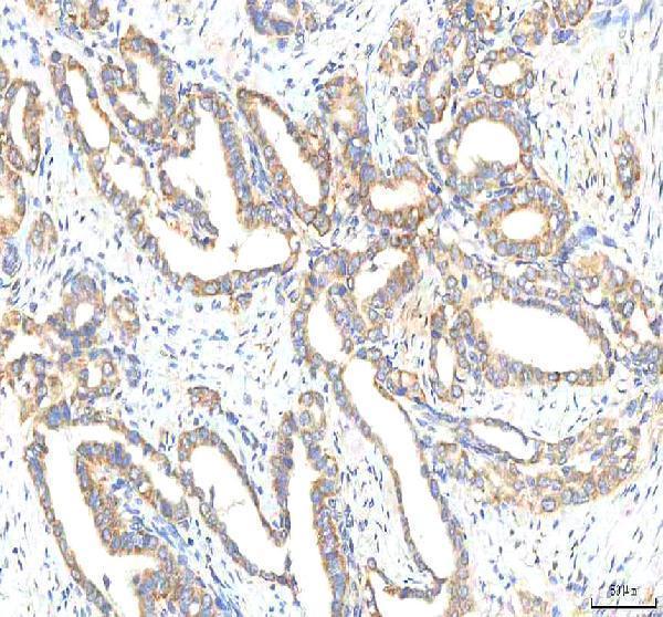
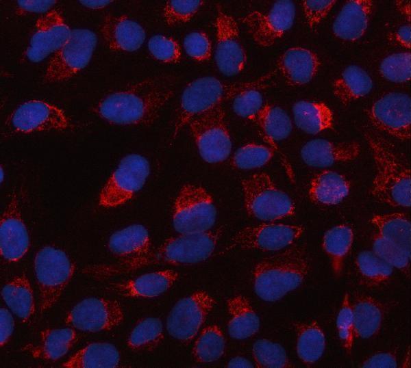
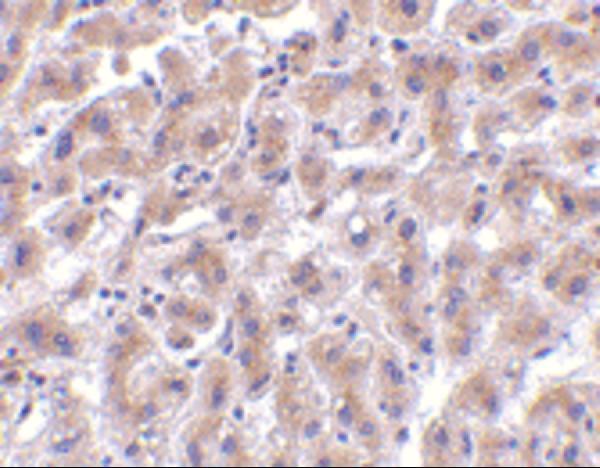
Catalog antibodies have real IHC images from human liver, liver cancer and brain tissue, plus IF images from human liver cells and U2OS cells (catalog image captions).
A01941 shows IHC in human liver tissue and IF in human liver cells; A01941-1 shows IHC in paraffin-embedded human brain tissue with a peptide-blocked comparison (catalog image captions). A01941-3 shows IHC in paraffin-embedded human liver cancer tissue and IF/ICC in U2OS cells (catalog image captions).
Which to pick: For tissue IHC, start with A01941-3 at 2–5 μg/mL: its paraffin-section image documents EDTA retrieval at pH 8.0 and DAB detection (A01941-3 datasheet and IHC caption). For IF/ICC, choose A01941-3 at 5 μg/mL because its application list includes both and its IF image shows U2OS cells; for human liver-cell IF, A01941 has an image at 20 μg/mL (A01941-3 application list and IF caption; A01941 IF caption). For human, mouse or rat reactivity, consider the polyclonal A01941-1, but its IHC image documents only paraffin-embedded human brain tissue; the fixative is unreported in the IHC captions (A01941-1 catalog reactivity, dilution notes and IHC caption; A01941 and A01941-3 IHC captions).