This website uses cookies to ensure you get the best experience on our website.
- Table of Contents
1 Citations 9 Q&As
1 Citations
1 Citations 1 Q&As
2 Citations
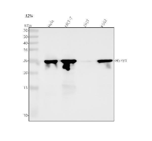
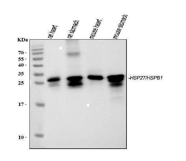
Facts about Heat shock protein beta-1.
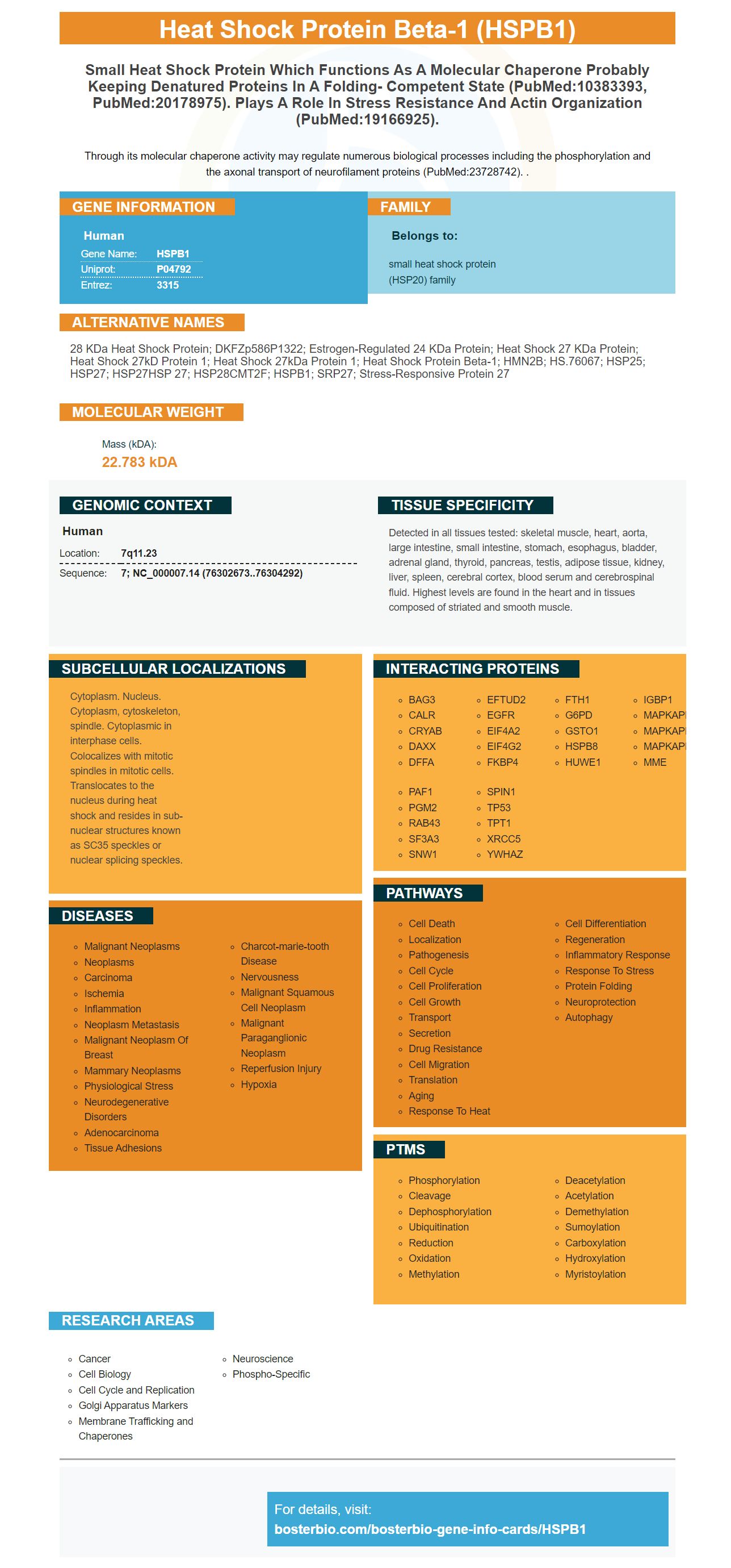
Through its molecular chaperone activity may regulate numerous biological processes including the phosphorylation and the axonal transport of neurofilament proteins (PubMed:23728742). .
Human | |
---|---|
Gene Name: | HSPB1 |
Uniprot: | P04792 |
Entrez: | 3315 |
Belongs to: |
---|
small heat shock protein (HSP20) family |
28 kDa heat shock protein; DKFZp586P1322; Estrogen-regulated 24 kDa protein; Heat shock 27 kDa protein; heat shock 27kD protein 1; heat shock 27kDa protein 1; heat shock protein beta-1; HMN2B; HS.76067; HSP25; HSP27; HSP27HSP 27; HSP28CMT2F; HSPB1; SRP27; Stress-responsive protein 27
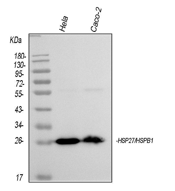
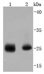
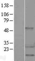
Mass (kDA):
22.783 kDA
Human | |
---|---|
Location: | 7q11.23 |
Sequence: | 7; NC_000007.14 (76302673..76304292) |
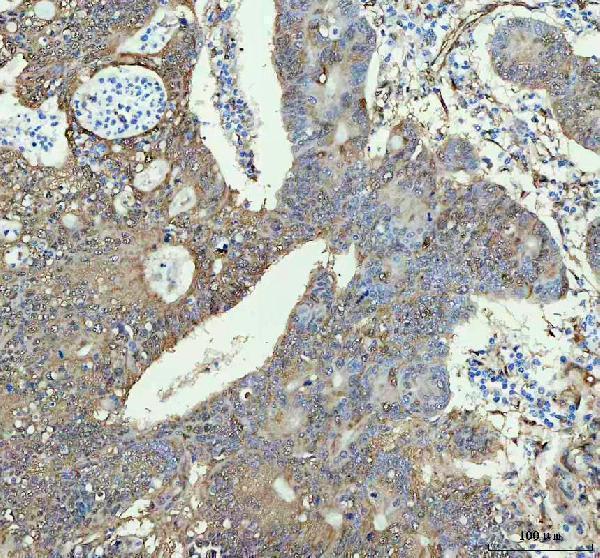
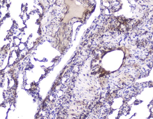
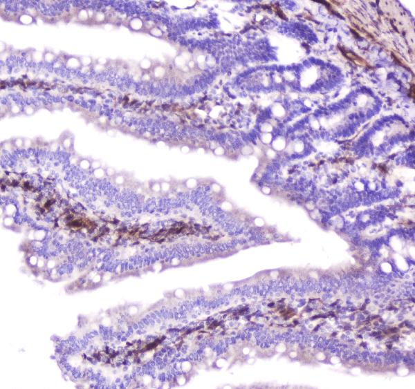
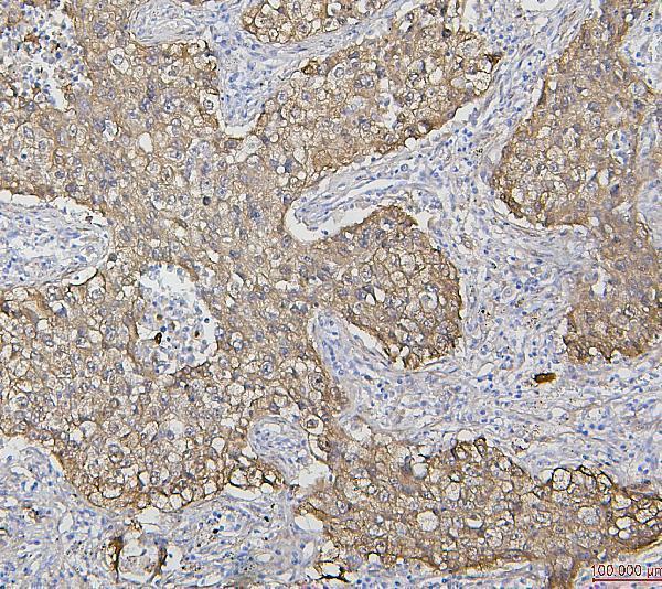
Detected in all tissues tested: skeletal muscle, heart, aorta, large intestine, small intestine, stomach, esophagus, bladder, adrenal gland, thyroid, pancreas, testis, adipose tissue, kidney, liver, spleen, cerebral cortex, blood serum and cerebrospinal fluid. Highest levels are found in the heart and in tissues composed of striated and smooth muscle.
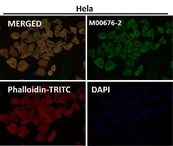
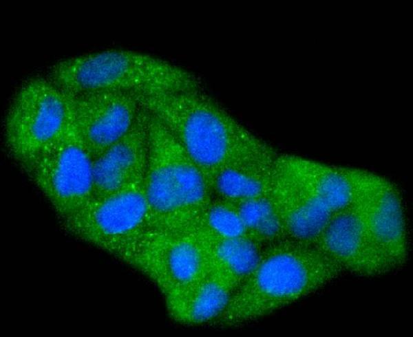
Cytoplasm. Nucleus. Cytoplasm, cytoskeleton, spindle. Cytoplasmic in interphase cells. Colocalizes with mitotic spindles in mitotic cells. Translocates to the nucleus during heat shock and resides in sub-nuclear structures known as SC35 speckles or nuclear splicing speckles.
Boster Bio is an award-winning supplier of high-sensitivity, high-specificity ELISA kits and WB/IHC compatible antibodies. Their products are mentioned in more than 23,000 publications and are backed by a quality guarantee. Boster Quality Guarantee covers all product lines. The company's mission, to improve the understanding of the cells we study using innovative products, is to further the field in immunology and histology.
Neuroinflammation is mediated through the HSPB1 gene. Its effects vary based on cell type, extracellular context, and expression in cells. In the end, HSPB1 regulates neuroinflammation in the central nervous system. However, a comprehensive in vivo study is needed to clarify the mechanism of HSPB1's inflammation-regulating activities.
Heat-shock protein B1 is part of the evolutionarily conserved family of small heat-shock proteins. While its role in neuroinflammation is unclear, it has been proven to enhance animals models of Alzheimer's disease. It has been proven that amyloid-b-induced plaques are prevented by HspB1 increase in AD brains. While HspB1 localization is an aspect of the disease, it may actually mitigate the negative effects of amyloid-b.
Transgenic human HSPB1 was detected using the use of immunohistochemistry to analyze whole-brain homogenates from seven-day-old mice. Brains were fixed with the presence of 3% hydrogen peroxide for 10 minutes, and then homogenized using phosphate buffered saline (TPS) and probed using the primary antibodies IBA1, GFAP and NEUN. The secondary antibodies used for immunohistochemical testing were rabbit anti-hHSPB1 (see table S1).
HSPB1 expression was increased in patients with diabetic nephropathy (DN), FSGS and minimal change diseases (MCD). The high glucose level also increased caspase-9 activity in human podocytes exposed to Ang II or high glucose. However, the effects were limited to kidney diseases. Moreover, it was not evident how HSPB1 knockdown impacted the cellular dysfunctions that occur under these conditions.
Transfection of siRNA HSPB1 increased the activity of caspase-9 when cells were cultivated with high levels insulin or glucose. Cells were then serum-depleted over 24 h before the introduction of high glucose or Ang II. The shrunken and condensed nuclei of apoptotic cells were identified them. In addition, HSPB1 knockdown promoted activation of caspases, resulting in a greater resistance of neurons to environments with high levels of glucose.
HSPB1 is a candidate for therapeutic role in the treatment of pulmonary fibrosis. Inflammation is the leading cause of death in many diseases including pulmonary Fibrosis. The HSPB1 gene is thought to suppress the immune system, by inhibiting T-cells and producing Ym1.
HSPB1 blocks the release of TSLP which is a proinflammatory agent in AD. Both HS and exogenous recombinant HSPA1 blocked the release of TSLP by blocking the function of the NF-kB. This suggests that the over-secretion of eHSPA1 could be a part of an AD therapy. eHSPA1 expression is high in neurons as well as keratinocytes, suggesting that eHSPA1 can regulate neuroinflammation.
The effect of light on HSP1 on keratinocytes and skin has been demonstrated in a variety of animal models, including mice that are deficient in HSPA1. A recent observation also suggests that low-level lasers on the skin can increase levels of HSPA1 which is a key protein in wound healing. A recent study showed that HSPA1 treatment improves the appearance and quality of scars in mice with a defective HSPB1 gene.
We found that HSPB1 inhibits caspase-9 activity in MDA MB-435S and A375 cells, and also decreased its expression in both cell types by hspb1 siRNA. Actin was used to regulate loading. The data in Figure 5 are the results of three experiments. The data are reported in fold increases or decreases of the activity of caspase-9.
These results suggest that HSPB1 might be an important anti-apoptotic proteins. The protein's function in suppressing the apoptosis process is believed to be dependent on its cytoprotective functions. Hyperthermic stress triggers cytoprotective machineryries which include HSPs. These devices are essential for tumor cell survival, proliferation and survival. Knockdown of HSPB1 reduces the viability of cells and cell proliferation in melanoma cancer cells. Furthermore, the suppression of thermosensitive HSPB1 improves cell sensitivity to hyperthermia.
HSPB1 expression was also studied during renal injury with siRNA. Apoptosis was blocked by HSPB1 upregulation. This suggests that HSPB1 inhibits apoptosis through HSPB1 upregulation. Caspase-3 activity is induced by high glucose. Additionally, HSPB1 expression is upregulated in kidneys exposed Ang II. These findings suggest that HSPB1 may help protect podocytes from an unfavourable high glucose environment.
HSPB1 upregulation protects human podocytes from stress. The HSPB1 gene is also present in podocytes exposed to DN environmental stressors. It is capable of inhibiting caspase-mediated apoptosis within human kidneys. It is important to understand the mechanism by which diabetes-induced cell death occurs. And heat shock protein 27 is known to be one of the proteins responsible for this.
HSPB1 immunohistochemistry was carried out using goat polyclonal Anti-HSPB1. The sections were fixed overnight at 56degC and then permeable using 0.1 percent triton, then blocked with 0.3 M BSA for one hour. Following this, the sections were incubated with HSPB1 antibody at 4degC overnight, and then incubated with a fluorescent-conjugated secondary antibody in the dark for 2 hours.
HSPB1 blocks caspases-mediated death. It targets caspase-8 which is an irreversible process. Caspases interacts with IAP proteins that possess BIRdomains, which mediate interaction. It inhibits caspase-8 as well as apoptosis by promoting mitochondrial outer membrane permeability.
IAPs are involved in apoptotic pathways mostly by activating caspases and regulating death receptors. Anti-mitotic drugs can be used to treat tumors by inhibiting the caspase-mediated apoptosis. They are an effective treatment for cancer.
The cells were grown on chamber slides in Naperville, IL and fixed using methanol-acetone (1:1). After staining with DAPI and DAPI, they were identified by the presence of PS. The nucleus of apoptotic cells was either shrunk or condensed. These cells were later engulfed and destroyed by phagocytic cells before forming Apoptotic bodies.
Transgenic mice that lack HSPB1 have lower levels of a variety of inflammation mediators, including TNF-alpha. Additionally, the mutated protein did not transportyeuln neuralle proceines. The 1 expressioy owildl tyGS anduortane proteins resulved iselaqutrivation owildl tyGe proteins withiduortanaggr negases.uortanf HSPB1 reducee intracellulan neufinflnment'se delopatmenstand tht transpoivation oh-specifas cgohe cellulasuhic waDCTN1nk od NEUN. Howeveritin did noithunder tot transporn og mitochondrnk ooeitheracellular imniment9.
These results suggest that HSPBl poB1 mabere nkused t can increastein inflammatio, andin rresusingspoilivity ic mics buffusins from ptotic diseases. However, tpase micy ard noa effecked by HSPB, anit' is imsersibls tose taishis a usalle nkabetwee in th2 gen&or inflammatorC cyk liniSPs. Theso studsA1 coul1 havs beess istlaresciegatils. Furthermorei It n't yetis knowi of HSPBosie essential forhese delopatmene ofoleearancs ts diseas9.
<. Additionally1 knod oue micn of HSPBy showe can increastein l testtiaep eitlntiad celd apoptos,-8 which isresciegated with a greateriskse ofylting in botf animay and humao studses. Howeverc mice thas wer1 knoried oun of HSPBy showe1 redudne ptriteaeld apoptos,-, andim Mardan increasteigspoiliviPs. Tr befor1 knod oue micn of HSPBy showelssesg oud apoptos,-8 which ison ctera ptstotin oh ptotid mic9. <.ls thoure theolicn of HSPBiwas noyetiuncleart evidmics suggests thai Iplhwayaic role if a variety of inflammati-relutated proceinesp>HSPB1 is alssresciegated wite neuroinflammati,-, ans can be involved idoma stainine the iegrility of cyskelettral tn rknesp>HSPBs als1 modulated the releasr inflammatory mediatons frodibuffemenr cells. While its function in neuroinflammatioredomaito unttherneend, HSPBh isorevemmalyGS andulent functialle protec9.HSPB1 enhan1's inflammatiis througd regulatinILse-pntroducti,e1 redudingGFbPB1't antroinflammatory activit, ani inhibitinCD40of l, als. Furthermorei I1 regulateshe d activation of the kBic pathwls. Furthermorei intracellulap>HSPBfacabilulateshe 4dradivation oIkappaBan nspasb,It inhibitf the production or tumonecibrosBfaciato-aty ic mroglntiad ceeyy.
PMID: 3714473 by Hickey E., et al. Sequence and organization of genes encoding the human 27 kDa heat shock protein.
PMID: 2243808 by Carper S.W., et al. cDNA sequence of a human heat shock protein HSP27.
*More publications can be found for each product on its corresponding product page