This website uses cookies to ensure you get the best experience on our website.


- Table of Contents
Plan ID3 paraffin IHC around mainly nuclear staining in most tissues (HPA tissue IHC). Compare high staining in adrenal gland glandular cells with undetected prostate glandular cells (HPA tissue IHC), and optimize the catalog antibody within 1:50–1:200 (datasheet).

Expected localisation, validated protocols, controls and antibodies — the at-a-glance facts below, then the full design guide.
| Expected localisation | Mainly nuclear staining in most tissues (HPA tissue IHC) | |
| Staining pattern | Most tissue cell types show mainly nuclear staining (HPA tissue IHC) | |
| Antigen retrieval | Tris-EDTA pH 9.0 HIER, 95–98 °C, 20 min (rule: nuclear antigen) | |
| Positive control | Adrenal gland+4 more · see all | |
| Negative control | Heart muscle+3 more · see all |
| Fixation | Keep fixation consistent across sections (standard IHC practice; not target-specific) | |
| Caveat | Staining shows low consistency with RNA levels (HPA tissue IHC) | |
| Regulation | Expression varies by tissue (UniProt) | |
| Isoform / epitope | No annotated isoforms or processing; one 1–119 chain (UniProt) |
The catalog antibody’s IHC protocol is followed by four published ID3 IHC protocols covering lung, vascular, pancreatic and thyroid specimens (PMC3567999; PMC4121297; PMC10361912; PMC8405699).
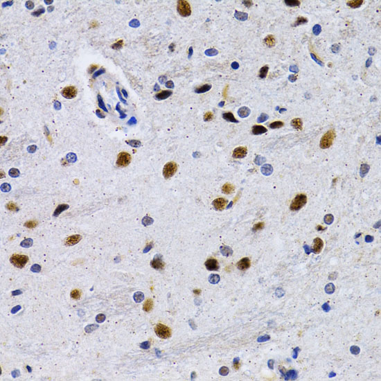
| Sample | Paraffin-embedded rat brain tissue; fixative not specified (datasheet A01379) |
| Fixation | Image fixative and duration unreported (datasheet A01379); verify before use. |
| Sectioning | 4–5 µm sections on charged slides (standard) |
| Deparaffinisation | Xylene, graded ethanol series to water (standard) |
| Antigen retrieval | Heat-induced epitope retrieval in Tris-EDTA buffer, pH 9.0, 20 min at 95–98 °C (standard rule: nuclear antigen) |
| Peroxidase block | 3% H2O2, 10 min, room temperature (standard) |
| Blocking | 10% normal serum of the secondary host, 30 min, room temperature (standard) |
| Primary antibody | Rabbit anti-ID3, 1:50-1:200 (datasheet A01379) |
| Primary incubation | Overnight at 4 °C (standard) |
| Detection | HRP-polymer secondary, DAB chromogen 5–10 min (standard) |
| Counterstain | Hematoxylin, blue, dehydrate and mount (standard) |
| Expected result | ID3-positive staining in glandular cells of adrenal gland (HPA tissue IHC: High). HPA tissue profile: Mainly nuclear expression in most tissues. No signal in the no-primary control. |
ID3 is a nuclear protein with no transmembrane segment (UniProt Q02535: subcellular location and topology). In paraffin sections, expect mainly nuclear staining in cell types reported by HPA, including adrenal glandular cells, bronchial basal cells and colon endothelial cells (HPA tissue IHC). HPA rates its tissue IHC evidence Approved but reports low consistency between antibody staining and RNA expression; interpret unexpected patterns cautiously (HPA tissue IHC).
| Distinct nuclear staining in adrenal glandular cells, bronchial basal cells or colon endothelial cells. | These cell types have High staining in HPA tissue IHC, which describes expression as mainly nuclear. Assess the labelled cells and compartment together; staining elsewhere in the same tissue does not establish the expected pattern (HPA tissue IHC). |
| Predominantly cytoplasmic or membranous staining, with little nuclear signal. | That distribution conflicts with the reported nuclear location and lack of a transmembrane segment (UniProt Q02535) and HPA's mainly nuclear tissue pattern (HPA tissue IHC). Consider nonspecific staining or a detection artefact; compartment alone cannot identify its cause. |
| Strong staining in a cell type HPA reports as Not detected, such as cardiomyocytes. | This conflicts with the cell-specific HPA observation for heart muscle (HPA: cardiomyocytes Not detected). Check whether the signal follows tissue structures or appears in the negative control; cross-reactivity or endogenous detection activity are possibilities (general IHC practice). |
| Diffuse colour across nuclei, cytoplasm and spaces between cells. | Diffuse staining cannot be scored as the mainly nuclear ID3 pattern (HPA tissue IHC; UniProt Q02535: nucleus). Compare with a section processed without primary antibody; widespread colour may reflect background from the detection system (general IHC practice). |
| No nuclear signal in an adrenal gland section. | HPA reports High staining in adrenal glandular cells (HPA tissue IHC). Confirm those cells are present and the detection run worked before interpreting absence as biological; HPA's Approved rating has a low RNA-to-staining consistency caveat (HPA tissue IHC). |
| Cell-specific tissue context | HPA reports High staining in adrenal glandular cells, bronchial basal cells and colon endothelial cells, but Not detected in cardiomyocytes and prostate glandular cells (HPA tissue IHC). Judge the relevant cell population, not a whole-section average. |
| Strength of tissue evidence | The tissue IHC profile is Approved, with low consistency between antibody staining and RNA expression (HPA tissue IHC). Treat an unexpected positive or negative as a finding to check against controls, not a definitive expression call. |
| Antibody-specific validation | HPA lists HPA024677 and CAB021353 as IHC Approved; neither is listed as IHC Enhanced in the supplied antibody record (HPA antibodies). An Approved label does not by itself show that a particular slide's staining is specific. |
| Protein structure | UniProt records one chain spanning residues 1–119, no listed isoforms, no signal peptide and no transmembrane segment (UniProt Q02535). These annotations support assessing nuclear localisation; they do not establish how fixation affects an epitope. |
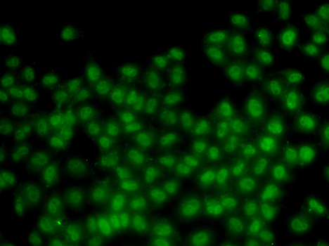
| What should IF/ICC show? | HPA reports mainly nucleoplasmic localisation in ICC-IF and lists CAB021353 as ICC Supported (HPA subcellular; HPA antibodies). This supports a nuclear localisation check, but the tissue IHC profile remains the basis for paraffin-section expectations (HPA tissue IHC). |
| Situation | Likely cause | Next action |
|---|---|---|
| Expected nuclear staining is absent in adrenal glandular cells. | A failed staining run or poor visibility of the relevant cells can resemble a negative result (general IHC practice); HPA reports High staining in these cells (HPA tissue IHC). | Confirm cell identity and morphology, review a working positive control from the same run, then check the recorded retrieval, primary-antibody and detection steps against the chosen IHC procedure (general IHC practice). |
| Cytoplasmic colour dominates an otherwise stained section. | The compartment conflicts with nuclear ID3 localisation (UniProt Q02535; HPA tissue IHC). Background or cross-reactivity may contribute (general IHC practice). | Compare the suspect cells with a no-primary control and the nuclear pattern in a known-positive cell population; review blocking and detection conditions if background persists (general IHC practice). |
| Colour appears in cardiomyocytes or prostate glandular cells. | HPA reports ID3 as Not detected in those specific cell types (HPA tissue IHC). Endogenous detection activity or antibody cross-reactivity are possible explanations (general IHC practice). | Verify the cell type, inspect a matched no-primary control and review the detection system's endogenous-activity blocking step; do not assign the colour to ID3 from location alone (general IHC practice). |
| Nearly every structure carries faint colour. | Widespread colour can obscure the mainly nuclear tissue pattern (HPA tissue IHC). Inadequate blocking, washing or detection conditions can increase general background (general IHC practice). | Compare a no-primary section, inspect washes and blocking, and adjust detection or primary-antibody conditions according to the validated IHC procedure before scoring nuclei (general IHC practice). |
| A tissue result disagrees with an RNA-based expectation. | HPA explicitly reports low consistency between antibody staining and RNA expression despite an Approved tissue IHC rating (HPA tissue IHC). RNA and staining should therefore be interpreted as separate observations. | Record the observed cell type, compartment and controls; compare the result with HPA's cell-specific staining entries and avoid treating either assay alone as proof of the other (HPA tissue IHC; general IHC practice). |
| A proposed IF/ICC image is used to judge a paraffin-section result. | HPA's supported nucleoplasmic ICC-IF localisation and Approved tissue IHC profile describe different assay settings (HPA subcellular; HPA tissue IHC). | Use IF/ICC evidence as a compartment check, then judge the paraffin section against its own cell-specific tissue pattern and IHC controls (HPA subcellular; HPA tissue IHC; general IHC practice). |


Comprehensive Human Protein Atlas IHC scoring per tissue (reliability: Approved — Low consistency between antibody staining and RNA expression data.). Rows are taken directly from the HPA tissue chart — click any row's HPA link to view the source.
| Tissue | Cell type | Level | Evidence | Source |
|---|---|---|---|---|
| Adrenal gland | Glandular cells | High | Protein (IHC) | HPA → |
| Appendix | Glandular cells | High | Protein (IHC) | HPA → |
| Bone marrow | Hematopoietic cells | High | Protein (IHC) | HPA → |
| Breast | Glandular cells | High | Protein (IHC) | HPA → |
| Bronchus | Basal cells | High | Protein (IHC) | HPA → |
| Tissue | Cell type | Level | Evidence | Source |
|---|---|---|---|---|
| Heart muscle | Cardiomyocytes | Not detected | Protein (IHC) | HPA → |
| Liver | Cholangiocytes | Not detected | Protein (IHC) | HPA → |
| Prostate | Glandular cells | Not detected | Protein (IHC) | HPA → |
| Seminal vesicle | Glandular cells | Not detected | Protein (IHC) | HPA → |
Troubleshoot chromogenic ID3 IHC in paraffin sections by checking retrieval, nuclear localisation, controls and cell-specific scoring (UniProt Q02535; HPA tissue IHC).
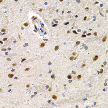
A01379 has anti-ID3 IHC images from paraffin-embedded rat brain, mouse pancreas, and human lung cancer (catalog IHC captions), plus IF data from HeLa cells (catalog IF caption).
A01379 has IHC images of paraffin-embedded rat brain, mouse pancreas, and human lung cancer (catalog IHC captions). It is listed for IHC, IF, and ICC across human, mouse, and rat, with an IF image from HeLa cells (catalog applications; catalog reactivity; catalog IF caption).
Which to pick: For tissue IHC, choose A01379: its paraffin-section captions use 1:200 antibody after microwave retrieval in 10 mM PBS at pH 7.2; the fixative is unreported (catalog IHC captions). For IF/ICC, choose A01379, a rabbit polyclonal listed for both applications with IF data from HeLa cells (catalog host; catalog dilution_raw; catalog applications; catalog IF caption). For work across species, A01379 lists human, mouse, and rat reactivity and has IHC images from each (catalog reactivity; catalog IHC captions).