This website uses cookies to ensure you get the best experience on our website.


- Table of Contents
Plan IDH2 staining in paraffin sections around the granular cytoplasmic pattern observed in tissue (HPA tissue IHC) and its mitochondrial location (UniProt). Compare cell types using matched controls; colon glandular cells provide a high-staining reference (HPA tissue IHC).

Expected localisation, validated protocols, controls and antibodies — the at-a-glance facts below, then the full design guide.
| Expected localisation | Granular cytoplasm (HPA tissue IHC) | |
| Staining pattern | Granular cytoplasmic staining across multiple cell types (HPA tissue IHC) | |
| Antigen retrieval | EDTA pH 8.0 HIER, heat-mediated (datasheet M00510-3) | |
| Positive control | Appendix+4 more · see all | |
| Negative control | Epididymis+4 more · see all |
| Fixation | Matched tissue-IHC evidence does not establish the fixation claim. Validate the specimen-specific method before use. (selected-SKU IHC image M00510-3) | |
| Caveat | Colon glands stain high; smooth muscle cells are undetected (HPA tissue IHC) | |
| Regulation | No expression regulator annotated (UniProt) | |
| Isoform / epitope | 2 isoforms; mature chain spans 40–452; verify epitope (UniProt) |
The catalog antibody’s IHC-P protocol (datasheet: M00510-3) is accompanied by four published protocols using mutation-specific IDH2 antibodies (PMC7286791; PMC7429919; PMC3892385; PMC7721993).
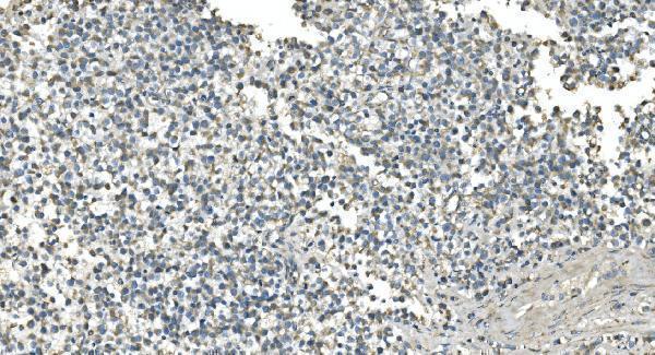
| Sample | Paraffin-embedded human lung cancer tissue; fixative not specified (datasheet M00510-3) |
| Fixation | Image fixative and duration unreported (datasheet M00510-3); verify before use. |
| Sectioning | 4–5 µm sections on charged slides (standard) |
| Deparaffinisation | Xylene, graded ethanol series to water (standard) |
| Antigen retrieval | Heat retrieval: EDTA pH 8.0 (datasheet M00510-3); 20 min, 95–100 °C (standard) |
| Peroxidase block | 3% H2O2, 10 min, room temperature (standard) |
| Blocking | 10% goat serum (datasheet M00510-3) |
| Primary antibody | Mouse monoclonal (clone 2H4) anti-IDH2, 2μg/ml (datasheet M00510-3) |
| Primary incubation | Overnight at 4 °C (datasheet M00510-3) |
| Detection | Streptavidin-biotin complex (SABC), DAB chromogen (datasheet M00510-3) |
| Counterstain | Hematoxylin, blue, dehydrate and mount (standard) |
| Expected result | IDH2-positive staining in glandular cells of appendix (HPA tissue IHC: High). HPA tissue profile: General cytoplasmic expression with a granular pattern. No signal in the no-primary control. |
IDH2 is mitochondrial and has no transmembrane segment (UniProt P48735). In tissue IHC, expect granular cytoplasmic staining, including high signal in appendix and colon glandular cells, bronchial respiratory epithelium, and bone marrow hematopoietic cells (HPA: tissue IHC). HPA rates the tissue pattern Enhanced, while reporting medium consistency between staining and RNA expression (HPA: tissue IHC). Interpret intensity by cell type, not by whole-tissue label (HPA: tissue IHC).
| Granular cytoplasmic signal in appendix or colon glandular cells. | This matches the reported distribution and high staining level in those cells (HPA: tissue IHC). Mitochondrial localization supports a punctate or granular cytoplasmic interpretation (UniProt P48735; HPA: subcellular ICC-IF). |
| Predominantly nuclear or sharply plasma-membrane staining. | Treat this as a localization mismatch requiring verification, because IDH2 is mitochondrial and has no transmembrane segment (UniProt P48735; HPA: subcellular ICC-IF). Review morphology and detection controls before attributing it to IDH2 (general IHC practice). |
| Strong signal in liver cholangiocytes, ovarian stroma cells, or smooth muscle cells. | Those specified cell populations were reported as not detected (HPA: tissue IHC). Unexpected staining may reflect cross-reactivity, endogenous detection activity, or a technical difference; the HPA observations alone cannot identify the cause (HPA: tissue IHC; general IHC practice). |
| Uniform haze across cells and tissue spaces, with little cellular granularity. | The appearance is less consistent with the reported granular cytoplasmic pattern (HPA: tissue IHC). Check background controls and tissue morphology before scoring weak haze as IDH2 positive (general IHC practice). |
| No signal in appendix or colon glandular cells while tissue morphology is preserved. | These are high-staining cell populations in the HPA tissue set (HPA: tissue IHC). A blank result warrants a detection and tissue-control check; differences between antibodies or preparations can still affect whether that reference pattern is reproduced (HPA: tissue IHC; general IHC practice). |
| Cell-resolved tissue reference | HPA reports high staining in appendix, cervix, colon, and duodenum glandular cells, bronchial respiratory epithelial cells, and bone marrow hematopoietic cells; it reports cholangiocytes and ovarian stroma cells as not detected (HPA: tissue IHC). Compare like cell populations when selecting controls. |
| Localization and processing | IDH2 is mitochondrial, lacks a transmembrane segment, and has an annotated mature chain at residues 40–452 (UniProt P48735). These facts guide compartment interpretation but do not specify the antibody epitope or predict antigen-retrieval performance. |
| Evidence and antibody scope | HPA labels tissue IHC reliability Enhanced and notes medium consistency with RNA data (HPA: tissue IHC). Its listed rabbit polyclonal antibody, HPA007831, has Enhanced IHC and Supported ICC validation (HPA: antibodies). Those ratings describe the assessed evidence, not every antibody or specimen. |
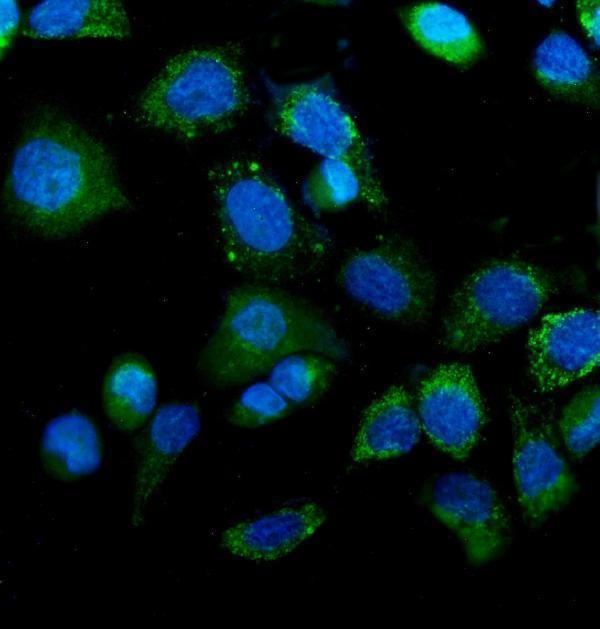
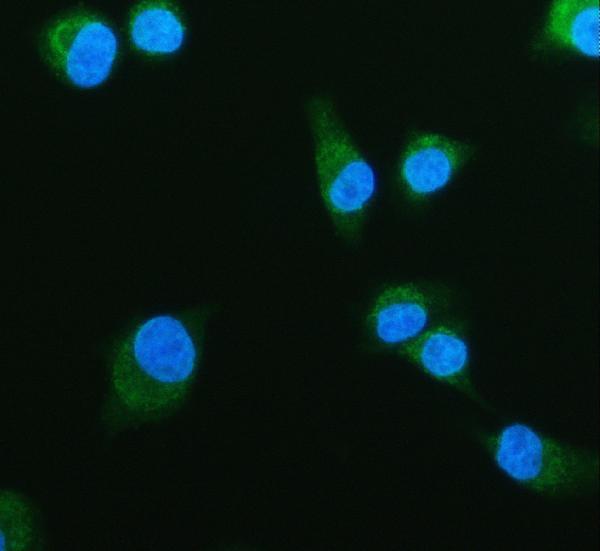
| IF/ICC Q: What localization should an IF image show? | A: Mitochondrial staining is supported by HPA ICC-IF images from A-431, U-251MG, and U2OS, consistent with UniProt localization (HPA: subcellular ICC-IF; UniProt P48735). This supports a compartment check; the paraffin-section IHC pattern remains the primary reference here (HPA: tissue IHC). |
| Situation | Likely cause | Next action |
|---|---|---|
| Known high-staining glandular cells are blank. | The observed result conflicts with the HPA reference for appendix or colon glands, but does not by itself identify a failed step (HPA: tissue IHC). | Confirm the intended cell population and tissue preservation; review the IHC-validated antibody's stated IHC-P conditions and run an appropriate positive control (general IHC practice). |
| Signal is diffuse throughout cytoplasm without visible granularity. | That pattern is less specific than HPA's general granular cytoplasmic description; diffuse background is one possibility (HPA: tissue IHC; general IHC practice). | Check a no-primary control, blocking, washes, and detection conditions; score localization only where cellular detail remains clear (general IHC practice). |
| Nuclei dominate the chromogenic signal. | Nuclear dominance disagrees with mitochondrial localization (UniProt P48735; HPA: subcellular ICC-IF). Counterstain overlap or nonspecific detection may complicate interpretation (general IHC practice). | Compare with the no-primary control and inspect whether granular cytoplasmic signal is independently visible before calling cells positive (general IHC practice). |
| A cell population reported as not detected stains strongly. | For example, cholangiocytes are listed as not detected, so strong signal needs review; HPA does not establish its mechanism (HPA: tissue IHC). | Verify cell identity, inspect a no-primary control for endogenous detection activity, and compare a known high-staining population on the same run (HPA: tissue IHC; general IHC practice). |
| Color develops broadly, including outside identifiable cells. | A widespread reaction can arise from detection-system background or endogenous activity (general IHC practice); it does not resemble HPA's cellular granular pattern (HPA: tissue IHC). | Review no-primary and detection controls, blocking, and wash steps; do not assign extracellular color to IDH2 (general IHC practice). |
| The chosen reference tissue gives mixed positive and negative cells. | HPA levels are attached to specified cell types, and its tissue summary describes general rather than universal cytoplasmic expression (HPA: tissue IHC). | Score the annotated cell population separately and compare its location and intensity with the relevant HPA entry; avoid treating the entire section as one control (HPA: tissue IHC; general IHC practice). |


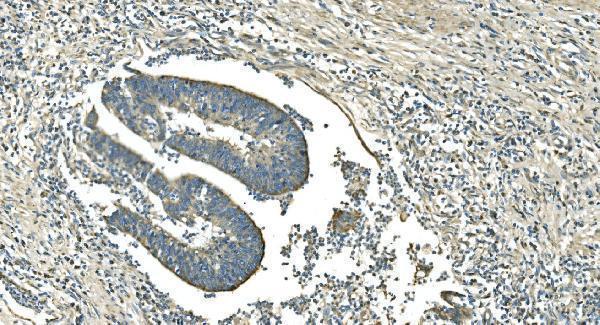
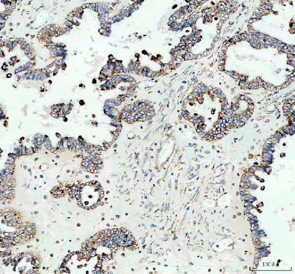
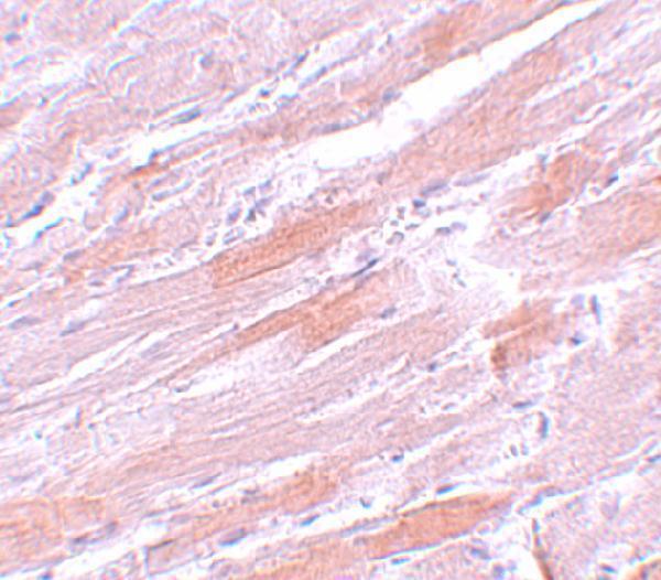
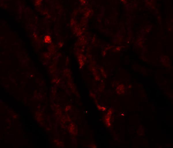


Comprehensive Human Protein Atlas IHC scoring per tissue (reliability: Enhanced — Medium consistency between antibody staining and RNA expression data.). Rows are taken directly from the HPA tissue chart — click any row's HPA link to view the source.
| Tissue | Cell type | Level | Evidence | Source |
|---|---|---|---|---|
| Appendix | Glandular cells | High | Protein (IHC) | HPA → |
| Bone marrow | Hematopoietic cells | High | Protein (IHC) | HPA → |
| Bronchus | Respiratory epithelial cells | High | Protein (IHC) | HPA → |
| Cerebellum | GLUC cells - cytoplasm/membrane | High | Protein (IHC) | HPA → |
| Cervix | Glandular cells | High | Protein (IHC) | HPA → |
| Tissue | Cell type | Level | Evidence | Source |
|---|---|---|---|---|
| Epididymis | Glandular cells | Not detected | Protein (IHC) | HPA → |
| Liver | Cholangiocytes | Not detected | Protein (IHC) | HPA → |
| Ovary | Ovarian stroma cells | Not detected | Protein (IHC) | HPA → |
| Smooth muscle | Smooth muscle cells | Not detected | Protein (IHC) | HPA → |
| Spleen | Cells in red pulp | Not detected | Protein (IHC) | HPA → |
Troubleshoot IDH2 staining by checking retrieval, cell compartment and background before comparing signal across paraffin sections (UniProt P48735; HPA tissue IHC).
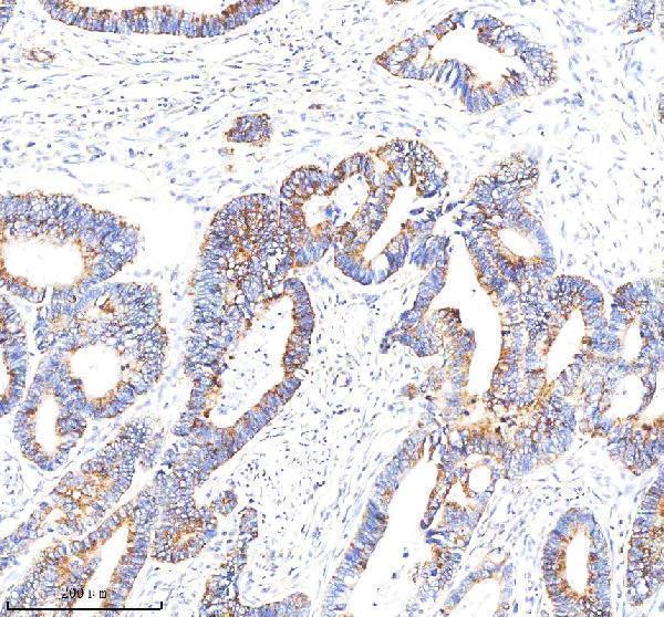
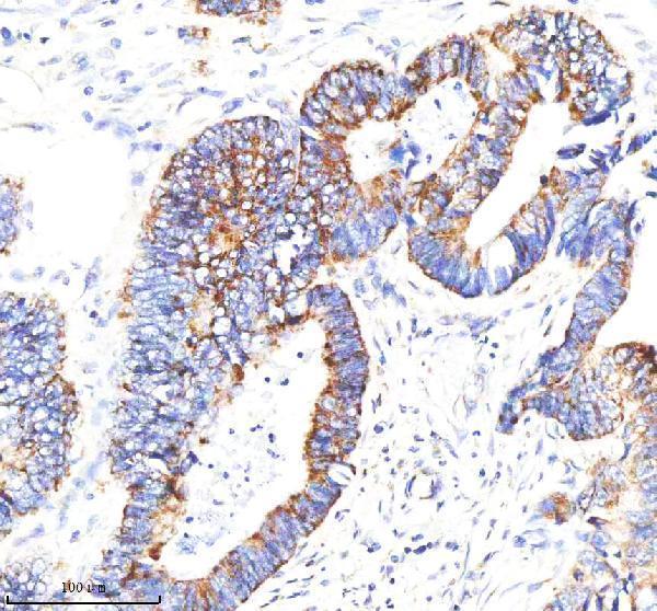
Anti-IDH2 antibodies have IHC images from human cancer sections and mouse heart, plus IF images from A431 cells, A549 cells, and mouse heart (catalog image captions).
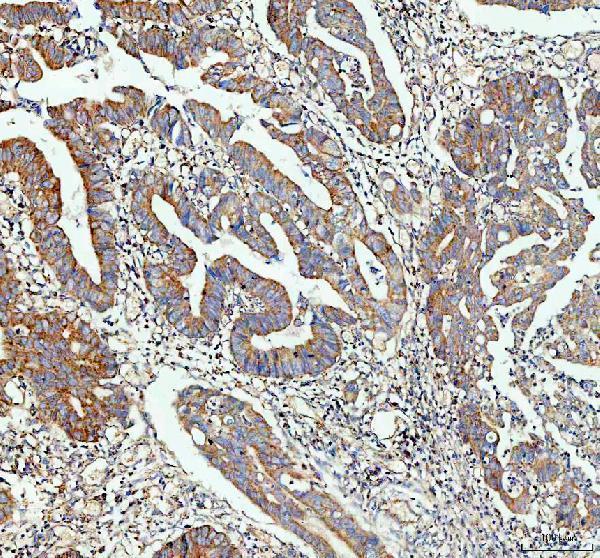
M00510-3 has IHC images from human lung cancer sections, M00510-4 from human testicular cancer sections, PB9602 and M00510 from human colon cancer sections, and A00510 from mouse heart (each SKU’s IHC image caption). M00510-2 has an IF image from A431 cells (M00510-2 IF image caption); M00510-4 also has an A431 IF image, PB9602 has an A549 IF image, and A00510 has a mouse heart IF image (each SKU’s IF image caption).
Which to pick: For tissue IHC, PB9602 is a useful starting choice because its own images show paraffin sections of human colon cancer and mouse and rat heart (PB9602 IHC image captions). For IF/ICC, choose M00510-2 for its A431 cell IF example (M00510-2 IF image caption), or M00510-4 if both IHC and IF/ICC are needed (M00510-4 application list and image captions). For work across the three listed species, PB9602 has human, mouse, and rat IHC images (PB9602 IHC image captions); its paraffin section captions specify EDTA pH 8.0 retrieval, but do not report the fixative (PB9602 IHC image captions).