Click image to see more details
-

-
-
-
-
+2
Product Info Summary
| SKU: | PB9602 |
|---|---|
| Size: | 100 μg/vial |
| Reactive Species: | Human, Mouse, Rat |
| Host: | Rabbit |
| Application: | IF, IHC, ICC, WB |
Customers Who Bought This Also Bought

Product info
Product Name
Anti-IDH2 Antibody Picoband®
SKU/Catalog Number
PB9602
Size
100 μg/vial
Form
Lyophilized
Description
Boster Bio Anti-IDH2 Antibody Picoband® catalog # PB9602. Tested in IF, IHC, ICC, WB applications. This antibody reacts with Human, Mouse, Rat. The brand Picoband indicates this is a premium antibody that guarantees superior quality, high affinity, and strong signals with minimal background in Western blot applications. Only our best-performing antibodies are designated as Picoband, ensuring unmatched performance.
Storage & Handling
Store at -20˚C for one year from date of receipt. After reconstitution, at 4˚C for one month. It can also be aliquotted and stored frozen at -20˚C for six months. Avoid repeated freeze-thaw cycles.
Cite This Product
Anti-IDH2 Antibody Picoband® (Boster Biological Technology, Pleasanton CA, USA, Catalog # PB9602)
Host
Rabbit
Contents
Each vial contains 4 mg Trehalose, 0.9 mg NaCl and 0.2 mg Na2HPO4.
Clonality
Polyclonal
Isotype
Rabbit IgG
Immunogen
A synthetic peptide corresponding to a sequence at the C-terminus of human IDH2, identical to the related mouse and rat sequences.
Cross-reactivity
No cross-reactivity with other proteins
Reactive Species
PB9602 is reactive to IDH2 in Human, Mouse, Rat
Observed Molecular Weight
45 kDa
Calculated molecular weight
50.9 kDa
Background of IDH2
Isocitrate dehydrogenase [NADP], mitochondrial is an enzyme that in humans is encoded by the IDH2 gene. Isocitrate dehydrogenases catalyze the oxidative decarboxylation of isocitrate to 2-oxoglutarate. These enzymes belong to two distinct subclasses, one of which utilizes NAD (+) as the electron acceptor and the other NADP (+). Five isocitrate dehydrogenases have been reported: three NAD (+)-dependent isocitrate dehydrogenases, which localize to the mitochondrial matrix, and two NADP (+)-dependent isocitrate dehydrogenases, one of which is mitochondrial and the other predominantly cytosolic. Each NADP (+)-dependent isozyme is a homodimer. The protein encoded by this gene is the NADP (+)-dependent isocitrate dehydrogenase found in the mitochondria. It plays a role in intermediary metabolism and energy production. This protein may tightly associate or interact with the pyruvate dehydrogenase complex. Alternative splicing results in multiple transcript variants.
Antibody Validation
Boster validates all antibodies on WB, IHC, ICC, Immunofluorescence, and ELISA with known positive control and negative samples to ensure specificity and high affinity, including thorough antibody incubations.
Application & Images
Applications
PB9602 is guaranteed for IF, IHC, ICC, WB Boster Guarantee
Recommend Dilution
| Application | Dilution | Species |
|---|---|---|
| Western blot | 0.1-0.5μg/ml | |
| Immunohistochemistry (Paraffin-embedded Section) | 2-5μg/ml | |
| Immunocytochemistry/Immunofluorescence | 5μg/ml |
Tested application
Suggested blocking solution with 5% non-fat milk or BSA; (*)Recommended protein loading: 20-40 µg per lane
Use TE buffer pH 9.0 for antigen retrieval; (*) citrate buffer pH 6.0 is an alternative.
Validation Images & Assay Conditions
Click image to see more details
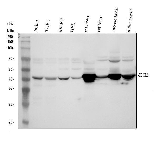
Western blot analysis of IDH2 using anti-IDH2 antibody (PB9602).
Electrophoresis was performed on a 10% SDS-PAGE gel at 80V (Stacking gel) / 120V (Resolving gel) for 2-3 hours. The sample well of each lane was loaded with 30 ug of sample under reducing conditions.
Lane 1: human Jurkat whole cell lysates,
Lane 2: human THP-1 whole cell lysates,
Lane 3: human MCF-7 whole cell lysates,
Lane 4: human HEL whole cell lysates,
Lane 5: rat heart tissue lysates,
Lane 6: rat liver tissue lysates,
Lane 7: mouse heart tissue lysates,
Lane 8: mouse liver tissue lysates.
After electrophoresis, proteins were transferred to a nitrocellulose membrane at 150 mA for 50-90 minutes. Blocked the membrane with 5% non-fat milk/TBS for 1.5 hour at RT. The membrane was incubated with rabbit anti-IDH2 antigen affinity purified polyclonal antibody (Catalog # PB9602) at 0.5 μg/mL overnight at 4°C, then washed with TBS-0.1%Tween 3 times with 5 minutes each and probed with a goat anti-rabbit IgG-HRP secondary antibody at a dilution of 1:5000 for 1.5 hour at RT. The signal is developed using an ECL Plus Western Blotting Substrate (Catalog # AR1196-200) with Tanon 5200 system. A specific band was detected for IDH2 at approximately 45 kDa. The expected band size for IDH2 is at 45, 50 kDa.

Click image to see more details
IHC analysis of IDH2 using anti-IDH2 antibody (PB9602).
IDH2 was detected in a paraffin-embedded section of human colon cancer tissue. Heat mediated antigen retrieval was performed in EDTA buffer (pH 8.0, epitope retrieval solution). The tissue section was blocked with 10% goat serum. The tissue section was then incubated with 2 μg/ml rabbit anti-IDH2 Antibody (PB9602) overnight at 4°C. Peroxidase Conjugated Goat Anti-rabbit IgG was used as secondary antibody and incubated for 30 minutes at 37°C. The tissue section was developed using HRP Conjugated Rabbit IgG Super Vision Assay Kit (Catalog # SV0002) with DAB as the chromogen.

Click image to see more details
IHC analysis of IDH2 using anti-IDH2 antibody (PB9602).
IDH2 was detected in a paraffin-embedded section of human ovarican cancer tissue. Heat mediated antigen retrieval was performed in EDTA buffer (pH 8.0, epitope retrieval solution). The tissue section was blocked with 10% goat serum. The tissue section was then incubated with 2 μg/ml rabbit anti-IDH2 Antibody (PB9602) overnight at 4°C. Peroxidase Conjugated Goat Anti-rabbit IgG was used as secondary antibody and incubated for 30 minutes at 37°C. The tissue section was developed using HRP Conjugated Rabbit IgG Super Vision Assay Kit (Catalog # SV0002) with DAB as the chromogen.

Click image to see more details
IHC analysis of IDH2 using anti-IDH2 antibody (PB9602).
IDH2 was detected in a paraffin-embedded section of mouse heart tissue. Heat mediated antigen retrieval was performed in EDTA buffer (pH 8.0, epitope retrieval solution). The tissue section was blocked with 10% goat serum. The tissue section was then incubated with 2 μg/ml rabbit anti-IDH2 Antibody (PB9602) overnight at 4°C. Peroxidase Conjugated Goat Anti-rabbit IgG was used as secondary antibody and incubated for 30 minutes at 37°C. The tissue section was developed using HRP Conjugated Rabbit IgG Super Vision Assay Kit (Catalog # SV0002) with DAB as the chromogen.

Click image to see more details
IHC analysis of IDH2 using anti-IDH2 antibody (PB9602).
IDH2 was detected in a paraffin-embedded section of rat heart tissue. Heat mediated antigen retrieval was performed in EDTA buffer (pH 8.0, epitope retrieval solution). The tissue section was blocked with 10% goat serum. The tissue section was then incubated with 2 μg/ml rabbit anti-IDH2 Antibody (PB9602) overnight at 4°C. Peroxidase Conjugated Goat Anti-rabbit IgG was used as secondary antibody and incubated for 30 minutes at 37°C. The tissue section was developed using HRP Conjugated Rabbit IgG Super Vision Assay Kit (Catalog # SV0002) with DAB as the chromogen.

Click image to see more details
IF analysis of IDH2 using anti-IDH2 antibody (PB9602).
IDH2 was detected in an immunocytochemical section of A549 cells. Enzyme antigen retrieval was performed using IHC enzyme antigen retrieval reagent (AR0022) for 15 mins. The cells were blocked with 10% goat serum. And then incubated with 5 μg/mL rabbit anti-IDH2 Antibody (PB9602) overnight at 4°C. DyLight®488 Conjugated Goat Anti-Rabbit IgG (BA1127) was used as secondary antibody at 1:500 dilution and incubated for 30 minutes at 37°C. The section was counterstained with DAPI. Visualize using a fluorescence microscope and filter sets appropriate for the label used.
Specific Publications For Anti-IDH2 Antibody Picoband® (PB9602)
Loading publications
Recommended Resources
Here are featured tools and databases that you might find useful.
- Boster's Pathways Library
- Protein Databases
- Bioscience Research Protocol Resources
- Data Processing & Analysis Software
- Photo Editing Software
- Scientific Literature Resources
- Research Paper Management Tools
- Molecular Biology Software
- Primer Design Tools
- Bioinformatics Tools
- Phylogenetic Tree Analysis
Customer Reviews
Have you used Anti-IDH2 Antibody Picoband®?
Share your experimental results or join a short interview to earn up to $1,000 in product credits or other rewards.
0 Reviews For Anti-IDH2 Antibody Picoband®
Customer Q&As
Have a question?
Find answers in Q&As, reviews.
Can't find your answer?
Submit your question
5 Customer Q&As for Anti-IDH2 Antibody Picoband®
Question
We purchased anti-IDH2 antibody for ICC on liver in the past. I am using mouse, and I plan to use the antibody for WB next. We want examining liver as well as muscle of leg in our next experiment. Could you please give me some suggestion on which antibody would work the best for WB?
Verified Customer
Verified customer
Asked: 2019-06-20
Answer
I have checked the website and datasheets of our anti-IDH2 antibody and it seems that PB9602 has been tested on mouse in both ICC and WB. Thus PB9602 should work for your application. Our Boster satisfaction guarantee will cover this product for WB in mouse even if the specific tissue type has not been validated. We do have a comprehensive range of products for WB detection and you can check out our website bosterbio.com to find out more information about them.
Boster Scientific Support
Answered: 2019-06-20
Question
Our lab were content with the WB result of your anti-IDH2 antibody. However we have observed positive staining in brain cortex mitochondrion. using this antibody. Is that expected? Could you tell me where is IDH2 supposed to be expressed?
G. Taylor
Verified customer
Asked: 2017-09-11
Answer
Based on literature, brain cortex does express IDH2. Generally IDH2 expresses in mitochondrion. Regarding which tissues have IDH2 expression, here are a few articles citing expression in various tissues:
Brain cortex, Heart, and Testis, Pubmed ID: 14702039
Colon, and Skin, Pubmed ID: 15489334
Liver, Pubmed ID: 24275569
Boster Scientific Support
Answered: 2017-09-11
Question
We have been able to see staining in human heart. Any tips? Is anti-IDH2 antibody supposed to stain heart positively?
K. Collins
Verified customer
Asked: 2017-09-07
Answer
According to literature heart does express IDH2. According to Uniprot.org, IDH2 is expressed in muscle of leg, heart, brain cortex, heart testis, colon skin, liver, among other tissues. Regarding which tissues have IDH2 expression, here are a few articles citing expression in various tissues:
Brain cortex, Heart, and Testis, Pubmed ID: 14702039
Colon, and Skin, Pubmed ID: 15489334
Liver, Pubmed ID: 24275569
Boster Scientific Support
Answered: 2017-09-07
Question
We are interested in using your anti-IDH2 antibody for citric acid cycle (tca cycle) studies. Has this antibody been tested with western blotting on hela whole cell lysate? We would like to see some validation images before ordering.
G. Anderson
Verified customer
Asked: 2017-03-06
Answer
I appreciate your inquiry. This PB9602 anti-IDH2 antibody is validated on rat liver tissue, cardiac muscle tissue, tissue lysate, nih3t3 whole cell lysate, sw620 whole cell lysate, hela whole cell lysate, 22rv1 whole cell lysate, mouse intestine tissue, intestinal cancer tissue, human placenta tissue, small intestine tissue. It is guaranteed to work for IHC-P, IHC-F, ICC, WB in human, mouse, rat. Our Boster guarantee will cover your intended experiment even if the sample type has not been be directly tested.
Boster Scientific Support
Answered: 2017-03-06
Question
We are currently using anti-IDH2 antibody PB9602 for human tissue, and we are satisfied with the ICC results. The species of reactivity given in the datasheet says human, mouse, rat. Is it likely that the antibody can work on feline tissues as well?
K. Bhatt
Verified customer
Asked: 2013-04-30
Answer
The anti-IDH2 antibody (PB9602) has not been validated for cross reactivity specifically with feline tissues, though there is a good chance of cross reactivity. We have an innovator award program that if you test this antibody and show it works in feline you can get your next antibody for free. Please contact me if I can help you with anything.
Boster Scientific Support
Answered: 2013-04-30
