This website uses cookies to ensure you get the best experience on our website.


- Table of Contents
Plan chromogenic IHC for IFT74 in paraffin sections using ciliated epithelia and late spermatids as reference patterns (HPA tissue IHC). The guide covers fixation consistency and scoring membranous and cytoplasmic staining (HPA tissue IHC), with a catalog antibody tested at 2–5 μg/ml (datasheet A08931-2).

Expected localisation, validated protocols, controls and antibodies — the at-a-glance facts below, then the full design guide.
| Expected localisation | Cilia and basal bodies (UniProt); membranous and cytoplasmic tissue staining (HPA tissue IHC) | |
| Staining pattern | Membranous in ciliated epithelia; cytoplasmic in other tissues (HPA tissue IHC) | |
| Antigen retrieval | EDTA pH 8.0 HIER, heat-mediated (datasheet A08931-2) | |
| Positive control | Testis+4 more · see all | |
| Negative control | Adipose tissue+4 more · see all |
| Fixation | Keep fixation consistent across sections (standard IHC practice; not target-specific) | |
| Caveat | Cytoplasmic staining also occurs in other tissues (HPA tissue IHC) | |
| Regulation | Staining varies by tissue and cell type (HPA tissue IHC) | |
| Isoform / epitope | Two isoforms; no transmembrane segment; epitope coverage unknown (UniProt) |
The catalog antibody has an IHC-P protocol (datasheet A08931-2). The published zebrafish retinal cryosection method reports staining conditions but no IFT74 primary-antibody dilution (PMC8431285).
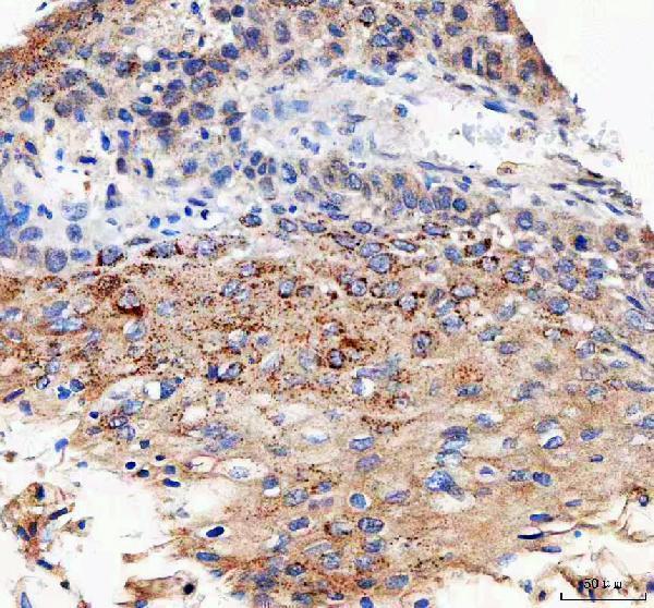
| Sample | Paraffin-embedded human esophageal squamous carcinoma tissue; fixative not specified (datasheet A08931-2) |
| Fixation | Image fixative and duration unreported (datasheet A08931-2); verify before use. |
| Sectioning | 4–5 µm sections on charged slides (standard) |
| Deparaffinisation | Xylene, graded ethanol series to water (standard) |
| Antigen retrieval | Heat retrieval: EDTA pH 8.0 (datasheet A08931-2); 20 min, 95–100 °C (standard) |
| Peroxidase block | 3% H2O2, 10 min, room temperature (standard) |
| Blocking | 10% goat serum (datasheet A08931-2) |
| Primary antibody | Rabbit anti-IFT74, 2-5 μg/ml (datasheet A08931-2) |
| Primary incubation | Overnight at 4 °C (datasheet A08931-2) |
| Detection | HRP-conjugated secondary, DAB chromogen (datasheet A08931-2) |
| Counterstain | Hematoxylin, blue, dehydrate and mount (standard) |
| Expected result | IFT74-positive staining in elongated or late spermatids of testis (HPA tissue IHC: High). HPA tissue profile: Membranous expression in ciliated cells in fallopian tube and respiratory epithelia. Cytoplasmic expression in several other tissues. No signal in the no-primary control. |
IFT74 is a ciliary and cytoplasmic vesicle protein with no transmembrane segment (UniProt Q96LB3). In paraffin sections, expect membranous staining in ciliated respiratory and fallopian tube cells, cytoplasmic staining in several other tissues, and strong staining in late spermatids (HPA tissue IHC). HPA rates its tissue IHC profile Approved, pending external verification (HPA tissue IHC).
| Strong staining in elongated or late spermatids; medium staining in bronchial respiratory epithelium or kidney tubules (HPA tissue IHC). | This distribution fits the reported positive cells and levels (HPA tissue IHC). In ciliated epithelia, look for membranous staining; other positive tissues may show cytoplasmic staining (HPA tissue IHC). Judge each compartment against its own tissue context rather than expecting one pattern everywhere. |
| Predominantly nuclear staining, with no corresponding ciliated-cell or cytoplasmic pattern. | A dominant nuclear pattern does not match the reported ciliary, basal body, or vesicular locations (UniProt Q96LB3) or the tissue IHC profile (HPA tissue IHC). Treat it as suspect, review morphology and detection controls, and seek independent confirmation before scoring it as IFT74. |
| Strong staining in adipocytes, marrow hematopoietic cells, or esophageal squamous epithelium. | These cell types are reported as not detected (HPA tissue IHC). Check whether staining follows the cells or instead pools over pigment, debris, or tissue edges. Persistent cell-associated signal could reflect cross-reactivity or endogenous detection activity; confirm with appropriate controls. |
| Diffuse color across the section, including stroma and areas without recognizable positive cells. | The reported IHC pattern is cell and compartment dependent (HPA tissue IHC). Uniform haze prevents reliable scoring of those features. Inspect the negative reagent control and the distribution of background before interpreting weak cytoplasmic color as target staining. |
| No staining in elongated or late spermatids, or no discernible signal in a positive bronchial section. | Late spermatids are reported High and bronchial respiratory epithelial cells Medium (HPA tissue IHC). A blank positive specimen leaves assay performance unresolved. Confirm that the relevant cells are present and intact, then review detection, antibody dilution, and the controls. |
| Cell and tissue context (HPA tissue IHC) | Reported levels range from High in late spermatids to Medium in bronchial epithelium and kidney tubules, while several sampled cell types are not detected (HPA tissue IHC). Score the expected cells rather than averaging color across an entire section. |
| Subcellular context (UniProt Q96LB3; HPA tissue IHC) | IFT74 localizes along primary cilia and near the basal body or centriole (UniProt Q96LB3). Tissue IHC also reports membranous staining in ciliated cells and cytoplasmic staining elsewhere (HPA tissue IHC). These patterns need not look identical. |
| Antibody evidence (HPA antibody validation; HPA tissue IHC) | Two listed rabbit polyclonal antibodies have Approved IHC status, while the overall tissue profile remains pending external verification (HPA antibody validation; HPA tissue IHC). Approval supports using the reported pattern as a reference, not treating every colored structure as confirmed IFT74. |
| IF/ICC interpretation (HPA subcellular; HPA antibody validation) | For the separate IF/ICC guide, HPA reports supported Golgi, centriolar satellite, and basal body localization plus approved cytosolic localization (HPA subcellular). The listed ICC assessment for HPA026684 is Uncertain (HPA antibody validation), so an IF image needs its own validation. |
| Situation | Likely cause | Next action |
|---|---|---|
| Known-positive testis section is blank. | The reported High signal in elongated or late spermatids is absent (HPA tissue IHC); the reason cannot be assigned from the slide alone. | Check that late spermatids are present, then review the reagent control, chromogenic detection, antibody dilution, and the assay's documented IHC procedure. Repeat with an appropriate positive section if the run control failed. |
| Kidney tubules look weak or variable. | Tubular cells are reported at a Medium level (HPA tissue IHC); weak signal alone does not establish assay failure. | Score intact tubular cells against background and a stronger reported positive, such as late spermatids (HPA tissue IHC). Confirm the tissue and cell identity before changing staining conditions. |
| Diffuse chromogen obscures ciliated-cell borders. | Section-wide background can hide the reported membranous pattern in ciliated epithelia (HPA tissue IHC). | Review negative reagent controls, blocking, wash stringency, and detection exposure as general IHC checks. Reassess the ciliated cells after background is reduced; do not score diffuse haze as localization. |
| Color is concentrated in nuclei or tissue edges. | That distribution is inconsistent with reported IFT74 locations (UniProt Q96LB3; HPA tissue IHC) and may represent nonspecific deposition. | Inspect section morphology and negative controls, then compare another reported positive compartment (HPA tissue IHC). Require reproducible cell-associated staining before assigning the signal to IFT74. |
| A reported negative cell type stains strongly. | Adipocytes, marrow hematopoietic cells, and esophageal squamous cells are listed as not detected (HPA tissue IHC); cross-reactivity or endogenous detection activity is possible. | Confirm the cell type and compare negative reagent and detection controls. If signal persists, seek confirmation with an independently validated antibody before changing the expected pattern. |
| What should an IF/ICC image show? | IF/ICC has separate localization evidence: supported Golgi, centriolar satellite, and basal body signals, with approved cytosolic signal (HPA subcellular). | Use the separate IF/ICC guide for imaging decisions. Interpret an image alongside the antibody's ICC status; HPA026684 is marked Uncertain for ICC (HPA antibody validation). |


Comprehensive Human Protein Atlas IHC scoring per tissue (reliability: Approved — Pending external verification.). Rows are taken directly from the HPA tissue chart — click any row's HPA link to view the source.
| Tissue | Cell type | Level | Evidence | Source |
|---|---|---|---|---|
| Testis | Elongated or late spermatids | High | Protein (IHC) | HPA → |
| Appendix | Glandular cells | Medium | Protein (IHC) | HPA → |
| Bronchus | Respiratory epithelial cells | Medium | Protein (IHC) | HPA → |
| Colon | Glandular cells | Medium | Protein (IHC) | HPA → |
| Duodenum | Glandular cells | Medium | Protein (IHC) | HPA → |
| Tissue | Cell type | Level | Evidence | Source |
|---|---|---|---|---|
| Adipose tissue | Adipocytes | Not detected | Protein (IHC) | HPA → |
| Bone marrow | Hematopoietic cells | Not detected | Protein (IHC) | HPA → |
| Cervix | Glandular cells | Not detected | Protein (IHC) | HPA → |
| Esophagus | Squamous epithelial cells | Not detected | Protein (IHC) | HPA → |
| Hippocampus | Glial cells | Not detected | Protein (IHC) | HPA → |
Troubleshoot IFT74 staining in paraffin sections by assessing retrieval, compartment, tissue context and controls before interpreting chromogenic signal.
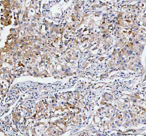
A08931-2 has IHC images from paraffin sections of human tumors and mouse testis (catalog IHC captions); its IHC listing covers human, mouse, and rat (catalog dilution_raw).
A08931-2 has IHC images from paraffin sections of human esophageal squamous carcinoma, lung cancer, rectum adenocarcinoma, and mouse testis (catalog IHC captions). Its listed IHC concentration is 2–5 μg/ml for human, mouse, and rat (catalog dilution_raw).
Which to pick: Choose A08931-2 for paraffin-section tissue IHC: its own captions document that preparation, while the fixative is unreported (catalog IHC captions); it is rabbit-hosted, with no clone specified (catalog host; catalog clone). No IF/ICC-validated SKU is present, so A08931-2 would require separate IF/ICC validation (catalog applications; catalog IF image alts). For cross-species IHC, A08931-2 lists human, mouse, and rat; monkey appears in overall reactivity but not in the application-specific IHC listing (catalog reactivity; catalog dilution_raw).