This website uses cookies to ensure you get the best experience on our website.


- Table of Contents
Plan IGF2BP1 IHC-P around cytoplasmic staining in testis duct and ovarian follicle cells (HPA tissue IHC). Use preleptotene spermatocytes in testis as a positive reference; their staining is reported as high (HPA tissue IHC).

Expected localisation, validated protocols, controls and antibodies — the at-a-glance facts below, then the full design guide.
| Expected localisation | Cytoplasmic staining in testis and ovary (HPA tissue IHC) | |
| Staining pattern | Cytoplasmic signal in testis duct and ovarian follicle cells (HPA tissue IHC) | |
| Antigen retrieval | Citrate pH 6 HIER, heat-mediated (datasheet A02007-1) | |
| Positive control | Testis+2 more · see all | |
| Negative control | Adipose tissue+4 more · see all |
| Fixation | Use formaldehyde-fixed paraffin sections (selected-SKU IHC image A02007-1); keep fixation consistent (standard IHC practice; not target-specific) | |
| Caveat | Signal varies by tissue and cell type (HPA tissue IHC) | |
| Regulation | Embryonic; expressed in some cancers (UniProt) | |
| Isoform / epitope | 2 isoforms; verify C-terminal epitope coverage (UniProt; datasheet A02007-1) |
The catalog antibody’s IHC-P protocol is accompanied by published IGF2BP1 staining protocols for leiomyoma (PMC9154092), lung adenocarcinoma (PMC12967950), and osteosarcoma (PMC6449306).
| Sample | Formaldehyde-fixed, paraffin-embedded human testis tissue (datasheet A02007-1) |
| Fixation | Image formalin-fixed; duration unreported (datasheet A02007-1); verify before use. |
| Sectioning | 4–5 µm sections on charged slides (standard) |
| Deparaffinisation | Xylene, graded ethanol series to water (standard) |
| Antigen retrieval | Heat retrieval: Citrate pH 6 (datasheet A02007-1); 20 min, 95–100 °C (standard) |
| Peroxidase block | 3% H2O2, 10 min, room temperature (standard) |
| Blocking | 10% normal serum of the secondary host, 30 min, room temperature (standard) |
| Primary antibody | Rabbit anti-IGF2BP1, 1:25 (datasheet A02007-1) |
| Primary incubation | 1 hours at 37°C (datasheet A02007-1) |
| Detection | HRP-polymer secondary, DAB chromogen 5–10 min (standard) |
| Counterstain | Hematoxylin, blue, dehydrate and mount (standard) |
| Expected result | IGF2BP1-positive staining in preleptotene spermatocytes of testis (HPA tissue IHC: High). HPA tissue profile: Cytoplasmic expression mainly in cells in seminiferous ducts of testis and follicle cells in ovary. No signal in the no-primary control. |
IGF2BP1 is mainly cytoplasmic in tissue IHC, especially in testicular preleptotene spermatocytes and ovarian follicle cells; kidney collecting ducts can also stain (HPA: tissue IHC). Nuclear foci and peripheral RNA granules are possible localizations, although HPA ICC-IF supports mainly cytosolic signal (UniProt Q9NZI8: subcellular location; HPA: subcellular). It has no transmembrane segment (UniProt Q9NZI8: topology). HPA rates the tissue IHC pattern Enhanced, with medium consistency against RNA expression (HPA: tissue IHC).
| Strong cytoplasmic signal in preleptotene spermatocytes; moderate signal in follicle cells or collecting ducts. | This matches the reported cell types and relative levels: testis High, ovary Medium, kidney Medium (HPA: tissue IHC). Compare stained cells with the surrounding unstained cells, rather than treating the whole section as uniformly positive (standard IHC interpretation). |
| Predominantly nuclear staining, with little cytoplasmic signal, across many cells. | Reassess specificity and scoring: HPA describes mainly cytoplasmic tissue staining and supported cytosolic ICC-IF staining (HPA: tissue IHC; HPA: subcellular). Discrete nuclear foci remain biologically plausible, so a limited nuclear component alone is not proof of artefact (UniProt Q9NZI8: subcellular location). |
| Prominent signal in adipocytes or other cells reported as not detected. | Consider cross-reactivity or endogenous chromogen-generating activity before calling those cells IGF2BP1-positive (HPA: adipose tissue IHC; standard IHC interpretation). Check cell identity, staining boundaries and detection controls; HPA's negative finding applies to the specified cell population, not every cell in that tissue (HPA: tissue IHC). |
| Diffuse colour covers tissue, extracellular space or most cell types without clear cytoplasmic boundaries. | A widespread haze does not resemble HPA's cell-selective cytoplasmic pattern (HPA: tissue IHC). Background from detection reagents, incomplete blocking or excess antibody is possible (standard IHC practice); interpret the slide only after a matched negative detection control and tissue morphology are reviewed. |
| No visible staining in preleptotene spermatocytes on a testis section. | The expected High signal is missing (HPA: testis IHC). First verify that the relevant cells are present and identifiable; then check antibody validation, retrieval and detection controls (standard IHC practice). A blank slide alone cannot establish true absence of IGF2BP1. |
| Compartment and topology (UniProt Q9NZI8: topology; HPA: subcellular) | IGF2BP1 lacks a transmembrane segment, and supported ICC-IF localization is cytosolic (UniProt Q9NZI8: topology; HPA: subcellular). Score intracellular staining by compartment; do not treat membrane outlining as the expected target pattern (standard IHC interpretation). |
| Choice of comparison cells (HPA: tissue IHC) | Preleptotene spermatocytes provide a reported High reference; collecting ducts and follicle cells are Medium references (HPA: tissue IHC). Adipocytes and several listed cell populations are reported as not detected, making cell-level comparison useful within the stated tissue context (HPA: tissue IHC). |
| Evidence strength (HPA: tissue IHC; HPA: antibodies) | HPA calls the tissue result Enhanced but reports only medium agreement between antibody staining and RNA expression (HPA: tissue IHC). Two listed antibodies, HPA021367 and HPA062273, have Enhanced IHC validation (HPA: antibodies). These ratings support the reported pattern without validating every unexpected stained cell. |
| Isoforms and epitope coverage (UniProt Q9NZI8: isoforms) | UniProt lists 2 isoforms (UniProt Q9NZI8: isoforms). The supplied evidence gives no epitope location or isoform coverage for the IHC-validated antibodies; a staining difference cannot be assigned to an isoform from these records alone. |
| IF/ICC Q&A: what localization should be expected? (HPA: subcellular) | HPA reports mainly cytosolic ICC-IF signal, with images from CACO-2, Hep-G2 and U2OS; HPA021367 has Supported ICC validation (HPA: subcellular; HPA: antibodies). UniProt also lists nuclear foci and cytoplasmic granules (UniProt Q9NZI8: subcellular location). Use the separate IF/ICC guide for that application's procedure. |
| Situation | Likely cause | Next action |
|---|---|---|
| Testis positive-control cells appear blank. | The relevant preleptotene spermatocytes may be absent from the viewed area, or an IHC workflow step may have failed (HPA: testis IHC; standard IHC practice). | Locate the named cells on the counterstained section, then review the catalog antibody's IHC-P instructions and run a positive tissue and detection control (standard IHC practice). Do not infer a target-specific retrieval defect from HPA's staining level. |
| Colour is widespread in adipocytes or glandular cells reported as not detected. | Cross-reactivity or endogenous detection activity is possible (HPA: tissue IHC; standard IHC interpretation). The HPA negative calls refer to particular cell populations. | Compare morphology and cell identity with HPA's named populations; inspect an appropriate reagent-only detection control and endogenous-activity control for the chosen chemistry (standard IHC practice). |
| Nuclei dominate while the expected cytoplasmic pattern is weak. | The compartment pattern differs from HPA's mainly cytoplasmic tissue result, although UniProt allows discrete nuclear foci (HPA: tissue IHC; UniProt Q9NZI8: subcellular location). | Score nuclear and cytoplasmic compartments separately, compare with a known-positive section and review detection controls (standard IHC practice). Avoid calling every nuclear focus nonspecific solely from compartment. |
| A diffuse haze obscures cell boundaries. | Background from antibody concentration, blocking or chromogenic detection can obscure cell-selective staining (standard IHC practice). | Review the catalog antibody's IHC-P dilution guidance, blocking and reagent-only control; adjust the general workflow only after comparing control and test sections (standard IHC practice). No target-specific dilution is supplied here. |
| Ovary or kidney staining is weaker than testis. | That ordering is compatible with HPA's Medium follicle-cell and collecting-duct findings versus High preleptotene-spermatocyte staining (HPA: tissue IHC). | Compare the named cells, section morphology and controls before treating the lower intensity as failure (standard IHC interpretation). HPA's levels are qualitative and do not define a numeric scoring threshold. |
| IF/ICC shows cytosolic puncta or a small nuclear component. | UniProt places IGF2BP1 in cytoplasmic mRNP granules and describes discrete nuclear foci; HPA reports mainly cytosolic ICC-IF localization (UniProt Q9NZI8: subcellular location; HPA: subcellular). | Interpret those features against the supported cytosolic pattern and appropriate IF controls (HPA: subcellular; standard IF practice). The IHC tissue ratings do not themselves validate an IF finding (HPA: tissue IHC; HPA: antibodies). |
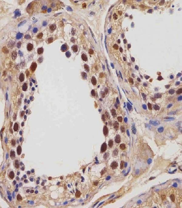
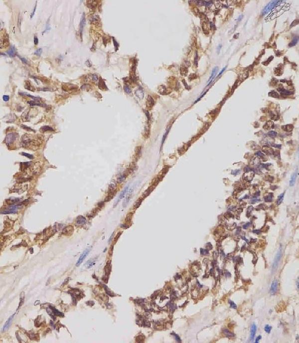


Comprehensive Human Protein Atlas IHC scoring per tissue (reliability: Enhanced — Medium consistency between antibody staining and RNA expression data.). Rows are taken directly from the HPA tissue chart — click any row's HPA link to view the source.
| Tissue | Cell type | Level | Evidence | Source |
|---|---|---|---|---|
| Testis | Preleptotene spermatocytes | High | Protein (IHC) | HPA → |
| Kidney | Collecting ducts | Medium | Protein (IHC) | HPA → |
| Ovary | Follicle cells | Medium | Protein (IHC) | HPA → |
| Tissue | Cell type | Level | Evidence | Source |
|---|---|---|---|---|
| Adipose tissue | Adipocytes | Not detected | Protein (IHC) | HPA → |
| Adrenal gland | Glandular cells | Not detected | Protein (IHC) | HPA → |
| Appendix | Glandular cells | Not detected | Protein (IHC) | HPA → |
| Bone marrow | Hematopoietic cells | Not detected | Protein (IHC) | HPA → |
| Breast | Adipocytes | Not detected | Protein (IHC) | HPA → |
Use matched controls and compartment-aware scoring to troubleshoot IGF2BP1 staining in paraffin sections; the immunofluorescence question addresses the separate IF/ICC workflow.
Real IGF2BP1 IHC data show staining in human testis and lung adenocarcinoma paraffin sections (A02007-1 image captions). Mouse reactivity is listed, but no IF data are supplied (catalog).
A02007-1 has IHC-P images from human testis and lung adenocarcinoma paraffin sections (A02007-1 image captions). It lists human and mouse reactivity and IHC-P as an application; no IF image is supplied (catalog).
Which to pick: Choose A02007-1 for tissue IHC-P: it is rabbit polyclonal, lists a 1:25 dilution, and its own captions describe paraffin sections with citrate retrieval at pH 6 (catalog; A02007-1 image captions). Its captions describe fixation as both paraformaldehyde and formaldehyde (A02007-1 image captions). No listed IF/ICC application or IF image supports an IF/ICC choice; for cross-species work, A02007-1 lists human and mouse reactivity, although its IHC images show human samples only (catalog; A02007-1 image captions).