Click image to see more details
Product Info Summary
| SKU: | A02007-1 |
|---|---|
| Size: | 80 µl |
| Reactive Species: | Human, Mouse |
| Host: | Rabbit |
| Application: | Flow Cytometry, IHC-P, WB |
Customers Who Bought This Also Bought

Product info
Product Name
Anti-IGF2BP1 Antibody (C-term)
SKU/Catalog Number
A02007-1
Size
80 µl
Description
Boster Bio Anti-IGF2BP1 Antibody (C-term) (Catalog # A02007-1). Tested in WB, IHC-P, Flow Cytometry application(s). This antibody reacts with Human, Mouse.
Storage & Handling
Maintain refrigerated at 2-8°C for up to 2 weeks. For long-term storage, store at -20°C in small aliquots to prevent freeze-thaw cycles.
Cite This Product
Anti-IGF2BP1 Antibody (C-term) (Boster Biological Technology, Pleasanton CA, USA, Catalog # A02007-1)
Host
Rabbit
Contents
Purified polyclonal antibody supplied in PBS with 0.09% (W/V) sodium azide.
Clonality
Polyclonal
Isotype
Rabbit IgG
Immunogen
This IGF2BP1 antibody is generated from rabbits immunized with a KLH conjugated synthetic peptide between 508-534 amino acids from the C-terminal region of human IGF2BP1.
Reactive Species
A02007-1 is reactive to IGF2BP1 in Human, Mouse
Calculated molecular weight
63.5 kDa
Background of IGF2BP1
IGF2BP1 is a member of the insulin-like growth factor 2 mRNA-binding protein family. The protein encoded by this gene contains four K homology domains and two RNA recognition motifs. It functions by binding to the mRNAs of certain genes, including insulin-like growth factor 2, beta-actin and beta-transducin repeat-containing protein, and regulating their translation. Two transcript variants encoding different isoforms have been found for this gene.
Antibody Validation
Boster validates all antibodies on WB, IHC, ICC, Immunofluorescence, and ELISA with known positive control and negative samples to ensure specificity and high affinity, including thorough antibody incubations.
Application & Images
Applications
A02007-1 is guaranteed for Flow Cytometry, IHC-P, WB Boster Guarantee
Recommend Dilution
WB: 1:2000
IHC-P: 1:25
FC: 1:25
Validation Images & Assay Conditions
Click image to see more details
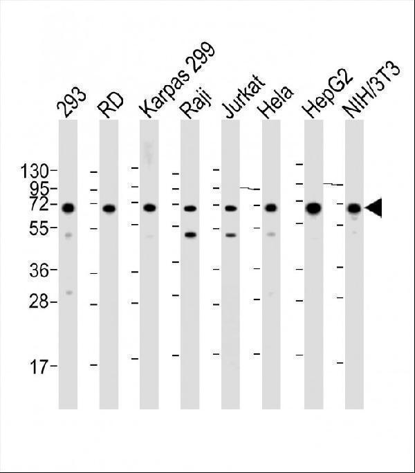
All lanes : Anti-IGF2BP1 Antibody (C-term) at 1:2000 dilution
Lane 1: 293 whole cell lysate
Lane 2: RD whole cell lysate
Lane 3: Karpas 299 whole cell lysate
Lane 4: Raji whole cell lysate
Lane 5: Jurkat whole cell lysate
Lane 6: Hela whole cell lysate
Lane 7: HepG2 whole cell lysate Lane 8: NIH/3T3 whole cell lysate Lysates/proteins at 20 µg per lane.
Secondary
Goat Anti-Rabbit IgG, (H+L), Peroxidase conjugated at 1/10000 dilution.
Predicted band size : 63 kDa
Blocking/Dilution buffer: 5% NFDM/TBST.

Click image to see more details
A02007-1 staining IGF2BP1 in human testis tissue sections by Immunohistochemistry (IHC-P -paraformaldehyde-fixed, paraffin-embedded sections). Tissue was fixed with formaldehyde and blocked with 3% BSA for 0. 5 hour at room temperature; antigen retrieval was by heat mediation with a citrate buffer (pH6). Samples were incubated with primary antibody (1/25) for 1 hours at 37°C. A undiluted biotinylated goat polyvalent antibody was used as the secondary antibody.

Click image to see more details
A02007-1 staining IGF2BP1 in human lung adenocarcinoma sections by Immunohistochemistry (IHC-P -paraformaldehyde-fixed, paraffin-embedded sections). Tissue was fixed with formaldehyde and blocked with 3% BSA for 0. 5 hour at room temperature; antigen retrieval was by heat mediation with a citrate buffer (pH6). Samples were incubated with primary antibody (1/25) for 1 hours at 37°C. A undiluted biotinylated goat polyvalent antibody was used as the secondary antibody.

Click image to see more details
Overlay histogram showing HepG2 cells stained with A02007-1 (green line). The cells were fixed with 2% paraformaldehyde (10 min) and then permeabilized with 90% methanol for 10 min. The cells were then icubated in 2% bovine serum albumin to block non-specific protein-protein interactions followed by the antibody (A02007-1, 1:25 dilution) for 60 min at 37ºC. The secondary antibody used was Goat-Anti-Rabbit IgG, DyLight® 488 Conjugated Highly Cross-Adsorbed at 1/200 dilution for 40 min at 37ºC. Isotype control antibody (blue line) was rabbit IgG1 (1μg/1x10^6 cells) used under the same conditions. Acquisition of >10, 000 events was performed.
Specific Publications For Anti-IGF2BP1 Antibody (C-term) (A02007-1)
Loading publications
Recommended Resources
Here are featured tools and databases that you might find useful.
- Boster's Pathways Library
- Protein Databases
- Bioscience Research Protocol Resources
- Data Processing & Analysis Software
- Photo Editing Software
- Scientific Literature Resources
- Research Paper Management Tools
- Molecular Biology Software
- Primer Design Tools
- Bioinformatics Tools
- Phylogenetic Tree Analysis
Customer Reviews
Have you used Anti-IGF2BP1 Antibody (C-term)?
Share your experimental results or join a short interview to earn up to $1,000 in product credits or other rewards.
0 Reviews For Anti-IGF2BP1 Antibody (C-term)
Customer Q&As
Have a question?
Find answers in Q&As, reviews.
Can't find your answer?
Submit your question
