IKBKB / Inhibitor of nuclear factor kappa-B kinase subunit beta · IHC design guide

Design Immunohistochemistry for IKBKB

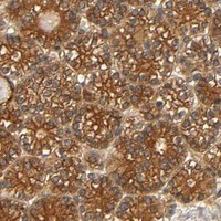
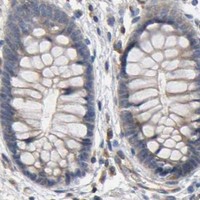
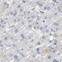
Plan paraffin IHC for IKBKB around the cytoplasmic staining seen in most tissues (HPA tissue IHC). Colon glandular cells show medium staining, while liver cholangiocytes have no detectable staining (HPA tissue IHC); keep fixation consistent and include matched controls (standard IHC practice).

Evidence assembled Oct 2026 · For research use; verify linked source records and product datasheet before use
Immunohistochemistry protocol sheet for IKBKB (IHC for IKBKB): expected localisation Cytoplasmic staining in most tissues (HPA tissue IHC), antibody M00118, validated IHC image, and IHC protocol steps
Printable IKBKB IHC protocol sheet — expected localisation Cytoplasmic staining in most tissues (HPA tissue IHC), antibody M00118, controls and protocol steps. Open the full IKBKB IHC guide →

IKBKB Immunohistochemistry Experimental Design Guide

Expected localisation, validated protocols, controls and antibodies — the at-a-glance facts below, then the full design guide.

Must know before staining
Expected localisation Cytoplasmic staining in most tissues (HPA tissue IHC)
Staining pattern Cytoplasmic signal in most tissues, including colon glands (HPA tissue IHC)
Antigen retrieval Tris-EDTA pH 9.0 HIER, 95–98 °C, 20 min (rule: nuclear antigen)
Positive control ⓘ Appendix+4 more · see all
Negative control ⓘ Liver
Important caveats
Reasons your staining may differ from the expected pattern.
Fixation Keep fixation consistent across sections (standard IHC practice; not target-specific); Matched tissue-IHC evidence does not establish the fixation claim. Validate the specimen-specific method before use. (selected-SKU IHC image M00118)
Caveat Liver cholangiocytes have no detectable staining (HPA tissue IHC)
Regulation Expression regulation unreported (UniProt)
Isoform / epitope 4 isoforms; epitope coverage is unknown (UniProt)
Section 1

Recommended IKBKB IHC & IF Protocols

The catalog antibody’s IHC-P protocol is accompanied by three published IKBKB IHC protocols covering rat brain, tumor specimens, and human ovarian tissue arrays (PMC10707860; PMC3164321; PMC6191368).

Recommended immunohistochemistry (IHC-P) protocol parameters
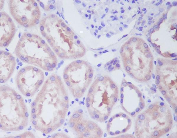
SampleParaffin-embedded human kidney tissue; fixative not specified (datasheet M00118)
FixationImage fixative and duration unreported (datasheet M00118); verify before use.
Sectioning4–5 µm sections on charged slides (standard)
DeparaffinisationXylene, graded ethanol series to water (standard)
Antigen retrievalHeat-induced epitope retrieval in Tris-EDTA buffer, pH 9.0, 20 min at 95–98 °C (standard rule: nuclear antigen)
Peroxidase block3% H2O2, 10 min, room temperature (standard)
Blocking10% normal serum of the secondary host, 30 min, room temperature (standard)
Primary antibodyRabbit monoclonal (clone AOOA-9) anti-IKBKB, 1:50 (datasheet M00118)
Primary incubationOvernight at 4 °C (standard)
DetectionHRP-polymer secondary, DAB chromogen 5–10 min (standard)
CounterstainHematoxylin, blue, dehydrate and mount (standard)
Expected resultIKBKB-positive staining in lymphoid tissue of appendix (HPA tissue IHC: Medium). HPA tissue profile: Cytoplasmic expression in most tissues. No signal in the no-primary control.
💡Decision noteStart with Tris-EDTA pH 9.0 HIER at 95–98 °C for 20 min for the catalog antibody (page retrieval rule). The published protocols used different antibodies and retrieval conditions (PMC3164321; PMC6191368).
Section 2

What Is the Expected IKBKB Staining Pattern?

IKBKB is cytoplasmic in most tissues, including glandular cells (HPA IHC: Enhanced); it has no TM span (UniProt O14920).

What am I looking at on my slide?
Medium cytoplasmic stain in colon glands (HPA IHC).Matches the observed colon pattern (HPA IHC: glandular cells, Medium).
Membrane-only rim stain (HPA IHC: chiefly cytoplasmic).Atypical; assess specificity, though raft localisation is reported (UniProt O14920).
Strong cholangiocyte stain (HPA IHC: not detected).Unexpected (HPA IHC); assess cross-reactivity or endogenous detection activity (general IHC).
Diffuse acellular haze (HPA IHC: cytoplasmic pattern).Suggests background; check blocking and a no-primary control (general IHC).
No colon gland stain (HPA IHC: Medium).Possible assay failure; check a positive control and detection steps (general IHC).
💡Expected IKBKB appearanceIn colon glands, expect Medium cytoplasmic stain; acellular haze is suspect (HPA IHC).
How each factor affects the staining
Tissue variation (HPA IHC)Colon glands stain Medium; cholangiocytes are not detected (HPA IHC).
Subcellular distribution (UniProt O14920)Cytoplasm and nucleus are listed; membrane raft localisation is also reported (UniProt O14920).
Isoforms (UniProt O14920)Four isoforms are listed; antibody coverage cannot be inferred without an epitope (UniProt O14920).
Antibody validation (HPA antibodies)CAB004447 is IHC Enhanced; HPA001249 is IHC Approved (HPA antibodies).
Why is my staining missing, weak or wrong?
SituationLikely causeNext action
Weak colon gland stain (HPA IHC: Medium).Retrieval or primary concentration may be suboptimal (general IHC).Optimize retrieval and concentration against a positive control (general IHC).
No colon gland stain (HPA IHC: Medium).A detection step may have failed (general IHC).Check the positive control and detection reagents (general IHC).
Diffuse DAB haze (general IHC).Inadequate blocking or washing is possible (general IHC).Run a no-primary control; adjust blocking and washes (general IHC).
Cholangiocyte stain (HPA IHC: not detected).Cross-reactivity or endogenous activity is possible (general IHC).Check a no-primary control and compare an HPA-positive tissue (HPA IHC).
Membrane-only stain (HPA IHC: cytoplasmic).Specificity is uncertain; raft localisation is reported (UniProt O14920).Compare with a second IHC-listed antibody (HPA antibodies).
IF/ICC: expected compartment?Cytosol is the supported location (HPA ICC-IF).Use the separate IF/ICC guide for its protocol.

Sample controls for IKBKB IHC & IF

🧪Run appendix first: its lymphoid tissue should stain at a Medium level (HPA: appendix, lymphoid tissue). Run liver cholangiocytes as the negative comparator (HPA: not detected); on the appendix slide, use cells without specific staining as an internal background reference, without assuming a particular cell type is IKBKB-negative.
Positive control tissue: Appendix (Lymphoid tissue, HPA Medium)
Negative control tissue: Liver (HPA Not detected)
ICC-IF cell lines (HPA subcellular resource): HPA ICC-IF images show IKBKB in A-431, U-251MG, U2OS, with annotated localisation: Cytosol (supported) (HPA subcellular).
Technical controls: Include a no-primary (secondary-only) slide, an irrelevant immunoglobulin matched to the primary antibody’s host species and isotype, and IKBKB-knockout tissue or cells as a biological negative (standard IHC controls). In appendix, block endogenous peroxidase before chromogenic detection and check inflammatory cells for residual background (standard IHC practice).
⚠️Feasibility: A target-specific fixation window or fixation effect is unreported in the supplied evidence; the selected paraffin-section kidney caption also leaves the fixative unreported (M00118 caption). Optimize antigen retrieval empirically for the IHC-validated antibody; the supplied evidence does not establish whether frozen sections or IF are easier (M00118 caption; HPA: cytosol supported in ICC-IF). In appendix, endogenous peroxidase in inflammatory cells can complicate chromogenic scoring (standard IHC practice).

HPA tissue IHC evidence for IKBKB

Comprehensive Human Protein Atlas IHC scoring per tissue (reliability: Enhanced — Medium consistency between antibody staining and RNA expression data.). Rows are taken directly from the HPA tissue chart — click any row's HPA link to view the source.

Positive expression · recommended positive controls

TissueCell typeLevelEvidenceSource
Appendix Lymphoid tissue Medium Protein (IHC) HPA →
Bone marrow Hematopoietic cells Medium Protein (IHC) HPA →
Breast Glandular cells Medium Protein (IHC) HPA →
Cerebral cortex Neuronal cells Medium Protein (IHC) HPA →
Cervix Glandular cells Medium Protein (IHC) HPA →

Undetected expression · recommended negative controls

TissueCell typeLevelEvidenceSource
Liver Cholangiocytes Not detected Protein (IHC) HPA →
Section 3

Advanced IKBKB IHC Tips

Troubleshooting IKBKB staining in paraffin sections, with one entry on related IF/ICC validation.

What should I change if IKBKB staining is weak after retrieval?
Start with Tris-EDTA at pH 9.0 for heat-induced epitope retrieval at 95–98 °C for 20 min (page-specified retrieval). If staining is weak, compare a second retrieval condition on adjacent sections while keeping antibody concentration, detection chemistry and development time fixed; record tissue preservation alongside signal. A paraffin-embedded human kidney image exists for the catalog antibody, but its caption does not state the fixative or retrieval method (catalog antibody M00118 caption). Assess whether signal is predominantly cytoplasmic in viable cells, as expected from tissue staining data (HPA tissue IHC: cytoplasmic expression in most tissues). Excessive retrieval can damage morphology, so reject any condition that increases staining while making cell boundaries difficult to score (standard IHC practice).
Could fixation explain variable IKBKB staining between paraffin blocks?
Target-specific fixation sensitivity is unknown from the supplied evidence; the selected kidney image reports paraffin embedding but does not identify a fixative (catalog antibody M00118 caption). Compare blocks using matched section thickness, retrieval, antibody incubation and chromogen development, then document each block’s known fixation history (standard IHC practice). Include an internal positive compartment or a separately processed reference section in every run so weak staining can be distinguished from a run-level failure (standard IHC practice). Score only preserved, viable regions because damaged morphology makes cellular staining difficult to interpret (standard IHC practice). Do not infer an IKBKB-specific fixation effect from its tissue distribution or modified residues (HPA tissue IHC; UniProt O14920).
Is nuclear or membrane staining plausible for IKBKB in IHC?
Predominantly cytoplasmic staining is the practical starting expectation for tissue sections (HPA tissue IHC: cytoplasmic expression in most tissues). Nuclear and membrane-raft localisation are also annotated, so a convincing minority pattern deserves validation rather than automatic rejection (UniProt O14920: subcellular location). IKBKB has no transmembrane segment, and its membrane-raft association should not be interpreted as a continuous cell-surface outline (UniProt O14920: topology and subcellular location). Compare the suspect pattern across adjacent sections, tissue regions and controls processed with the same retrieval and chromogen conditions (standard IHC practice). Diffuse staining over empty spaces or section edges lacks the cellular distribution needed to support localisation (standard IHC practice).
How can epitope choice affect interpretation across IKBKB isoforms?
IKBKB has 4 annotated isoforms, so isoform coverage depends on where the antibody binds (UniProt O14920: isoforms). Check the catalog antibody’s stated immunogen or epitope against the sequences of isoforms 1–4 before treating differences in staining as differences in total IKBKB (UniProt O14920: isoforms; standard antibody-validation practice). The protein kinase domain spans residues 15–300, and annotated modifications include residues 177 and 181; an epitope overlapping modified residues could require separate validation (UniProt O14920: domains and modified residues). No epitope sequence or isoform reactivity is supplied here, so the cause of any staining difference remains unresolved (supplied catalog antibody evidence). Compare independent epitopes, if available, on matched sections (standard IHC practice).
How should I cross-check the chromogenic IHC pattern by multiplex IF?
Use IF/ICC as a companion localisation check: cytosol is the supported main location in cell images, while tissue IHC is generally cytoplasmic (HPA subcellular; HPA tissue IHC). In a tissue selected for glandular cells, multiplex IKBKB with a validated glandular-cell marker and check whether both signals occupy the same cells (HPA tissue IHC: medium staining in breast glandular cells; standard IF practice). Choose spectrally separated fluorophores and a channel away from the tissue’s strongest autofluorescence, using single-stain controls to assess bleed-through (standard IF practice). Because IKBKB lacks a transmembrane segment and includes cytosolic localisation, permeabilise sufficiently to access an intracellular epitope, while optimising detergent strength for morphology (UniProt O14920: topology and subcellular location; standard IF practice). An IF result alone does not establish the chromogenic stain’s specificity (standard antibody-validation practice).
What causes diffuse brown background in an IKBKB section?
First compare a no-primary control with the stained section under identical retrieval and DAB development conditions to locate detection-system background (standard IHC practice). Apply an appropriate peroxidase block before chromogenic detection, and assess whether residual colour follows tissue components rather than intact cells (standard IHC practice). If background rises with primary antibody concentration, titrate the antibody and adjust blocking or wash conditions while retaining the same positive reference tissue (standard IHC practice). Cytoplasmic staining across many tissues can be biologically plausible, so widespread colour alone is insufficient to call background (HPA tissue IHC: cytoplasmic expression in most tissues). Treat precipitate, section folds and edge-concentrated colour as technical patterns until controls support a cellular signal (standard IHC practice).
How should I quantify heterogeneous IKBKB chromogenic staining? ⚠ ANSWER MARKED FOR VERIFICATION
Define the cell population and region before scoring, then use an H-score combining intensity with the percentage of positive cells when both dimensions matter (standard IHC quantification practice). Alternatively, report percent positive cells or positive-cell density per mm² when intensity cannot be scored reproducibly (standard IHC quantification practice). Normalise counts to viable cells of the chosen type, or density to measured viable tissue area, excluding folds and necrosis (standard IHC quantification practice). Keep retrieval, imaging and DAB development consistent across slides, and set thresholds using reference and no-primary controls (standard IHC practice). Report cytoplasmic and nuclear scores separately because cytoplasmic tissue expression predominates but nuclear localisation is annotated (HPA tissue IHC; UniProt O14920: subcellular location).
How can I distinguish true IKBKB staining from artefact?
A credible result places staining within identifiable viable cells and usually shows a cytoplasmic component, consistent with the reported tissue pattern (HPA tissue IHC: cytoplasmic expression in most tissues). Check the stained cell type against the section’s morphology; for example, medium staining is reported in breast glandular cells, whereas liver cholangiocytes were not detected in the cited survey (HPA tissue IHC). Nuclear signal can be plausible, but an isolated nuclear pattern needs an independent specificity check because cytosol is the supported main location in cell images (UniProt O14920: subcellular location; HPA subcellular). Disregard edge effects, necrotic areas and colour reproduced by a no-primary control or residual endogenous enzyme activity (standard IHC practice). Neither staining intensity nor localisation alone proves kinase activation (UniProt O14920: function; standard IHC interpretation practice).
Boster reagents

Best IKBKB / Inhibitor of nuclear factor kappa-B kinase subunit beta IHC Antibodies

IHC and IF image data cover human kidney paraffin sections, human lung cancer tissue, and human HeLa cells (catalog image captions); M00118 also lists mouse and rat reactivity (catalog applications/reactivity).

Real IHC data Immunohistochemical analysis of paraffin-embedded human kidney, using IKK beta Antibody.
Anti-IKK beta IKBKB Rabbit Monoclonal Antibody
Cat # M00118
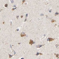
Real IHC data Anti-IKK beta antibody, PB9221,IHC(P) IHC(P): Human Lung Cancer Tissue
Anti-IKK beta/IKBKB Antibody ®
Cat # PB9221
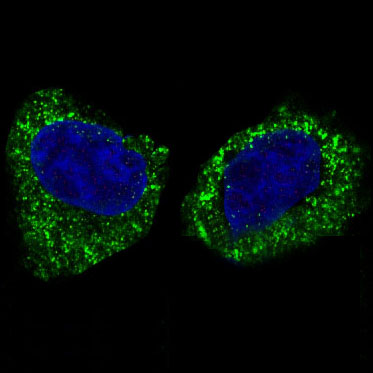
Real IF data Immunocytochemistry staining of HeLa cells using IKK beta antibody at 10 μg/mL.
Anti-IKK beta IKBKB Antibody
Cat # A00118-1

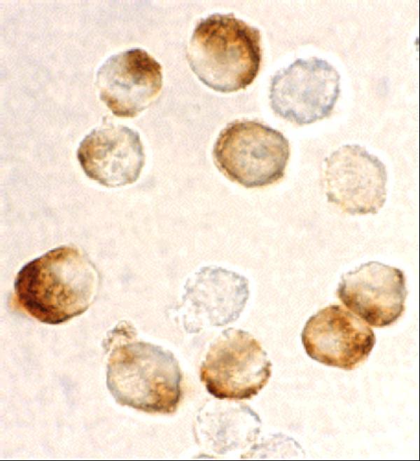
M00118 shows IHC on paraffin-embedded human kidney and lists IHC and IF applications (M00118 image caption; catalog applications); PB9221 shows IHC(P) on human lung cancer tissue and lists IHC and IF (PB9221 image caption; catalog applications). A00118-1 shows ICC staining of HeLa cells and lists IF and ICC, with no IHC application listed (A00118-1 image caption; catalog applications).

Which to pick: For tissue IHC, choose M00118 for human paraffin sections or PB9221 for human IHC(P): their respective images show kidney paraffin sections and lung cancer tissue, and neither caption reports the fixative (M00118 and PB9221 IHC image captions). For IF/ICC, A00118-1 has a HeLa ICC image and lists both applications (A00118-1 image caption; catalog applications). For mouse or rat samples, M00118 is the rabbit monoclonal with listed mouse and rat reactivity, although its IHC image shows human tissue (M00118 catalog host/clone/reactivity; M00118 IHC image caption).

Each figure is that product's own IHC / IF validation image from its datasheet.

References

  1. UniProt Consortium. UniProt entry O14920 (IKKB_HUMAN, Inhibitor of nuclear factor kappa-B kinase subunit beta).
  2. Human Protein Atlas. IKBKB tissue IHC expression (reliability: Enhanced).
  3. Human Protein Atlas. IKBKB subcellular location (ICC-IF): Localized to the cytosol..
  4. Human Protein Atlas. IKBKB antibody validation summary (2 antibodies).
  5. Network Analysis and Experimental Verification of the Mechanisms of Hydroxysafflor Yellow A in Ischemic Stroke Following Atherosclerosis. Molecules (Basel, Switzerland) 2023 — PMC10707860.
  6. Genetic disruption of KEAP1/CUL3 E3 ubiquitin ligase complex components is a key mechanism of NF-kappaB pathway activation in lung cancer. Journal of thoracic oncology : official publication of the International Association for the Study of Lung Cancer 2011 — PMC3164321.
  7. Molecular map of cGAS-STING pathway-related genes in bladder cancer: the perspective toward immune microenvironment and prognosis. Aging 2024 — PMC10866408.
  8. Paclitaxel Sensitivity of Ovarian Cancer Can be Enhanced by Knocking Down Pairs of Kinases that Regulate MAP4 Phosphorylation and Microtubule Stability. Clinical cancer research : an official journal of the American Association for Cancer Research 2018 — PMC6191368.
  9. PubMed PMID:9346484 — UniProt-cited evidence.
  10. PubMed PMID:9346485 — UniProt-cited evidence.
  11. PubMed PMID:9813230 — UniProt-cited evidence.