This website uses cookies to ensure you get the best experience on our website.


- Table of Contents
Plan IKBKE IHC-P around cytoplasmic tissue staining (HPA tissue IHC) and the catalog antibody’s 10 μg/mL starting concentration (datasheet A01816). Score glandular and hematopoietic cells separately, and interpret intensity with the reported medium staining–RNA consistency in mind (HPA tissue IHC).

Expected localisation, validated protocols, controls and antibodies — the at-a-glance facts below, then the full design guide.
| Expected localisation | Cytoplasmic staining in most tissues (HPA tissue IHC) | |
| Staining pattern | Glandular and hematopoietic cells: cytoplasmic stain (HPA tissue IHC) | |
| Antigen retrieval | Citrate pH 6 HIER, heat-mediated (datasheet A01816) | |
| Positive control | Adrenal gland+4 more · see all | |
| Negative control | None in HPA (detected in all 45 tissues); use no-primary + isotype controls |
| Fixation | Formaldehyde-fixed paraffin sections (selected-SKU IHC image A01816); keep fixation consistent (standard IHC practice; not target-specific) | |
| Caveat | Staining and RNA show medium consistency (HPA tissue IHC) | |
| Regulation | Tissue-enhanced RNA in lymphoid tissue (HPA RNA) | |
| Isoform / epitope | 2 isoforms; no extracellular domain to target (UniProt) |
The catalog antibody protocol uses citrate pH 6 retrieval (datasheet: A01816). Three published IKBKE IHC protocols provide additional tissue and staining details (PMC7418810; PMC5243176; PMC5846766).
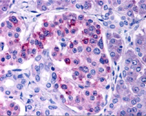
| Sample | Formaldehyde-fixed, paraffin-embedded human pancreas tissue (datasheet A01816) |
| Fixation | Image formalin-fixed; duration unreported (datasheet A01816); verify before use. |
| Sectioning | 4–5 µm sections on charged slides (standard) |
| Deparaffinisation | Xylene, graded ethanol series to water (standard) |
| Antigen retrieval | Heat retrieval: Citrate pH 6 (datasheet A01816); 20 min, 95–100 °C (standard) |
| Peroxidase block | 3% H2O2, 10 min, room temperature (standard) |
| Blocking | 10% serum (datasheet A01816) |
| Primary antibody | Rabbit anti-IKBKE, 10 μg/mL (datasheet A01816) |
| Primary incubation | Overnight at 4 °C (standard) |
| Detection | HRP-polymer secondary, DAB chromogen 5–10 min (standard) |
| Counterstain | Hematoxylin, blue, dehydrate and mount (standard) |
| Expected result | IKBKE-positive staining in glandular cells of adrenal gland (HPA tissue IHC: High). HPA tissue profile: Cytoplasmic expression in most tissues. No signal in the no-primary control. |
IKBKE is primarily cytoplasmic: HPA reports cytoplasmic staining in most tissues, and ICC-IF supports cytosolic localisation (HPA). Expect staining in glandular and hematopoietic cells in the listed positive tissues, with intensity varying by tissue and cell type (HPA). Nuclear localisation can occur under specific conditions (UniProt Q14164). IKBKE has no transmembrane segment (UniProt Q14164 topology). HPA rates its tissue IHC evidence Approved, with medium staining–RNA consistency and external verification pending (HPA).
| Distinct cytoplasmic staining in adrenal or breast glandular cells, or bone marrow hematopoietic cells. | This fits the expected compartment and cells: HPA records High staining in each of these tissue–cell combinations (HPA). Score the identified cells and their cytoplasm, since a tissue-wide impression can obscure which cells carry signal (standard IHC practice). Agreement with these examples supports interpretation but does not independently establish antibody specificity; HPA's tissue evidence still awaits external verification (HPA). |
| Predominantly nuclear or membrane-like staining, with little cytoplasmic signal. | An isolated nuclear pattern conflicts with HPA's usual cytoplasmic tissue profile and supported cytosolic ICC-IF location (HPA). Membrane-like staining also lacks support from IKBKE topology, which has no transmembrane segment (UniProt Q14164 topology). Nuclear signal is not automatically artefactual: UniProt describes TOPORS-dependent targeting to PML nuclear bodies after DNA damage (UniProt Q14164). Interpret that exception only when the experimental context supports it. |
| Strong staining in cells outside the expected positive population, while the listed cells remain faint. | For example, HPA records High staining in bone marrow hematopoietic cells, but Low staining in ovarian stromal cells and adipocytes (HPA). A reversed pattern raises concern about cross-reactivity, endogenous detection activity, or cell identification (standard IHC practice). HPA's listed levels describe observed examples, not a rule that every unlisted cell must be negative; compare morphology and controls before calling a new distribution. |
| Diffuse colour covers cells, extracellular spaces, and the slide without a clear cytoplasmic boundary. | That appearance does not reproduce HPA's cellular cytoplasmic profile (HPA). Broad deposit can arise from background binding or detection chemistry, so it should not be scored as IKBKE-positive cells without an interpretable intracellular pattern (standard IHC practice). Inspect the no-primary control and tissue morphology, then compare staining confined to plausible cells with colour spread across unrelated structures. |
| Little or no signal in a selected positive tissue, including its expected cell population. | HPA lists High staining in several candidate positive samples, including adrenal glandular cells and bone marrow hematopoietic cells (HPA). A blank section therefore warrants a run-level check of the antibody and chromogenic detection workflow (standard IHC practice). It does not by itself prove absent IKBKE: HPA calls its tissue IHC Approved but reports only medium staining–RNA consistency and pending external verification (HPA). |
| Tissue and cell selection | HPA reports High staining in adrenal, breast, duodenal, epididymal, fallopian-tube and gallbladder glandular cells, and bone marrow hematopoietic cells; endometrial glandular cells are Medium (HPA). HPA lists glial cells in hippocampus and caudate, ovarian stromal cells, and adipocytes as Low (HPA). Choose comparisons by cell population as well as tissue name, and treat those levels as observations rather than guarantees for every section. |
| Compartment and stimulus | Cytoplasmic staining is the usual tissue pattern, and cytosol is the supported ICC-IF location (HPA). UniProt also places IKBKE in the nucleus and PML bodies, with TOPORS-dependent PML targeting after DNA damage, and describes punctate cytoplasmic bodies with TRIM6 coexpression (UniProt Q14164). Those conditional patterns require matching experimental context; routine nuclear or punctate staining alone does not establish the condition. |
| Antibody evidence and isoforms | HPA labels tissue IHC for HPA015788 and CAB025983 Approved, and ICC for CAB025983 Supported (HPA). UniProt lists two IKBKE isoforms (UniProt Q14164). The supplied record gives no epitope positions, so isoform coverage cannot be inferred; verify the selected antibody's stated application and epitope information before interpreting discrepant staining (standard IHC practice). |
| Fixation evidence | Target-specific fixation sensitivity is unreported in the supplied UniProt and HPA records. Their topology, modification sites, and observed tissue staining do not establish how fixation or antigen retrieval affects IKBKE detection (UniProt Q14164; HPA). Record those workflow conditions when comparing sections, without attributing an observed difference to a documented IKBKE fixation effect. |
| Situation | Likely cause | Next action |
|---|---|---|
| The entire section is pale, including expected positive cells. | A detection-run problem is possible; HPA nevertheless records High staining in selected glandular and hematopoietic populations (HPA; standard IHC practice). | Check that a suitable positive section stains in the same run, then review the antibody's IHC validation and the detection reagents (HPA; standard IHC practice). Confirm the intended cells are present before treating an unstained section as biological absence. |
| Colour remains in a no-primary control or coats unrelated tissue structures. | Endogenous activity or nonspecific detection can produce chromogen independently of primary-antibody binding (standard IHC practice). | Review the detection system's endogenous-activity block and background controls, and score only cell-associated staining with plausible cytoplasmic localisation (standard IHC practice; HPA). Do not use widespread colour as evidence for the HPA tissue pattern. |
| Only nuclei stain strongly in an otherwise routine section. | That differs from HPA's predominant cytoplasmic profile, although conditional nuclear and PML-body localisation is reported by UniProt (HPA; UniProt Q14164). | Check morphology and compartment boundaries; assess whether DNA damage and TOPORS-dependent targeting are relevant to this experiment before assigning the nuclear signal to IKBKE (UniProt Q14164; standard IHC practice). |
| Unexpected cells stain more strongly than the HPA-listed target cells. | Cell misidentification, cross-reactivity, or background detection may explain the mismatch (standard IHC practice); HPA levels are tissue–cell observations with medium staining–RNA consistency (HPA). | Recheck cell identity on the counterstained section, compare a listed High population and a listed Low population, and inspect controls before extending the expected pattern to new cells (HPA; standard IHC practice). |
| Two antibodies give different tissue distributions. | HPA lists HPA015788 and CAB025983 as IHC Approved but describes overall tissue staining–RNA consistency as Medium, with external verification pending (HPA). The supplied record does not identify their epitopes. | Compare staining in the same identified cell populations and check each antibody's application and epitope documentation; do not resolve the disagreement by assuming either antibody detects both UniProt isoforms (HPA; UniProt Q14164; standard IHC practice). |
| Can IF/ICC help assess a disputed compartment? | HPA reports supported cytosolic ICC-IF localisation and marks CAB025983 ICC Supported; its imaged cell lines include A-431, U-251MG and U2OS (HPA). | Use that HPA localisation as an independent compartment comparison when interpreting IHC, while recognising that the listed ICC-IF evidence does not establish the staining pattern of every paraffin tissue (HPA). |


Comprehensive Human Protein Atlas IHC scoring per tissue (reliability: Approved — Medium consistency between antibody staining and RNA expression data. Pending external verification.). Rows are taken directly from the HPA tissue chart — click any row's HPA link to view the source.
| Tissue | Cell type | Level | Evidence | Source |
|---|---|---|---|---|
| Adrenal gland | Glandular cells | High | Protein (IHC) | HPA → |
| Bone marrow | Hematopoietic cells | High | Protein (IHC) | HPA → |
| Breast | Glandular cells | High | Protein (IHC) | HPA → |
| Duodenum | Glandular cells | High | Protein (IHC) | HPA → |
| Endometrium | Glandular cells | Medium | Protein (IHC) | HPA → |
| Tissue | Cell type | Level | Evidence | Source |
|---|---|---|---|---|
| None in HPA: IKBKE is detected in all 45 scored tissues. Use a no-primary (secondary-only) and an isotype control instead. | ||||
Troubleshoot IKBKE staining in paraffin sections by checking retrieval, cell type, compartment and controls before comparing staining intensity across samples.
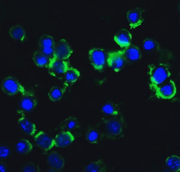
A01816 has human paraffin-section IHC and HeLa cell IF images; its catalog also lists human and mouse reactivity (A01816: IHC and IF captions; catalog reactivity).
A01816 is the only SKU with a rendered card: its images show IHC in human pancreas paraffin sections and IF in HeLa cells (A01816: IHC and IF captions). M01816 has IHC and ICC/IF in its application list but no IHC or IF image caption, so it has no rendered card (M01816: catalog applications and image alts).
Which to pick: Choose A01816 for tissue IHC: its own caption documents formaldehyde-fixed human pancreas paraffin sections, citrate retrieval at pH 6, and primary antibody at 10 μg/mL (A01816: IHC caption). For IF with image evidence, choose A01816 at 20 μg/mL in paraformaldehyde-fixed HeLa cells; M01816 lists ICC/IF and is monoclonal, but its fixative is unreported and it has no IF image caption (A01816: IF caption; M01816: catalog applications, clone and image alts). For human and mouse reactivity, choose A01816; its IHC and IF image evidence is human only (A01816: catalog reactivity, IHC and IF captions).