This website uses cookies to ensure you get the best experience on our website.


- Table of Contents
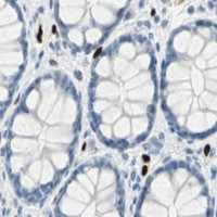
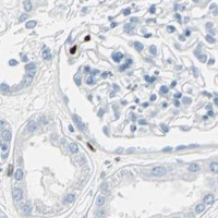
Plan IL33 paraffin-section IHC around selective nuclear staining in endothelial, immune and epithelial cells (HPA tissue IHC). Use bronchial respiratory epithelium as a positive reference and adrenal glandular cells as a negative reference (HPA tissue IHC).

Expected localisation, validated protocols, controls and antibodies — the at-a-glance facts below, then the full design guide.
| Expected localisation | Nuclear staining in select cells (HPA tissue IHC) | |
| Staining pattern | Nuclear endothelial staining; immune and epithelial subsets (HPA tissue IHC) | |
| Antigen retrieval | Citrate pH 6 HIER, heat-mediated (datasheet A00113) | |
| Positive control | Bronchus+4 more · see all | |
| Negative control | Adrenal gland+4 more · see all |
| Fixation | Keep fixation consistent across samples (standard IHC practice; not target-specific); validate locally. | |
| Caveat | Secreted IL33 may differ from the nuclear tissue pattern (UniProt) | |
| Regulation | High in high endothelial venules (UniProt) | |
| Isoform / epitope | 4 isoforms; cleavage can remove N-terminal epitopes (UniProt) |
The catalog antibody uses citrate pH 6 heat retrieval (datasheet: A00113). The four published IL33 IHC protocols below provide additional reported conditions (PMC13285577; PMC11794308; PMC10749886; PMC7060130).
| Sample | Paraffin-embedded human tonsil tissue; fixative not specified (datasheet A00113) |
| Fixation | Image fixative and duration unreported (datasheet A00113); verify before use. |
| Sectioning | 4–5 µm sections on charged slides (standard) |
| Deparaffinisation | Xylene, graded ethanol series to water (standard) |
| Antigen retrieval | Heat retrieval: Citrate pH 6, 20 min (datasheet A00113) |
| Peroxidase block | 3% H2O2, 10 min, room temperature (standard) |
| Blocking | 10% goat serum (datasheet A00113) |
| Primary antibody | Rabbit anti-IL33, 1:100–1:500 starting range (standard) |
| Primary incubation | Overnight at 4 °C (standard) |
| Detection | Streptavidin-biotin complex (SABC), DAB chromogen (datasheet A00113) |
| Counterstain | Hematoxylin, blue, dehydrate and mount (standard) |
| Expected result | IL33-positive staining in respiratory epithelial cells of bronchus (HPA tissue IHC: High). HPA tissue profile: High selective nuclear expression in endothelial cells, in a subset of immune cells in non-germinal center and in a subset of cells in epithelia. No signal in the no-primary control. |
Expect nuclear IL33 in endothelial and select epithelial and immune cells (HPA: Supported IHC; UniProt: no TM segment).
| Strong epithelial nuclear stain in bronchus (HPA: High). | Expected positive pattern (HPA: High in bronchial respiratory epithelium). |
| Nuclear stain in endothelial cells (HPA: tissue IHC profile). | Consistent with selective endothelial expression (HPA: tissue IHC profile). |
| Membrane-only staining (UniProt: no TM segment). | Unexpected for IL33; assess nonspecific staining (UniProt: no TM segment; general IHC practice). |
| Stain in adrenal glandular cells (HPA: Not detected). | Check cross-reactivity or endogenous detection activity (HPA: Not detected; general IHC practice). |
| Diffuse stain across the section (general IHC practice). | Suggests background; compare a no-primary control (general IHC practice). |
| IHC antibody validation (HPA: HPA024426 and CAB007057 Supported). | These antibodies have Supported IHC status (HPA: antibody validation). |
| Processing (UniProt: full-length and truncated IL33 chains). | Epitope location determines which chains an antibody can detect (UniProt: processing; general IHC practice). |
| Secretion (UniProt: extracellular release). | Secreted IL33 may extend beyond its producing cell (UniProt: secretion). |
| IF localisation (HPA: supported nucleoplasm and vesicles). | Vesicular signal can occur; assess it separately from the expected nuclear IHC pattern (HPA: subcellular). |
| Situation | Likely cause | Next action |
|---|---|---|
| No stain in bronchial epithelium (HPA: High). | Possible assay failure (general IHC practice). | Check IHC antibody validation and optimise retrieval with controls (HPA: antibodies; general IHC practice). |
| Only membrane outlines stain (UniProt: no TM segment). | Possible nonspecific staining (general IHC practice). | Compare no-primary and known-positive controls (general IHC practice). |
| Adrenal glandular cells stain (HPA: Not detected). | Possible cross-reactivity or endogenous activity (general IHC practice). | Check no-primary and detection controls (general IHC practice). |
| Diffuse slide-wide colour (general IHC practice). | Possible background from detection or insufficient blocking (general IHC practice). | Check no-primary control; optimise blocking and washes (general IHC practice). |
| Weak nuclear stain in a positive region (HPA: bronchus High). | Retrieval or antibody dilution may need optimisation (general IHC practice). | Titrate retrieval and dilution using a positive control (general IHC practice). |
| IF shows vesicles alongside nuclei (HPA: subcellular). | Both locations are supported in ICC-IF (HPA: subcellular). | Score nuclear and vesicular signal separately (HPA: subcellular; general IF practice). |



![HPA ICC-IF BJ [Human fibroblast] — IL33 (HPA022899)](https://images.proteinatlas.org/22899/1936_B1_1_blue_red_green.jpg)
![HPA ICC-IF BJ [Human fibroblast] — IL33 (HPA022899)](https://images.proteinatlas.org/22899/1936_B1_3_blue_red_green.jpg)
Comprehensive Human Protein Atlas IHC scoring per tissue (reliability: Supported — High consistency between antibody staining and RNA expression data. At least one protein variant secreted, tissue location of RNA and protein might differ and correlation is complex. Antibody staining in cells/structures not annotated, view images.). Rows are taken directly from the HPA tissue chart — click any row's HPA link to view the source.
| Tissue | Cell type | Level | Evidence | Source |
|---|---|---|---|---|
| Bronchus | Respiratory epithelial cells | High | Protein (IHC) | HPA → |
| Cervix | Glandular cells | High | Protein (IHC) | HPA → |
| Endometrium | Ciliated epithelial cells | High | Protein (IHC) | HPA → |
| Nasopharynx | Respiratory epithelial cells | High | Protein (IHC) | HPA → |
| Stomach | Glandular cells | High | Protein (IHC) | HPA → |
| Tissue | Cell type | Level | Evidence | Source |
|---|---|---|---|---|
| Adrenal gland | Glandular cells | Not detected | Protein (IHC) | HPA → |
| Duodenum | Glandular cells | Not detected | Protein (IHC) | HPA → |
| Epididymis | Glandular cells | Not detected | Protein (IHC) | HPA → |
| Fallopian tube | Glandular cells | Not detected | Protein (IHC) | HPA → |
| Gallbladder | Glandular cells | Not detected | Protein (IHC) | HPA → |
Troubleshoot IL33 staining in paraffin sections by checking retrieval, nuclear localisation, cell identity and controls before comparing staining scores (HPA tissue IHC; UniProt O95760).
A00113 has real IHC data from human tonsil paraffin sections (A00113 image caption); the catalog supplies no IF figure or IF validation (catalog).
A00113 is listed for human IHC (catalog: applications and reactivity). Its IHC figure shows staining in a human tonsil paraffin section (A00113 image caption).
Which to pick: Choose A00113 for human tissue IHC in paraffin sections; its caption reports citrate retrieval at pH 6 for 20 minutes, and does not report the fixative (A00113 image caption). There is no catalog supported IF/ICC or cross species choice here: A00113 lists human reactivity and IHC, but no IF/ICC application or other species (catalog: applications and reactivity).