Click image to see more details
Product Info Summary
| SKU: | A00113 |
|---|---|
| Size: | 100 μg/vial |
| Reactive Species: | Human |
| Host: | Rabbit |
| Application: | IHC, WB, ELISA (Cap) |
Customers Who Bought This Also Bought

Product info
Product Name
Anti-IL33 Antibody Picoband®
SKU/Catalog Number
A00113
Size
100 μg/vial
Form
Lyophilized
Description
Boster Bio Anti-IL33 Antibody Picoband® catalog # A00113. Tested in ELISA, IHC, WB applications. This antibody reacts with Human. The brand Picoband indicates this is a premium antibody that guarantees superior quality, high affinity, and strong signals with minimal background in Western blot applications. Only our best-performing antibodies are designated as Picoband, ensuring unmatched performance.
Storage & Handling
Store at -20˚C for one year from date of receipt. After reconstitution, at 4˚C for one month. It can also be aliquotted and stored frozen at -20˚C for six months. Avoid repeated freeze-thaw cycles.
Cite This Product
Anti-IL33 Antibody Picoband® (Boster Biological Technology, Pleasanton CA, USA, Catalog # A00113)
Host
Rabbit
Contents
Each vial contains antibody formulated with stabilizing components, 0.9 mg NaCl, 0.2 mg Na2HPO4, and 0.05 mg NaN3.
*This antibody is supplied in a stabilized formulation.
Compatibility with conjugation reactions depends on the chemistry of the conjugation method used.
For conjugation methods that are not compatible with the stabilizing components present in this formulation, a carrier-free antibody format is required.
Clonality
Polyclonal
Isotype
Rabbit IgG
Immunogen
E. coli-derived human IL33 recombinant protein (Position: A95-T270).
Cross-reactivity
No cross-reactivity with other proteins.
Reactive Species
A00113 is reactive to IL33 in Human
Observed Molecular Weight
18 kDa
Calculated molecular weight
30.8 kDa
Background of IL33
Interleukin 33 (IL-33) is a protein that in humans is encoded by the IL33 gene. The protein encoded by this gene is a cytokine that binds to the IL1RL1/ST2 receptor. The encoded protein is involved in the maturation of Th2 cells and the activation of mast cells, basophils, eosinophils and natural killer cells. Several transcript variants encoding different isoforms have been found for this gene.
Antibody Validation
Boster validates all antibodies on WB, IHC, ICC, Immunofluorescence, and ELISA with known positive control and negative samples to ensure specificity and high affinity, including thorough antibody incubations.
Application & Images
Applications
A00113 is guaranteed for IHC, WB, ELISA (Cap) Boster Guarantee
Recommend Dilution
| Application | Dilution | Species |
|---|---|---|
| Western blot | 0.1-0.5μg/ml | |
| Immunohistochemistry (Paraffin-embedded Section) | 0.5-1μg/ml | |
| ELISA (Cap) | 1-5μg/ml |
Tested application
Use TE buffer pH 9.0 for antigen retrieval; (*) citrate buffer pH 6.0 is an alternative.
Validation Images & Assay Conditions
Click image to see more details
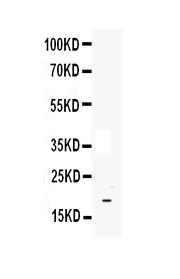
Western blot analysis of IL33 using anti-IL33 antibody (A00113).
Electrophoresis was performed on a 5-20% SDS-PAGE gel at 70V (Stacking gel) / 90V (Resolving gel) for 2-3 hours.
Lane 1: recombinant human IL33 protein 1ng.
After Electrophoresis, proteins were transferred to a Nitrocellulose membrane at 150mA for 50-90 minutes. Blocked the membrane with 5% Non-fat Milk/ TBS for 1.5 hour at RT. The membrane was incubated with rabbit anti-IL33 antigen affinity purified polyclonal antibody (Catalog # A00113) at 0.5 μg/mL overnight at 4°C, then washed with TBS-0.1%Tween 3 times with 5 minutes each and probed with a goat anti-rabbit IgG-HRP secondary antibody at a dilution of 1:10000 for 1.5 hour at RT. The signal is developed using an Enhanced Chemiluminescent detection (ECL) kit (Catalog # EK1002) with Tanon 5200 system. A specific band was detected for IL33 at approximately 18KD. The expected band size for IL33 is at 18KD.

Click image to see more details
Immunohistochemistry analysis of TNF-α, PPAR-γ, IL-6, and IL-33 in lesional and non-lesional skin samples of patients with dermatomyositis. (A) TNF-α: Left panel: Expression of TNF-α in lesional and non-lesional dermatomyositis skin samples. The cytoplasmic expression is similar in the keratinocytes, dendritic cells (black arrow), and endothelial cells (empty arrow). Top right and left corners: Negative controls were obtained by omitting the primary antibody. Right panel: Statistical analysis of semi-quantitative measurements; (independent sample t -test; p > 0.1). (B) PPAR-γ: Left panel: Expression of PPAR-γ in lesional and non-lesional dermatomyositis skin samples. There is no difference between the lesional and non-lesional DM skin samples in the expression of the PPAR-γ. Empty arrow: endothelial cells. Black arrow: dendritic cells. Top right and left corners: Negative controls were obtained by omitting the primary antibody. Right panel: Statistical analysis of semi-quantitative measurements; (independent sample t -test; p > 0.1). (C) IL-6: Left panel: Expression of IL-6 in lesional and non-lesional dermatomyositis skin samples. Strong cytoplasmic/nuclear reaction with IL-6 in the epidermal endothelial (empty arrow) and dendritic cells (black arrow). Top right and left corners: Negative controls were obtained by omitting the primary antibody. Right panel: Statistical analysis of semi-quantitative measurements; (independent sample t -test; p > 0.1). (D) IL-33: Expression of IL-33 in lesional and non-lesional dermatomyositis skin samples. Positive reaction was detected in the nucleus of endothelial cells with IL-33, but there was no significant difference between lesional and non-lesional DM skin samples. Top right and left corners: Negative controls were obtained by omitting the primary antibody. Right panel: Statistical analysis of semi-quantitative measurements; (independent sample t -test; p > 0.1).
Index in PubMed under a CC BY license. PMID: 37250649

Click image to see more details
IHC analysis of IL33 using anti-IL33 antibody (A00113).
IL33 was detected in paraffin-embedded section of human tonsil . Heat mediated antigen retrieval was performed in citrate buffer (pH6, epitope retrieval solution) for 20 mins. The tissue section was blocked with 10% goat serum. The tissue section was then incubated with 1ug/ml rabbit anti-IL33 Antibody (A00113) overnight at 4 Biotinylated goat anti-rabbit IgG was used as secondary antibody and incubated for 30 minutes at 37 The tissue section was developed using Strepavidin-Biotin-Complex (SABC)(Catalog # SA1022) with DAB as the chromogen.

Click image to see more details
Sandwich ELISA - Recombinant human IL33 protein standard curve.
Use in combination with reagents from Human IL33 ELISA Kit EZ-Set (DIY Antibody Pairs) (EZ0929).
Specific Publications For Anti-IL33 Antibody Picoband® (A00113)
Loading publications
Recommended Resources
Here are featured tools and databases that you might find useful.
- Boster's Pathways Library
- Protein Databases
- Bioscience Research Protocol Resources
- Data Processing & Analysis Software
- Photo Editing Software
- Scientific Literature Resources
- Research Paper Management Tools
- Molecular Biology Software
- Primer Design Tools
- Bioinformatics Tools
- Phylogenetic Tree Analysis
Customer Reviews
Have you used Anti-IL33 Antibody Picoband®?
Share your experimental results or join a short interview to earn up to $1,000 in product credits or other rewards.
0 Reviews For Anti-IL33 Antibody Picoband®
Customer Q&As
Have a question?
Find answers in Q&As, reviews.
Can't find your answer?
Submit your question
4 Customer Q&As for Anti-IL33 Antibody Picoband®
Question
Our team were happy with the WB result of your anti-IL33 antibody. However we have seen positive staining in left coronary artery nucleus using this antibody. Is that expected? Could you tell me where is IL33 supposed to be expressed?
A. Johnson
Verified customer
Asked: 2017-04-07
Answer
From what I have seen in literature, left coronary artery does express IL33. Generally IL33 expresses in nucleus. Regarding which tissues have IL33 expression, here are a few articles citing expression in various tissues:
Adrenal gland, and Trachea, Pubmed ID: 14702039
Brain, Pubmed ID: 15489334
Endothelial cell, Pubmed ID: 12819012
Boster Scientific Support
Answered: 2017-04-07
Question
We are currently using anti-IL33 antibody A00113 for human tissue, and we are content with the ELISA results. The species of reactivity given in the datasheet says human. Is it likely that the antibody can work on canine tissues as well?
B. Yang
Verified customer
Asked: 2015-11-23
Answer
The anti-IL33 antibody (A00113) has not been tested for cross reactivity specifically with canine tissues, though there is a good chance of cross reactivity. We have an innovator award program that if you test this antibody and show it works in canine you can get your next antibody for free. Please contact me if I can help you with anything.
Boster Scientific Support
Answered: 2015-11-23
Question
We have tried in the past anti-IL33 antibody for WB on brain in a previous experiment. I am using human, and I plan to use the antibody for ELISA next. I am interested in examining brain as well as endothelial cell in our next experiment. Could give a recommendation on which antibody would work the best for ELISA?
T. Jha
Verified customer
Asked: 2015-04-23
Answer
I have checked the website and datasheets of our anti-IL33 antibody and it appears that A00113 has been validated on human in both WB and ELISA. Thus A00113 should work for your application. Our Boster satisfaction guarantee will cover this product for ELISA in human even if the specific tissue type has not been validated. We do have a comprehensive range of products for ELISA detection and you can check out our website bosterbio.com to find out more information about them.
Boster Scientific Support
Answered: 2015-04-23
Question
We have seen staining in human adrenal gland trachea. What should we do? Is anti-IL33 antibody supposed to stain adrenal gland trachea positively?
C. Zhang
Verified customer
Asked: 2014-09-02
Answer
Based on literature adrenal gland trachea does express IL33. Based on Uniprot.org, IL33 is expressed in left coronary artery, endothelial cell, adrenal gland trachea, brain, among other tissues. Regarding which tissues have IL33 expression, here are a few articles citing expression in various tissues:
Adrenal gland, and Trachea, Pubmed ID: 14702039
Brain, Pubmed ID: 15489334
Endothelial cell, Pubmed ID: 12819012
Boster Scientific Support
Answered: 2014-09-02
