This website uses cookies to ensure you get the best experience on our website.


- Table of Contents
This guide maps cytoplasmic ILK staining and positive tissue controls for paraffin IHC (HPA tissue IHC). It covers antibody titration, consistent fixation, and chromogenic scoring (datasheet A02932-3; standard IHC practice).

Expected localisation, validated protocols, controls and antibodies — the at-a-glance facts below, then the full design guide.
| Expected localisation | Cytoplasmic staining in most organs (HPA tissue IHC) | |
| Staining pattern | Myocyte cytoplasm prominent; cytoplasm in most organs (HPA tissue IHC) | |
| Antigen retrieval | EDTA pH 8.0 HIER, heat-mediated (datasheet A02932-3) | |
| Positive control | Kidney+4 more · see all | |
| Negative control | Adipose tissue+4 more · see all |
| Fixation | Keep fixation conditions consistent across sections. (standard IHC practice; not target-specific) | |
| Caveat | Antibody staining and RNA show medium consistency (HPA tissue IHC) | |
| Regulation | No defined intensity regulator (UniProt) | |
| Isoform / epitope | 3 isoforms; epitope differences unspecified (UniProt) |
The catalog antibody’s IHC-P protocol is accompanied by 4 published ILK IHC protocols (PMC12826184; PMC2588856; PMC4878430; PMC546218).
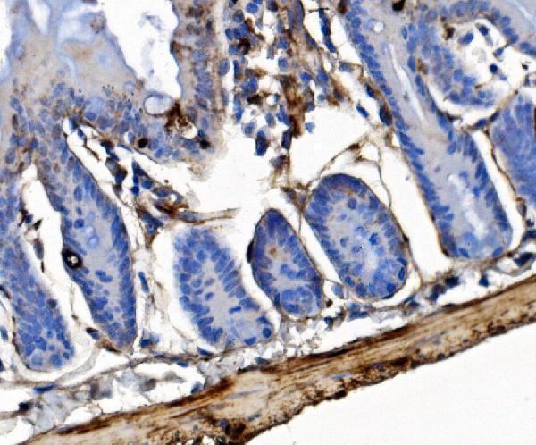
| Sample | Paraffin-embedded mouse colon tissue; fixative not specified (datasheet A02932-3) |
| Fixation | Image fixative and duration unreported (datasheet A02932-3); verify before use. |
| Sectioning | 4–5 µm sections on charged slides (standard) |
| Deparaffinisation | Xylene, graded ethanol series to water (standard) |
| Antigen retrieval | Heat retrieval: EDTA pH 8.0 (datasheet A02932-3); 20 min, 95–100 °C (standard) |
| Peroxidase block | 3% H2O2, 10 min, room temperature (standard) |
| Blocking | 10% goat serum (datasheet A02932-3) |
| Primary antibody | Rabbit anti-ILK, 2-5 μg/ml (datasheet A02932-3) |
| Primary incubation | Overnight at 4 °C (datasheet A02932-3) |
| Detection | Streptavidin-biotin complex (SABC), DAB chromogen (datasheet A02932-3) |
| Counterstain | Hematoxylin, blue, dehydrate and mount (standard) |
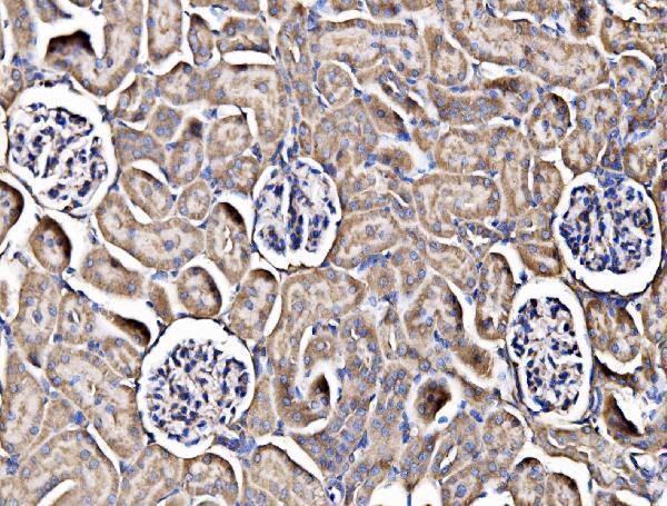
| Expected result | ILK-positive staining in cells in glomeruli of kidney (HPA tissue IHC: High). HPA tissue profile: Cytoplasmic expression in most organs, with highest abundance in muscle tissues. No signal in the no-primary control. |
ILK is a cytoplasmic scaffold associated with focal adhesions and other intracellular sites; it has no transmembrane segment (UniProt Q13418 topology and subcellular location). In tissue IHC, expect the clearest signal in skeletal muscle myocytes and kidney glomerular cells, both scored High (HPA tissue IHC). HPA rates its tissue profile Enhanced, while noting medium consistency between staining and RNA data (HPA tissue IHC).
| Strong cytoplasmic staining in skeletal muscle myocytes or kidney glomerular cells, with little diffuse slide background. | This matches two HPA High cell populations and the broad cytoplasmic tissue profile (HPA tissue IHC). Focal adhesion enrichment is plausible from ILK localization, but tissue IHC need not resolve individual adhesion sites (UniProt Q13418; HPA subcellular ICC-IF). |
| Staining appears mainly as a crisp outline around every cell, with little cytoplasmic signal. | Treat a uniform surface-only pattern as suspect: ILK has no transmembrane segment, although it associates with the cell membrane and cortex (UniProt Q13418 topology and subcellular location). Compare compartmental staining with a known-positive section before interpreting the outline as ILK. |
| Strong staining appears in adipocytes or liver cholangiocytes while known-positive cells are weak. | HPA reports ILK as Not detected in those specific cell populations, so this reversal raises cross-reactivity or endogenous detection activity as possibilities (HPA tissue IHC; general IHC practice). It does not establish that the entire adipose or liver section must be negative. |
| Brown color is spread across tissue, empty spaces, or the whole section without a clear cell-associated pattern. | Diffuse color is difficult to assign to ILK's reported cytoplasmic distribution (HPA tissue IHC). Check the no-primary detection control, washing, blocking, and chromogen development to distinguish background from cell-associated signal (general IHC practice). |
| No signal is visible in skeletal muscle myocytes or kidney glomerular cells. | Those are HPA High reference populations, so a blank result first calls for a run-level check (HPA tissue IHC). Confirm that a suitable positive section stained in the same run and review retrieval, primary antibody use, and detection steps (general IHC practice). |
| Tissue and cell selection | Use skeletal muscle myocytes or kidney glomerular cells as strong reference populations; HPA scores both High (HPA tissue IHC). Several glandular and hematopoietic populations score Medium, so weaker staining there can still fit the reported profile (HPA tissue IHC). |
| Compartment and topology | ILK is reported at focal adhesions, in cytoplasm, and at additional intracellular sites, with no transmembrane segment (UniProt Q13418). Interpret membrane-adjacent staining in its cellular context rather than requiring a continuous cell-surface rim (UniProt Q13418 topology and subcellular location). |
| HPA IHC evidence | The tissue profile is rated Enhanced but has medium staining–RNA consistency (HPA tissue IHC). CAB004041 carries an IHC Enhanced validation label; that supports pattern interpretation, while the supplied record does not establish equivalence for an unspecified catalog antibody (HPA antibodies). |
| Isoforms and processing | UniProt lists three ILK isoforms and one 1–452 chain, with no signal peptide or propeptide (UniProt Q13418). Without an antibody epitope in the supplied record, isoform coverage cannot be predicted; there is no basis here for a shedding-based staining pattern. |
| Antigen retrieval | For paraffin IHC, compare the catalog antibody's stated retrieval condition with the lab's run conditions and a known-positive section (general IHC practice). The supplied UniProt and HPA records do not report an ILK-specific retrieval requirement or fixation sensitivity. |
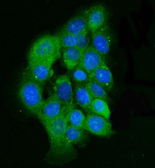
| IF/ICC Q&A: should puncta be expected? | Yes, focal adhesion sites are the supported main ICC-IF location; cytosol is also supported, actin filaments approved, and nucleoplasm uncertain (HPA subcellular ICC-IF). This is context for a separate IF/ICC guide, not an IHC protocol option. |
| Situation | Likely cause | Next action |
|---|---|---|
| Known-positive myocytes and glomerular cells are blank. | The expected High populations failed to stain (HPA tissue IHC); a technical failure is possible (general IHC practice). | Check a same-run positive section, the catalog antibody's IHC-P instructions, retrieval execution, reagent sequence, and detection performance (general IHC practice). Do not infer ILK absence from this run alone. |
| All cells show similar brown color, including HPA Not detected cell populations. | A uniform pattern conflicts with the HPA cell-specific observations; endogenous detection activity or nonspecific binding may contribute (HPA tissue IHC; general IHC practice). | Examine a no-primary control and assess blocking, washes, and detection chemistry; compare cell types within the same section (general IHC practice). |
| Only a sharp continuous cell border stains. | A surface-only pattern needs review because ILK lacks a transmembrane segment, though membrane and cortex association is reported (UniProt Q13418 topology and subcellular location). | Compare cytoplasmic staining in a known-positive section and inspect the no-primary control before assigning the border to ILK (HPA tissue IHC; general IHC practice). |
| Nuclear staining is present alongside cytoplasmic staining. | Nuclear localization is listed by UniProt, while HPA rates ICC-IF nucleoplasmic localization uncertain (UniProt Q13418; HPA subcellular ICC-IF). | Record nuclear and cytoplasmic staining separately, check the no-primary control, and avoid treating nuclear color alone as a definitive positive IHC result (general IHC practice). |
| A Medium-scoring glandular population stains less intensely than skeletal muscle. | That difference can follow HPA's cell-level ratings: several glandular populations are Medium, while skeletal muscle myocytes are High (HPA tissue IHC). | Score the named cell population and compartment rather than requiring equal intensity across organs; compare sections processed in the same run (HPA tissue IHC; general IHC practice). |
| A whole organ is called negative because one listed cell type has no staining. | HPA's Not detected entries refer to specified cells, such as adipocytes or liver cholangiocytes, rather than every cell in those organs (HPA tissue IHC). | Restrict the interpretation to the observed cell population and inspect other cell types individually; use a known-positive section to verify the run (HPA tissue IHC; general IHC practice). |


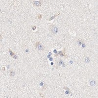
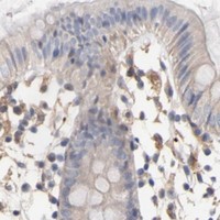
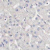
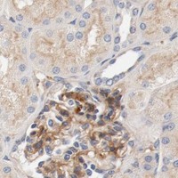


Comprehensive Human Protein Atlas IHC scoring per tissue (reliability: Enhanced — Medium consistency between antibody staining and RNA expression data. Antibody staining in cells/structures not annotated, view images.). Rows are taken directly from the HPA tissue chart — click any row's HPA link to view the source.
| Tissue | Cell type | Level | Evidence | Source |
|---|---|---|---|---|
| Kidney | Cells in glomeruli | High | Protein (IHC) | HPA → |
| Skeletal muscle | Myocytes | High | Protein (IHC) | HPA → |
| Adrenal gland | Glandular cells | Medium | Protein (IHC) | HPA → |
| Bone marrow | Hematopoietic cells | Medium | Protein (IHC) | HPA → |
| Endometrium | Glandular cells | Medium | Protein (IHC) | HPA → |
| Tissue | Cell type | Level | Evidence | Source |
|---|---|---|---|---|
| Adipose tissue | Adipocytes | Not detected | Protein (IHC) | HPA → |
| Caudate | Glial cells | Not detected | Protein (IHC) | HPA → |
| Cerebellum | Cells in granular layer | Not detected | Protein (IHC) | HPA → |
| Esophagus | Squamous epithelial cells | Not detected | Protein (IHC) | HPA → |
| Hippocampus | Glial cells | Not detected | Protein (IHC) | HPA → |
Use the validated paraffin-section workflow as a starting point, then judge ILK staining by cell type, compartment and appropriate controls.
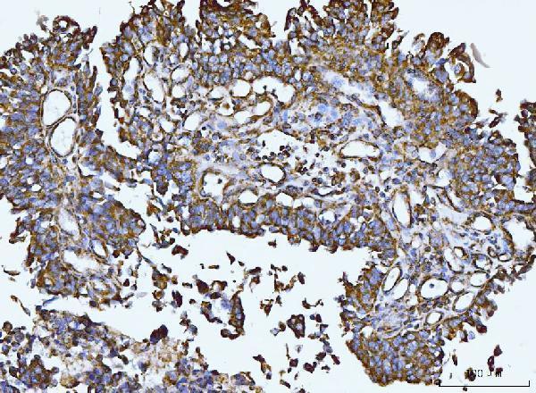
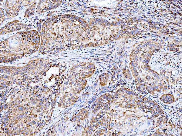
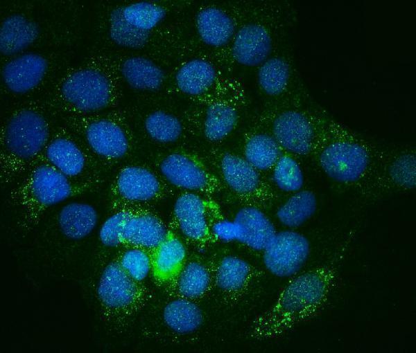
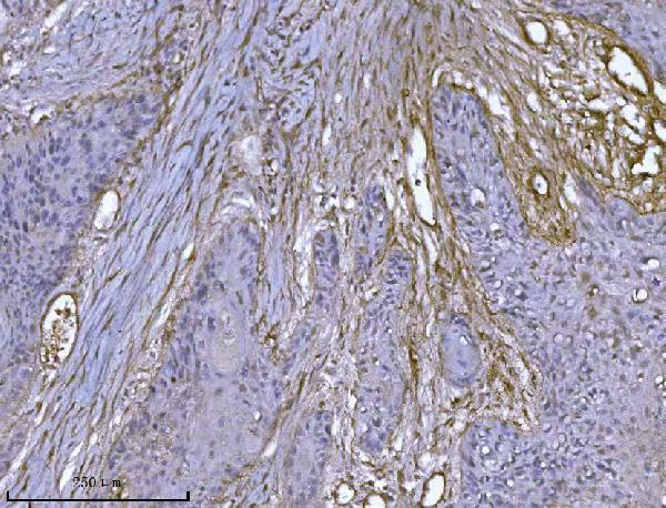
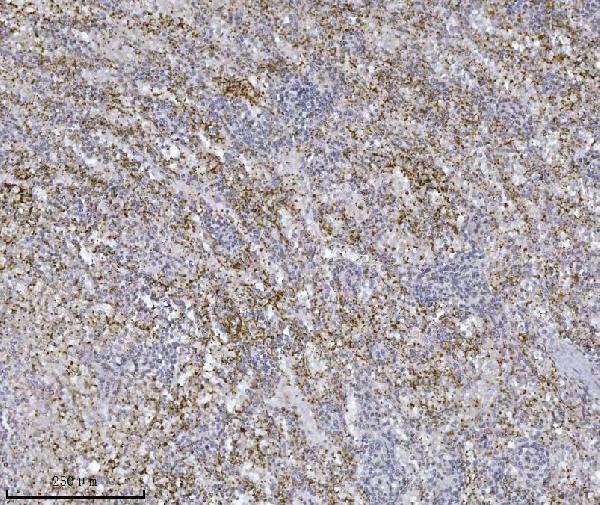
Anti-ILK antibodies have IHC images from human, mouse and rat paraffin sections and IF/ICC images from A431 and 293 cells (catalog: IHC and IF image captions).
A02932-3 has paraffin-section IHC images from mouse colon, rat kidney, human lung cancer and human renal clear cell carcinoma; A02932-2 has images from human bladder epithelial carcinoma, laryngeal squamous cell carcinoma and breast cancer (catalog: each SKU’s IHC captions). M02932-2 has paraffin-section IHC images from human lung carcinoma, human spleen and mouse kidney, while M02932 has a human stomach IHC image; A02932-2 and A02932-3 have A431 IF images, and M02932 has a 293-cell IF image (catalog: each SKU’s image captions).
Which to pick: For human tissue IHC, A02932-2 has several paraffin-section examples; choose M02932-2 if a mouse monoclonal is preferred (catalog: A02932-2 IHC captions; M02932-2 clone 3C7E1 and IHC captions). For IF/ICC, A02932-3 lists both applications and has an A431 IF image (catalog: A02932-3 applications and IF caption). For IHC across human, mouse and rat, A02932-3 has an image in each species; its paraffin-section captions do not report the fixative (catalog: A02932-3 IHC captions).