This website uses cookies to ensure you get the best experience on our website.


- Table of Contents
Plan chromogenic paraffin-section IHC for IRF1 using the catalog antibody’s tissue staining evidence (datasheet A00580-1). Assess nuclear and nucleolar staining, with placental endothelial cells and testis pachytene spermatocytes as high-staining references (HPA tissue IHC).

Expected localisation, validated protocols, controls and antibodies — the at-a-glance facts below, then the full design guide.
| Expected localisation | Nuclear and nucleolar tissue staining (HPA tissue IHC) | |
| Staining pattern | Nuclear/nucleolar; high in placental endothelial cells (HPA tissue IHC) | |
| Antigen retrieval | EDTA pH 8.0 HIER, heat-mediated (datasheet A00580-1) | |
| Positive control | Placenta+4 more · see all | |
| Negative control | Cerebellum+4 more · see all |
| Fixation | Keep fixation consistent across samples. (standard IHC practice; not target-specific) | |
| Caveat | MYD88 association can increase nuclear migration (UniProt) | |
| Regulation | No intensity regulator specified (UniProt) | |
| Isoform / epitope | No isoforms or processing variants annotated (UniProt) |
The catalog antibody’s IHC-P protocol is accompanied by published IRF1 staining methods for breast tissue (PMC4359953), human and mouse tissue (PMC13472745), and gastric tissue (PMC12799679).
| Sample | Paraffin-embedded human Lung cancer tissue; fixative not specified (datasheet A00580-1) |
| Fixation | Image fixative and duration unreported (datasheet A00580-1); verify before use. |
| Sectioning | 4–5 µm sections on charged slides (standard) |
| Deparaffinisation | Xylene, graded ethanol series to water (standard) |
| Antigen retrieval | Heat retrieval: EDTA pH 8.0 (datasheet A00580-1); 20 min, 95–100 °C (standard) |
| Peroxidase block | 3% H2O2, 10 min, room temperature (standard) |
| Blocking | 10% goat serum (datasheet A00580-1) |
| Primary antibody | Rabbit anti-IRF1, 0.5-1μg/ml (datasheet A00580-1) |
| Primary incubation | Overnight at 4 °C (datasheet A00580-1) |
| Detection | Streptavidin-biotin complex (SABC), DAB chromogen (datasheet A00580-1) |
| Counterstain | Hematoxylin, blue, dehydrate and mount (standard) |
| Expected result | IRF1-positive staining in endothelial cells of placenta (HPA tissue IHC: High). HPA tissue profile: Nuclear and nucleolar expression in several different tissue types. No signal in the no-primary control. |
IRF1 is expected mainly in nuclei, with nuclear and nucleolar staining reported across several tissues (HPA tissue IHC). Cytoplasmic localization is also recorded (UniProt P10914; HPA subcellular ICC-IF). For IHC controls, placental endothelial cells and testicular pachytene spermatocytes show high staining (HPA tissue IHC). The tissue pattern has Supported reliability, with medium consistency between staining and RNA data (HPA tissue IHC). IRF1 has no transmembrane segment (UniProt P10914 topology).
| Strong nuclear staining in placental endothelial cells or testicular pachytene spermatocytes (HPA tissue IHC). | This matches HPA's high-staining cell populations. Nuclear and nucleolar staining is reported in tissue IHC (HPA tissue IHC). Judge the named cells, rather than every cell in the section, when assessing a positive control. |
| Predominantly cytoplasmic staining with little nuclear staining in an otherwise strong section. | This differs from the reported nuclear and nucleolar tissue pattern (HPA tissue IHC). IRF1 can also be cytoplasmic (UniProt P10914), so compartment alone cannot prove artefact. Check the pattern against a known-positive section and detection controls (standard IHC practice). |
| Strong staining in a cell population listed as not detected by HPA. | For example, HPA did not detect staining in liver cholangiocytes or lymph-node germinal-center cells (HPA tissue IHC). Unexpected signal warrants checks for cross-reactivity or endogenous detection activity (standard IHC practice); an HPA 'not detected' entry is cell-specific, not a claim about the entire tissue. |
| Diffuse color over tissue and empty areas, with weak cellular boundaries. | This is difficult to interpret as the reported nuclear and nucleolar pattern (HPA tissue IHC). Excess primary antibody, incomplete blocking, inadequate washing, or detection background are general possibilities; assess a no-primary control (standard IHC practice). |
| No visible staining in placental endothelial cells or testicular pachytene spermatocytes. | Both are reported as high-staining populations (HPA tissue IHC), so a blank result raises a workflow concern. Confirm that those cells are present, then review retrieval, antibody dilution, detection, and counterstain against the assay controls (standard IHC practice). |
| Cell population and tissue | HPA reports high staining in placental endothelial cells and testicular pachytene spermatocytes, medium staining in several other listed populations, and no detection in specific cells such as liver cholangiocytes (HPA tissue IHC). Select and score controls by cell type. |
| Compartment | Tissue IHC reports nuclear and nucleolar expression (HPA tissue IHC). UniProt also lists cytoplasm and notes more efficient nuclear migration for MYD88-associated IRF1 (UniProt P10914). Cytoplasmic signal therefore needs context; it does not by itself establish specificity. |
| Validation limits | HPA rates the tissue pattern Supported and describes medium consistency with RNA expression (HPA tissue IHC). Its listed IHC antibodies HPA063131 and CAB011662 are Supported; that status supports interpretation but does not validate every antibody or staining condition (HPA antibodies; standard IHC practice). |
| Isoforms and processing | UniProt annotates no isoforms, signal peptide, or propeptide and identifies one chain spanning residues 1–325 (UniProt P10914). The supplied record provides no basis for predicting a distinct compartment-specific isoform or cleavage-product staining pattern. |
| IF/ICC Q&A | Q: What should IF/ICC show? A: Mainly nucleoplasmic staining, with additional cytosolic localization (HPA subcellular ICC-IF). This is a cross-check for localization; IF/ICC assay design belongs on its own guide page. |
| Fixation evidence | Target-specific fixation sensitivity is unreported in the supplied UniProt and HPA evidence. If staining fails, assess retrieval and detection as general IHC workflow variables (standard IHC practice), without attributing the result to a demonstrated IRF1 fixation effect. |
| Situation | Likely cause | Next action |
|---|---|---|
| Known-positive cells are blank. | The expected cells may be absent from the section, or retrieval, antibody concentration, or detection may be inadequate (HPA tissue IHC; standard IHC practice). | Locate placental endothelial cells or testicular pachytene spermatocytes, then verify the antibody's IHC conditions, retrieval step, and detection controls (HPA tissue IHC; standard IHC practice). |
| Most color is cytoplasmic. | IRF1 can occur in cytoplasm, but tissue IHC describes nuclear and nucleolar staining (UniProt P10914; HPA tissue IHC). The observed pattern needs a specificity check. | Compare nuclear and cytoplasmic signal in a known-positive cell population; inspect a no-primary control for detection background (HPA tissue IHC; standard IHC practice). |
| Unexpected cells stain strongly. | Cross-reactivity or endogenous detection activity is possible (standard IHC practice). HPA 'not detected' calls apply to the named cells, not every cell in the tissue (HPA tissue IHC). | Identify the stained cell type, compare its HPA entry, and examine no-primary and detection controls before assigning IRF1 positivity (HPA tissue IHC; standard IHC practice). |
| Color is diffuse across cells or empty areas. | Non-specific antibody binding, incomplete blocking, insufficient washing, or detection background can obscure the reported cellular pattern (standard IHC practice; HPA tissue IHC). | Inspect a no-primary control; review blocking, washing, primary-antibody dilution, and chromogen development using the assay instructions (standard IHC practice). |
| A presumed negative tissue shows focal staining. | A tissue-wide negative assumption may be wrong: HPA reports staining by cell population, including cells scored not detected within a tissue (HPA tissue IHC). | Match each stained cell to the named HPA population and use a cell-specific comparator; investigate signal outside that population with controls (HPA tissue IHC; standard IHC practice). |
| Nuclear staining is hard to distinguish from counterstain. | Heavy counterstain or weak chromogen can make nuclear localization difficult to score (standard IHC practice). | Review chromogen and counterstain balance alongside a known-positive section, and score only nuclei with distinguishable specific signal (standard IHC practice; HPA tissue IHC). |




Comprehensive Human Protein Atlas IHC scoring per tissue (reliability: Supported — Medium consistency between antibody staining and RNA expression data.). Rows are taken directly from the HPA tissue chart — click any row's HPA link to view the source.
| Tissue | Cell type | Level | Evidence | Source |
|---|---|---|---|---|
| Placenta | Endothelial cells | High | Protein (IHC) | HPA → |
| Testis | Pachytene spermatocytes | High | Protein (IHC) | HPA → |
| Adipose tissue | Adipocytes | Medium | Protein (IHC) | HPA → |
| Adrenal gland | Glandular cells | Medium | Protein (IHC) | HPA → |
| Appendix | Glandular cells | Medium | Protein (IHC) | HPA → |
| Tissue | Cell type | Level | Evidence | Source |
|---|---|---|---|---|
| Cerebellum | Cells in granular layer | Not detected | Protein (IHC) | HPA → |
| Liver | Cholangiocytes | Not detected | Protein (IHC) | HPA → |
| Lymph node | Germinal center cells | Not detected | Protein (IHC) | HPA → |
| Ovary | Ovarian stroma cells | Not detected | Protein (IHC) | HPA → |
| Prostate | Glandular cells | Not detected | Protein (IHC) | HPA → |
Troubleshoot IRF1 staining in paraffin sections by checking retrieval, cell type and nuclear signal before comparing chromogenic scores across samples.
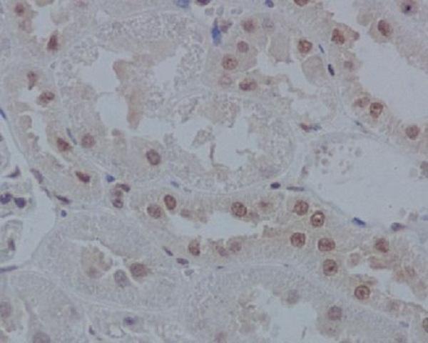
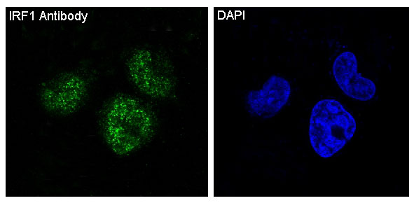
IHC images show IRF1 staining in human lung cancer, mouse intestine, and rat kidney paraffin sections; IF images show staining in A431 and Hela cells (catalog image captions).
A00580-1 has IHC images from human lung cancer and mouse intestine paraffin sections and an IF image from A431 cells (A00580-1 image captions). M00580 has an IHC image from a rat kidney paraffin section and an IF image from Hela cells (M00580 image captions).
Which to pick: For human or mouse tissue IHC, choose A00580-1 because its own captions document both species in paraffin sections; choose M00580 for rat tissue IHC, as shown in its rat kidney paraffin-section caption (A00580-1 and M00580 IHC image captions). For IF/ICC, both list those applications, with A00580-1 shown in A431 cells and M00580 shown in Hela cells; M00580 is a rabbit monoclonal and lists human, mouse, and rat reactivity, making it the broader cross-species option (catalog applications, reactivity, clone, and IF image captions). Neither IHC caption reports the fixative (A00580-1 and M00580 IHC image captions).