IRF2BP2 / Interferon regulatory factor 2-binding protein 2 · IHC design guide

Design Immunohistochemistry for IRF2BP2

Plan chromogenic IHC-P for IRF2BP2 using 5 μg/mL as the catalog antibody's starting concentration (datasheet: 5 μg/mL). Assess nuclear staining (HPA tissue IHC), using bone marrow hematopoietic cells as a high-staining reference and heart cardiomyocytes as a not-detected reference (HPA tissue IHC).

Evidence assembled Oct 2026 · For research use; verify linked source records and product datasheet before use
Immunohistochemistry protocol sheet for IRF2BP2 (IHC for IRF2BP2): expected localisation Nuclear tissue staining (HPA tissue IHC); cytoplasm also annotated (UniProt), antibody A05598, validated IHC image, and IHC protocol steps
Printable IRF2BP2 IHC protocol sheet — expected localisation Nuclear tissue staining (HPA tissue IHC); cytoplasm also annotated (UniProt), antibody A05598, controls and protocol steps. Open the full IRF2BP2 IHC guide →

IRF2BP2 Immunohistochemistry Experimental Design Guide

Expected localisation, validated protocols, controls and antibodies — the at-a-glance facts below, then the full design guide.

Must know before staining
Expected localisation Nuclear tissue staining (HPA tissue IHC); cytoplasm also annotated (UniProt)
Staining pattern General nuclear staining across tissues (HPA tissue IHC)
Antigen retrieval Tris-EDTA pH 9.0 HIER, 95–98 °C, 20 min (rule: nuclear antigen)
Positive control ⓘ Appendix+4 more · see all
Negative control ⓘ Heart muscle
Important caveats
Reasons your staining may differ from the expected pattern.
Fixation Keep fixation consistent across sections. (standard IHC practice; not target-specific)
Caveat Staining and RNA show medium consistency (HPA tissue IHC)
Regulation Expression regulation not annotated (UniProt)
Isoform / epitope 3 isoforms; no extracellular domain; epitope coverage unknown (UniProt)
Section 1

Recommended IRF2BP2 IHC & IF Protocols

The catalog antibody protocol is paired with two published IRF2BP2 IHC protocols (PMC6851004; PMC11683149). The rows below contain only details stated in each article’s excerpt.

Recommended immunohistochemistry (IHC-P) protocol parameters
SampleTissue sections; selected-image fixative not specified (standard IHC workflow)
FixationImage fixative and duration unreported (datasheet A05598); verify before use.
Sectioning4–5 µm sections on charged slides (standard)
DeparaffinisationXylene, graded ethanol series to water (standard)
Antigen retrievalHeat-induced epitope retrieval in Tris-EDTA buffer, pH 9.0, 20 min at 95–98 °C (standard rule: nuclear antigen)
Peroxidase block3% H2O2, 10 min, room temperature (standard)
Blocking10% normal serum of the secondary host, 30 min, room temperature (standard)
Primary antibodyRabbit anti-IRF2BP2, 5 μg/mL (datasheet A05598)
Primary incubationOvernight at 4 °C (standard)
DetectionHRP-polymer secondary, DAB chromogen 5–10 min (standard)
CounterstainHematoxylin, blue, dehydrate and mount (standard)
Expected resultIRF2BP2-positive staining in glandular cells of appendix (HPA tissue IHC: High). HPA tissue profile: General nuclear expression. No signal in the no-primary control.
💡Decision noteStart with Tris-EDTA pH 9.0 retrieval at 95–98 °C for 20 min (page retrieval rule; UniProt Q7Z5L9: nucleus). Neither published excerpt specifies retrieval conditions.
Section 2

What Is the Expected IRF2BP2 Staining Pattern?

IRF2BP2 should appear predominantly in nuclei across many tissue cell types (HPA: general nuclear expression; low tissue specificity). High staining is reported in appendix glandular cells, bone marrow hematopoietic cells, and bronchial respiratory epithelial cells (HPA: High). UniProt also lists cytoplasmic localization and no transmembrane segment (UniProt Q7Z5L9: subcellular location; topology). HPA rates its tissue IHC evidence Approved, with medium consistency between staining and RNA expression (HPA: reliability).

What am I looking at on my slide?
Distinct nuclear chromogen in glandular or respiratory epithelial cells, with identifiable unstained spaces between cells.This fits the reported general nuclear pattern and high staining in appendix glandular and bronchial respiratory epithelial cells (HPA: tissue IHC). Score nuclei in the named cell population; do not treat every stained area of a section as equivalent evidence.
Signal is mainly at cell borders, in lumens, or outside cells; nuclei are faint.That distribution conflicts with the reported general nuclear IHC pattern (HPA: tissue IHC) and the absence of a transmembrane segment (UniProt Q7Z5L9: topology). Check section morphology, detection deposits, and controls. Some cytoplasmic signal alone is inconclusive because UniProt also lists cytoplasm (UniProt Q7Z5L9: subcellular location).
Strong staining appears in cardiomyocytes or in an unexpected cell population.HPA reports cardiomyocytes as not detected, while also describing low tissue specificity overall (HPA: heart muscle IHC; RNA specificity). Compare the cell type with the relevant HPA entry before calling it unexpected. Investigate cross-reactivity or endogenous chromogenic activity when staining persists in a negative reagent control.
Broad haze or pigment obscures nuclear boundaries across the section.The slide cannot reliably establish the reported nuclear pattern (HPA: general nuclear expression). Uneven chromogen, insufficient washing, or detection background are general IHC possibilities; inspect a negative reagent control and reassess only where individual cells and nuclei remain distinguishable.
No nuclear signal appears in a section expected to contain positive cells.First confirm that the sampled section actually contains the named population: HPA reports high staining in bone marrow hematopoietic cells and breast glandular cells (HPA: tissue IHC). If those cells are present and controls also fail, review the general IHC workflow before concluding that IRF2BP2 is absent.
💡Expected IRF2BP2 appearanceCall a convincing positive when nuclear staining is clear in the expected cells, potentially strong in HPA High populations; dominant extracellular or border staining is suspect (HPA: general nuclear expression; tissue IHC; UniProt Q7Z5L9: topology).
How each factor affects the staining
Tissue and cell selectionUse a named HPA High population, such as appendix glandular or bone marrow hematopoietic cells, to judge whether the assay reveals the expected nuclear pattern (HPA: tissue IHC). Heart muscle cardiomyocytes are reported as not detected, but other cells in that section need separate interpretation (HPA: heart muscle IHC).
Compartment evidenceHPA summarizes tissue staining as generally nuclear and ICC-IF localization as enhanced in the nucleoplasm (HPA: tissue IHC; subcellular ICC-IF). UniProt lists both nucleus and cytoplasm, so a modest cytoplasmic component should be assessed with controls rather than dismissed solely by location (UniProt Q7Z5L9: subcellular location).
Antibody evidenceHPA027815 has Approved IHC status, whereas its ICC status is Enhanced; HPA062269 has Enhanced ICC status without an IHC status in the supplied record (HPA: antibody validation). These application labels should not be transferred from one antibody or assay to another.
Isoforms and epitope coverageThree IRF2BP2 isoforms are listed, but the supplied record gives no antibody epitope or isoform coverage (UniProt Q7Z5L9: isoforms; HPA: antibody validation). A difference between preparations therefore cannot be assigned to a particular isoform from this evidence.
Why is my staining missing, weak or wrong?
SituationLikely causeNext action
Expected positive cells are present, but nuclei are blank.The assay may have failed, or the sampled cells may differ from the HPA reference; a blank slide alone cannot distinguish these possibilities (HPA: tissue IHC).Verify cell identity and tissue integrity, then check a known positive section and the staining run. Review retrieval, primary antibody dilution, detection, and counterstain as general IHC steps; no IRF2BP2-specific retrieval setting is supplied.
Nuclei stain weakly while background is strong.Background can conceal the expected general nuclear pattern (HPA: tissue IHC). Excess detection activity or inadequate washing are general IHC possibilities.Compare a negative reagent control, inspect wash quality, and adjust blocking or detection conditions using the assay's control results. Judge improvement by whether nuclear boundaries become interpretable.
Chromogen clusters at edges, lumens, or vessel contents.Such distribution does not match the reported general nuclear pattern (HPA: tissue IHC). Deposits or endogenous detection activity are general chromogenic IHC possibilities.Inspect a negative reagent control and adjacent morphology. Review the detection and blocking steps, then rescore only intact cells with distinguishable nuclei.
Cardiomyocytes stain strongly.HPA reports cardiomyocytes as not detected in heart muscle IHC (HPA: heart muscle IHC). UniProt describes a role in cardiac muscle, but that functional annotation does not establish detectable cardiomyocyte staining (UniProt Q7Z5L9: function).Confirm the stained cells are cardiomyocytes, compare a negative reagent control, and investigate nonspecific or endogenous signal before interpreting the result as IRF2BP2.
A diffuse cytoplasmic component accompanies clear nuclear staining.UniProt includes cytoplasm as a location, while HPA describes the tissue IHC pattern as generally nuclear (UniProt Q7Z5L9: subcellular location; HPA: tissue IHC).Keep nuclear staining as the primary readout, compare controls, and record the cytoplasmic component separately. Escalate interpretation only if it is reproducible and resolves to cells.
Can IF/ICC images clarify an uncertain IHC compartment?HPA reports enhanced nucleoplasmic ICC-IF localization, including images from A-431 and U2OS cells (HPA: subcellular ICC-IF). ICC-IF and tissue IHC are different preparations.Use the ICC-IF finding as supporting localization evidence, then decide the tissue result from its own morphology and controls. The supplied ICC-IF evidence does not specify an IHC protocol.

Sample controls for IRF2BP2 IHC & IF

🧪Run appendix first: its glandular cells should stain (HPA: High in appendix glandular cells). Use heart muscle cardiomyocytes as the negative tissue (HPA: Not detected in cardiomyocytes); on the appendix slide, treat morphologically distinct neighboring cells as internal negative comparators only if they show little or no staining, since their IRF2BP2 status is not specified by the supplied HPA rows.
Positive control tissue: Appendix (Glandular cells, HPA High)
Negative control tissue: Heart muscle (HPA Not detected)
ICC-IF cell lines (HPA subcellular resource): HPA ICC-IF images show IRF2BP2 in A-431, U-251MG, U2OS, A-549, SK-MEL-30, U2OS, siRNA 1 (10x), U2OS, siRNA 1 (40x), U2OS, siRNA 2 (10x), U2OS, siRNA 2 (40x), U2OS, scrambled (10x), U2OS, scrambled (40x), with annotated localisation: Nucleoplasm (enhanced) (HPA subcellular).
Technical controls: Include a no-primary, secondary-only control; an isotype control matched to the primary antibody’s host species and clonality; and a biological specificity control using IRF2BP2 knockout material or peptide competition where a suitable immunizing peptide is available (standard IHC practice). Check appendix sections for endogenous peroxidase signal, particularly in inflammatory cells, before interpreting chromogenic staining (standard IHC practice).
⚠️Feasibility: A target-specific fixation window or fixation effect is unreported, and the selected A05598 placenta IHC caption does not state a fixative (selected tissue-IHC caption: fixative not stated). Retrieval dependence is unreported; optimize antigen retrieval on matched paraffin sections, and do not assume frozen sections or IF/ICC are easier from the supplied evidence. In appendix, inflammatory cells can contribute peroxidase background to chromogenic IHC (standard IHC practice).

HPA tissue IHC evidence for IRF2BP2

Comprehensive Human Protein Atlas IHC scoring per tissue (reliability: Approved — Medium consistency between antibody staining and RNA expression data.). Rows are taken directly from the HPA tissue chart — click any row's HPA link to view the source.

Positive expression · recommended positive controls

TissueCell typeLevelEvidenceSource
Appendix Glandular cells High Protein (IHC) HPA →
Bone marrow Hematopoietic cells High Protein (IHC) HPA →
Breast Glandular cells High Protein (IHC) HPA →
Bronchus Respiratory epithelial cells High Protein (IHC) HPA →
Cervix Glandular cells High Protein (IHC) HPA →

Undetected expression · recommended negative controls

TissueCell typeLevelEvidenceSource
Heart muscle Cardiomyocytes Not detected Protein (IHC) HPA →
Section 3

Advanced IRF2BP2 IHC Tips

Troubleshoot IRF2BP2 staining in paraffin sections by checking retrieval, nuclear localisation, controls, and cell type before comparing signal intensity.

Which retrieval conditions should I start with for IRF2BP2 paraffin sections?
Start with Tris-EDTA at pH 9.0 for heat-induced retrieval at 95–98 °C for 20 min (page retrieval setting: nuclear antigen). Keep section thickness, heating vessel, and cooling time consistent across cases so changes in nuclear staining can be attributed to the variable under test (standard IHC practice). If staining is weak, compare a citrate buffer at pH 6.0 on matched sections as a fallback, while checking tissue integrity and background (standard IHC practice). IRF2BP2 has nuclear and cytoplasmic annotations, so judge retrieval against crisp nuclear staining rather than total brown signal alone (UniProt Q7Z5L9; HPA: general nuclear expression).
How should I troubleshoot weak staining when fixation history varies?
Target-specific sensitivity of IRF2BP2 to fixation is unknown from the supplied evidence, so do not assign weak staining to a particular fixative or fixation duration (supplied IHC evidence). Record the available fixation history and compare sections processed under the same conditions before changing antibody concentration or retrieval (standard IHC practice). Include an internal tissue control when possible, and inspect morphology for poor preservation or uneven processing that could confound a nuclear readout (standard IHC practice; HPA: general nuclear expression). The catalog antibody has a human placenta IHC image at 5 μg/mL, but its caption does not state the fixative (A05598 IHC caption).
What staining compartment is credible for IRF2BP2?
Prioritise distinct nuclear staining when assessing chromogenic IRF2BP2 IHC, because the tissue profile describes general nuclear expression and cell imaging places it in the nucleoplasm (HPA: tissue IHC; HPA: nucleoplasm). Cytoplasmic staining is biologically possible because UniProt also annotates the cytoplasm, but diffuse cytoplasmic DAB alone needs independent validation (UniProt Q7Z5L9; standard IHC practice). Examine hematoxylin-counterstained nuclei at comparable magnification and exposure, separating cell-associated signal from pigment, debris, and section folds (standard IHC practice). IRF2BP2 has no annotated transmembrane segment, so a sharply membrane-restricted pattern warrants a specificity check rather than a membrane-localisation claim (UniProt Q7Z5L9 topology).
Can isoforms or epitope masking explain discordant IRF2BP2 staining?
IRF2BP2 has 3 annotated isoforms, so establish which sequence region the catalog antibody recognises before interpreting differences between samples as expression changes (UniProt Q7Z5L9 isoforms; standard IHC practice). The supplied evidence does not locate this antibody's epitope or establish which isoforms it detects, leaving isoform-specific IHC claims unsupported (supplied antibody evidence). UniProt lists modified residues including phosphoserines at 71, 175, 240, and 318, but their effect on this antibody's staining is unknown (UniProt Q7Z5L9). If staining diverges, compare retrieval conditions on adjacent sections and seek an independently validated antibody with a documented, distinct epitope (standard IHC practice).
How can IF help check a disputed IHC localisation pattern?
Use IF as an orthogonal localisation check, multiplexing IRF2BP2 with a marker appropriate to the cell population being evaluated and a nuclear counterstain (standard IF practice; HPA: nucleoplasm). Choose spectrally separated fluorophores and place the weaker signal in a channel with low measured tissue autofluorescence, using single-stain controls to assess bleed-through (standard IF practice). Because IRF2BP2 is annotated in the nucleus and cytoplasm and has no transmembrane segment, optimise permeabilisation for access to intracellular epitopes while preserving nuclear structure (UniProt Q7Z5L9; standard IF practice). Compare IF compartment patterns with chromogenic IHC, but assess antibody specificity in each application separately (standard IF/IHC practice).
How do I reduce diffuse or misleading DAB background?
First identify whether brown signal follows nuclei, tissue edges, vessels, or damaged areas; HPA describes general nuclear IRF2BP2 expression (HPA: tissue IHC; standard IHC practice). Check deparaffinisation, washing, blocking, and antibody concentration on matched sections, changing 1 variable at a time while preserving a positive control (standard IHC practice). Include a peroxidase block and a no-primary control to reveal endogenous enzyme activity or detection-reagent background during DAB development (standard chromogenic IHC practice). If background rises with stronger retrieval, shorten heating or compare the validated starting condition with a milder fallback, then judge nuclear contrast and morphology together (page retrieval setting; standard IHC practice).
How should I score IRF2BP2 staining across tissue samples? ⚠ ANSWER MARKED FOR VERIFICATION
Define the cell population and nuclear scoring rule before comparing sections, because HPA reports general nuclear expression but differing levels among cell types (HPA: tissue IHC). For a semiquantitative readout, report the percentage of positive nuclei and an H-score calculated as the sum of intensity category multiplied by its percentage, spanning 0–300 with 0–3 intensity categories (standard IHC practice). Alternatively, report positive nuclei per mm² when cell density itself matters, and normalise counts to viable tissue area or the total eligible nuclei (standard IHC practice). Hold retrieval, DAB development, imaging, and thresholds constant, and score cytoplasmic signal separately if it is retained (standard IHC practice; UniProt Q7Z5L9 localisation).
How do I distinguish credible IRF2BP2 positivity from artefact?
Credible staining should align with intact cells and a predominantly nuclear pattern, consistent with the reported tissue profile and nucleoplasmic localisation (HPA: tissue IHC; HPA: nucleoplasm). Compare cell identity against tissue context: HPA reports high staining in appendix glandular cells and no detection in heart muscle cardiomyocytes, although these observations are reference patterns rather than universal controls (HPA: appendix; HPA: heart muscle). Discount signal confined to section edges, folds, necrotic areas, or pigment, and check no-primary and peroxidase-block controls when DAB appears in unexpected locations (standard IHC practice). Interpret a cytoplasmic component cautiously because UniProt permits cytoplasmic localisation, while the HPA tissue profile emphasises nuclei (UniProt Q7Z5L9; HPA: tissue IHC).
Boster reagents

Best IRF2BP2 / Interferon regulatory factor 2-binding protein 2 IHC Antibodies

The catalog includes human placenta IHC data (A05598: IHC image caption) and mouse A20-cell ICC data presented as IF (A05598-1: ICC image caption; datasheet: mouse ICC validation; figure tag: Real IF data).

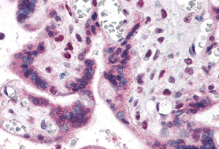
Real IHC data Immunohistochemistry of IRF2BP2 in human placenta with IRF2BP2 antibody at 5 μg/mL.
Anti-IRF2BP2 Antibody
Cat # A05598
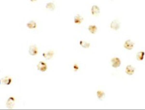
Real IF data Immunocytochemistry of IRF2BP2 in A20 cells with IRF2BP2 antibody at 2.5 μg/mL.
Anti-IRF2BP2 Antibody
Cat # A05598-1

A05598 is listed for human IHC-P, with an image of human placenta stained at 5 μg/mL (A05598: applications, reactivity, IHC image caption). A05598-1 is listed for human and mouse reactivity and ICC, with mouse ICC validation and an A20-cell image at 2.5 μg/mL (A05598-1: reactivity, applications, datasheet, ICC image caption).

Which to pick: For paraffin tissue IHC, choose the rabbit polyclonal A05598; its IHC-P listing and human placenta image support that choice, while the image caption leaves the fixative unreported (A05598: datasheet, applications, IHC image caption). For IF/ICC, choose A05598-1 based on its ICC listing and A20-cell IF figure; its clonality is unreported (A05598-1: applications, ICC image caption, figure tag, catalog clone field). For work involving human and mouse samples, A05598-1 lists reactivity with both species, although the supplied ICC validation is in mouse samples and IHC-P is absent from its application list (A05598-1: reactivity, datasheet, applications).

Each figure is that product's own IHC / IF validation image from its datasheet.

References

  1. UniProt Consortium. UniProt entry Q7Z5L9 (I2BP2_HUMAN, Interferon regulatory factor 2-binding protein 2).
  2. Human Protein Atlas. IRF2BP2 tissue IHC expression (reliability: Approved).
  3. Human Protein Atlas. IRF2BP2 subcellular location (ICC-IF): Localized to the nucleoplasm..
  4. Human Protein Atlas. IRF2BP2 antibody validation summary (2 antibodies).
  5. Down-regulation of interferon regulatory factor 2 binding protein 2 suppresses gastric cancer progression by negatively regulating connective tissue growth factor. Journal of cellular and molecular medicine 2019 — PMC6851004.
  6. Malate initiates a proton-sensing pathway essential for pH regulation of inflammation. Signal transduction and targeted therapy 2024 — PMC11683149.
  7. Broadening the spectrum of NTRK rearranged mesenchymal tumors and usefulness of pan-TRK immunohistochemistry for identification of NTRK fusions. Modern pathology : an official journal of the United States and Canadian Academy of Pathology, Inc 2021 — PMC7817523.
  8. PubMed PMID:12799427 — UniProt-cited evidence.
  9. PubMed PMID:16710414 — UniProt-cited evidence.
  10. PubMed PMID:15489334 — UniProt-cited evidence.