ITGB5 / Integrin beta-5 · IHC design guide

Design Immunohistochemistry for ITGB5

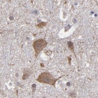
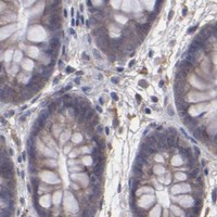
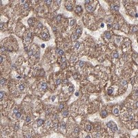
Plan paraffin-section IHC for ITGB5 using its expected membrane location (UniProt) and the cytoplasmic pattern reported in tissue (HPA tissue IHC). Compare staining in documented positive and undetected cell populations (HPA tissue IHC).

Evidence assembled Oct 2026 · For research use; verify linked source records and product datasheet before use
Immunohistochemistry protocol sheet for ITGB5 (IHC for ITGB5): expected localisation Membrane expected (UniProt); cytoplasm observed (HPA tissue IHC), antibody M04201, validated IHC image, and IHC protocol steps
Printable ITGB5 IHC protocol sheet — expected localisation Membrane expected (UniProt); cytoplasm observed (HPA tissue IHC), antibody M04201, controls and protocol steps. Open the full ITGB5 IHC guide →

ITGB5 Immunohistochemistry Experimental Design Guide

Expected localisation, validated protocols, controls and antibodies — the at-a-glance facts below, then the full design guide.

Must know before staining
Expected localisation Membrane expected (UniProt); cytoplasm observed (HPA tissue IHC)
Staining pattern General cytoplasmic staining in positive cell types (HPA tissue IHC)
Antigen retrieval EDTA pH 8.0 HIER, heat-mediated (datasheet M04201)
Positive control ⓘ Breast+4 more · see all
Negative control ⓘ Bone marrow+3 more · see all
Important caveats
Reasons your staining may differ from the expected pattern.
Fixation Keep fixation consistent across sections (standard IHC practice; not target-specific)
Caveat Membrane expectation differs from tissue staining (UniProt; HPA tissue IHC)
Regulation No specific expression regulator annotated (UniProt)
Isoform / epitope No isoforms listed; extracellular versus cytoplasmic epitope matters (UniProt)
Section 1

Recommended ITGB5 IHC & IF Protocols

The catalog antibody protocol uses heat-mediated EDTA pH 8.0 retrieval (datasheet M04201). The published ITGB5 IHC protocols below cover liver, tongue, and colorectal tissue (PMC13570297; PMC11344732; PMC11914080; PMC5793391).

Recommended immunohistochemistry (IHC-P) protocol parameters
SampleParaffin-embedded human lung cancer tissue; fixative not specified (datasheet M04201)
FixationImage fixative and duration unreported (datasheet M04201); verify before use.
Sectioning4–5 µm sections on charged slides (standard)
DeparaffinisationXylene, graded ethanol series to water (standard)
Antigen retrievalHeat retrieval: EDTA pH 8.0 (datasheet M04201); 20 min, 95–100 °C (standard)
Peroxidase block3% H2O2, 10 min, room temperature (standard)
Blocking10% goat serum (datasheet M04201)
Primary antibodyMouse monoclonal (clone 9E2) anti-ITGB5, 2μg/ml (datasheet M04201)
Primary incubationOvernight at 4 °C (datasheet M04201)
DetectionStreptavidin-biotin complex (SABC), DAB chromogen (datasheet M04201)
CounterstainHematoxylin, blue, dehydrate and mount (standard)
Expected resultITGB5-positive staining in glandular cells of breast (HPA tissue IHC: Medium). HPA tissue profile: General cytoplasmic expression. No signal in the no-primary control.
💡Decision noteStart with EDTA pH 8.0 retrieval for the catalog antibody (datasheet M04201); Tris-EDTA pH 9.0 is a published ITGB5 alternative (PMC5793391).
Section 2

What Is the Expected ITGB5 Staining Pattern?

ITGB5 is a cell-membrane integrin with an extracellular region at residues 24–719 and a cytoplasmic tail at 743–799 (UniProt P18084 topology). In tissue IHC, expect staining in selected glandular, endothelial, glomerular, cholangiocyte, macrophage and endocrine cells; HPA also describes general cytoplasmic expression (HPA tissue IHC). HPA rates tissue staining Enhanced, with medium consistency between antibody staining and RNA expression (HPA tissue IHC).

What am I looking at on my slide?
Staining follows cell outlines or appears cytoplasmic in expected cell populations, with little staining in the comparison tissue.Membrane-associated signal fits ITGB5 topology (UniProt P18084 topology). Cytoplasmic signal can also fit HPA's reported tissue profile; judge it alongside cell identity and background (HPA tissue IHC).
The main signal is confined to nuclei, away from cell outlines and cytoplasm.Nuclear-only staining conflicts with UniProt's cell-membrane assignment and HPA's cytoplasmic tissue profile; treat it as a possible artefact and check controls before scoring it as ITGB5 (UniProt P18084 subcellular location; HPA tissue IHC).
Strong staining appears in bone-marrow hematopoietic cells or esophageal squamous epithelium.HPA reports ITGB5 as not detected in those cell populations (HPA tissue IHC). Check for antibody cross-reactivity or endogenous detection activity; their presence is a troubleshooting possibility, not established by HPA (standard IHC practice).
Brown signal spreads across tissue and spaces without clear cell boundaries.A widespread deposit that does not resolve into cells is more consistent with background than an interpretable ITGB5 pattern (standard IHC practice). Compare the primary-antibody omission control and inspect washing, blocking and detection steps (standard IHC practice).
No staining appears in the chosen known-positive tissue, including its expected cell population.Recheck which cells are being assessed: HPA reports Medium staining in lung macrophages, liver cholangiocytes and breast glandular cells, among others (HPA tissue IHC). An absent result may reflect the assay or the sampled cells; HPA does not identify the cause in a given section (HPA tissue IHC).
💡Expected ITGB5 appearanceCall a result positive when identifiable expected cells show cell-associated membrane or cytoplasmic staining around HPA's reported Medium level; nuclear-only signal or similarly strong staining in HPA-negative cell populations warrants control review (UniProt P18084 topology; HPA tissue IHC).
How each factor affects the staining
Cell population and tissue choiceUse the named cells when comparing sections: cerebral-cortex endothelial cells and kidney glomerular cells are Medium, whereas bone-marrow hematopoietic cells are not detected (HPA tissue IHC). A whole-tissue judgment can miss this cell-level distinction (HPA tissue IHC).
Compartment and antibody epitopeITGB5 has an extracellular region and a shorter cytoplasmic tail (UniProt P18084 topology). The supplied evidence gives no epitope for the catalog antibody, so topology alone cannot predict which staining step will expose its epitope (UniProt P18084 topology; supplied antibody record).
Paraffin antigen retrievalRetrieval conditions are an assay variable in paraffin IHC (standard IHC practice). No ITGB5-specific retrieval condition or fixation sensitivity is supplied; assess any adjustment against a positive cell population and a negative control (HPA tissue IHC; standard IHC practice).
Antibody validationHPA lists HPA001820 as IHC Enhanced and CAB022050 as IHC Approved (HPA antibodies). Enhanced supports greater confidence in the reported tissue pattern, but the profile still has medium consistency with RNA data and does not guarantee every section will stain identically (HPA tissue IHC).
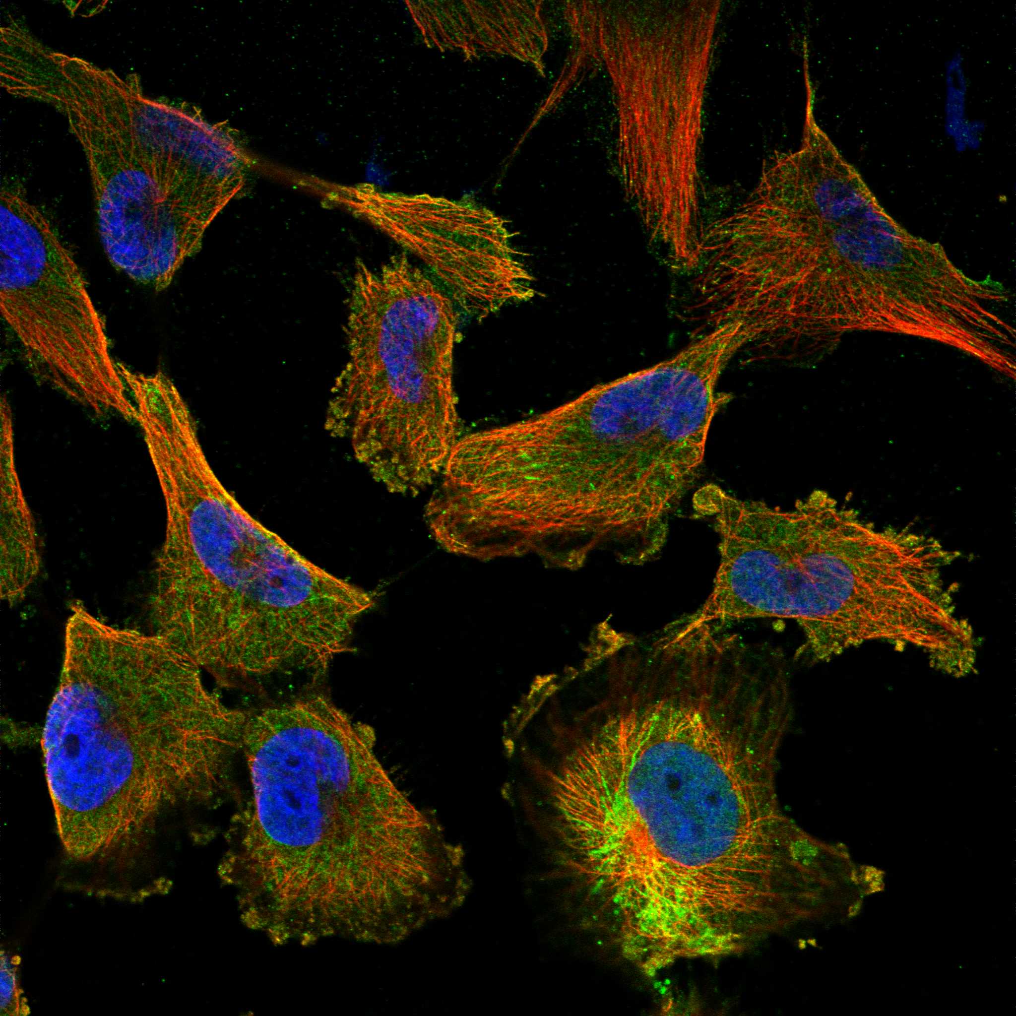
IF/ICC: what localisation is expected?HPA reports an approved plasma-membrane location and an uncertain mitochondrial location in ICC-IF images from U-251MG and U2OS (HPA subcellular ICC-IF). Interpret mitochondrial signal cautiously; this observation does not specify an IF/ICC protocol (HPA subcellular ICC-IF).
Why is my staining missing, weak or wrong?
SituationLikely causeNext action
Expected cells are unstained.The sampled field may lack the named positive cells, or an IHC step may have failed; the section alone cannot distinguish these causes (HPA tissue IHC; standard IHC practice).Confirm the target cell population in the section, then review retrieval, primary-antibody application and chromogenic detection against a concurrently processed positive control (HPA tissue IHC; standard IHC practice).
Signal is weak in an expected population.HPA reports Medium staining rather than uniformly high staining in its listed positive cells; weak signal may also reflect assay conditions (HPA tissue IHC; standard IHC practice).Compare like cells in a positive control and optimize retrieval or antibody dilution using the catalog antibody's IHC-P instructions where available (HPA tissue IHC; standard IHC practice).
Nuclear staining dominates.Nuclear-only staining does not match the assigned cell-membrane location or the reported general cytoplasmic tissue profile (UniProt P18084 subcellular location; HPA tissue IHC).Check the primary-antibody omission control, then reassess detection background and whether the expected cells show a separate cell-associated signal (standard IHC practice; HPA tissue IHC).
HPA-negative cells stain strongly.Cross-reactivity or endogenous detection activity is possible when staining contradicts a reported not-detected population (HPA tissue IHC; standard IHC practice).Check a primary-antibody omission control and the detection-system controls; compare the signal with a named HPA-positive cell population on a separately processed section (standard IHC practice; HPA tissue IHC).
Diffuse brown background obscures cells.Nonspecific antibody binding, insufficient washing or endogenous detection activity can contribute to chromogenic background (standard IHC practice).Inspect the omission control and review blocking, washing, antibody dilution and detection steps before scoring cellular staining (standard IHC practice).
Cytoplasmic staining appears without a crisp membrane rim.HPA describes general cytoplasmic tissue expression even though UniProt assigns ITGB5 to the cell membrane (HPA tissue IHC; UniProt P18084 subcellular location).Score the named cell population and background together; do not reject an otherwise cell-specific cytoplasmic pattern solely for lacking a sharp rim (HPA tissue IHC).

Sample controls for ITGB5 IHC & IF

🧪Run kidney first and assess glomerular cells for ITGB5 staining (HPA: kidney cells in glomeruli, Medium). Use bone marrow hematopoietic cells as the negative tissue (HPA: Not detected); on the kidney slide, nonglomerular cells without specific membrane staining should show only counterstain or background, but their ITGB5 status must be verified rather than assumed (UniProt P18084: cell membrane).
Positive control tissue: Breast (Glandular cells, HPA Medium)
Negative control tissue: Bone marrow (HPA Not detected)
ICC-IF cell lines (HPA subcellular resource): HPA ICC-IF images show ITGB5 in U-251MG, U2OS, with annotated localisation: Plasma membrane (approved), Mitochondria (uncertain) (HPA subcellular).
Technical controls: Include a no-primary, secondary-only control and a mouse IgG isotype control matched to the primary antibody’s subclass where known (caption: mouse primary); use an ITGB5 knockout sample or validated immunizing-peptide competition as a biological negative. Quench endogenous peroxidase and block endogenous biotin in kidney sections if using avidin–biotin/DAB detection (caption: SABC/DAB; standard IHC practice).
⚠️Feasibility: A target-specific fixation window or fixation effect is unreported, and the exact M04201 tissue-IHC caption does not state a fixative (caption: fixative not stated). The paraffin-section example uses heat retrieval in EDTA at pH 8.0; it does not establish that retrieval is required for every specimen (caption: EDTA pH 8.0). The supplied evidence does not establish whether frozen sections or IF are easier; renal tubular endogenous biotin can create background with avidin–biotin detection (standard IHC practice).

HPA tissue IHC evidence for ITGB5

Comprehensive Human Protein Atlas IHC scoring per tissue (reliability: Enhanced — Medium consistency between antibody staining and RNA expression data.). Rows are taken directly from the HPA tissue chart — click any row's HPA link to view the source.

Positive expression · recommended positive controls

TissueCell typeLevelEvidenceSource
Breast Glandular cells Medium Protein (IHC) HPA →
Cerebral cortex Endothelial cells Medium Protein (IHC) HPA →
Epididymis Glandular cells Medium Protein (IHC) HPA →
Gallbladder Glandular cells Medium Protein (IHC) HPA →
Kidney Cells in glomeruli Medium Protein (IHC) HPA →

Undetected expression · recommended negative controls

TissueCell typeLevelEvidenceSource
Bone marrow Hematopoietic cells Not detected Protein (IHC) HPA →
Esophagus Squamous epithelial cells Not detected Protein (IHC) HPA →
Nasopharynx Respiratory epithelial cells Not detected Protein (IHC) HPA →
Vagina Squamous epithelial cells Not detected Protein (IHC) HPA →
Section 3

Advanced ITGB5 IHC Tips

Troubleshoot ITGB5 staining in paraffin sections by checking retrieval, compartment, cell type, and controls before comparing chromogenic signal across samples.

What retrieval should I start with if ITGB5 staining is weak?
Start with heat-mediated retrieval in EDTA at pH 8.0 (datasheet M04201). The selected paraffin-section example used this retrieval before incubation with 2 μg/ml catalog antibody overnight at 4°C (datasheet M04201). If signal is weak, compare a shorter and longer heating period on adjacent sections while holding antibody concentration and detection conditions constant (standard IHC practice). Include a known positive cell population and a no-primary control in each run so increased signal can be distinguished from increased background (standard IHC practice). Record heating and cooling times because retrieval intensity can affect morphology and staining (standard IHC practice).
How should I assess whether fixation caused weak ITGB5 staining?
The selected ITGB5 image describes a paraffin-embedded section but does not state its fixative, so target-specific fixation sensitivity is unknown (datasheet M04201). Record the specimen’s actual fixative and fixation duration, then compare sections with documented processing histories under the same EDTA pH 8.0 retrieval and staining conditions (datasheet M04201; standard IHC practice). Evaluate morphology alongside signal, since poor preservation can make compartment assignment unreliable (standard IHC practice). Do not attribute weak staining to a particular fixation condition solely from tissue staining patterns or ITGB5 membrane topology; neither establishes a fixation effect for this antibody (HPA tissue IHC; UniProt P18084 topology).
Should ITGB5 appear at the membrane or in the cytoplasm?
Prioritize staining at cell boundaries when assessing ITGB5, a cell-membrane protein with an extracellular region spanning residues 24–719 and a transmembrane segment at 720–742 (UniProt P18084 topology). HPA tissue IHC reports general cytoplasmic expression, so cytoplasmic staining should be assessed in its cell and tissue context rather than dismissed automatically (HPA tissue IHC). HPA subcellular data approve the plasma membrane location but mark the mitochondrial location uncertain (HPA subcellular). Compare boundary and cytoplasmic signal separately in positive cells, and inspect adjacent negative tissue and no-primary controls before calling diffuse cytoplasmic staining specific (standard IHC practice).
How can epitope position affect staining in paraffin sections?
Check the antibody’s documented immunogen or epitope before interpreting discordant staining; none is specified in the supplied caption (datasheet M04201). ITGB5 has an extracellular region at residues 24–719 and a cytoplasmic tail at 743–799, so epitope location determines which side of the membrane the antibody targets (UniProt P18084 topology). The record lists 8 glycosylation sites and one example phosphoserine at 770, but it provides no evidence that either modification changes staining with this antibody (UniProt P18084). No isoforms are listed in the supplied record; investigate retrieval and antibody specificity before assigning discrepant staining to an isoform (UniProt P18084; standard IHC practice).
How would I check an ITGB5 IHC pattern by multiplex IF?
Use IF as a separate validation experiment and pair ITGB5 with a marker for the cell population being assessed; HPA reports medium staining in lung macrophages and cerebral-cortex endothelial cells (HPA tissue IHC). Choose fluorophores after examining unstained tissue for autofluorescence, and place the weaker signal in a channel with less tissue background (standard IF practice). If the antibody epitope is extracellular, compare staining with minimal permeabilisation; if it is cytoplasmic, test permeabilisation, because ITGB5 spans the membrane and its epitope is unspecified here (UniProt P18084 topology; datasheet M04201; standard IF practice). Include single-stain and no-primary controls to assess bleed-through and background before comparing IF with chromogenic IHC (standard IF practice).
What should I adjust when ITGB5 DAB staining is diffuse?
First inspect no-primary and tissue controls, then determine whether colour follows cell boundaries, tissue edges, damaged regions, or the entire section (standard IHC practice; UniProt P18084 subcellular location). The selected example blocked with 10% goat serum, applied 2 μg/ml primary antibody overnight at 4°C, and used a biotin-based DAB detection system (datasheet M04201). If background is widespread, compare a lower primary concentration and stronger washing while keeping retrieval fixed at EDTA pH 8.0 (datasheet M04201; standard IHC practice). For this chromogenic workflow, assess endogenous peroxidase and biotin-related background with appropriate controls before increasing detection intensity (standard IHC practice; datasheet M04201).
How should I quantify ITGB5 staining across paraffin sections? ⚠ ANSWER MARKED FOR VERIFICATION
Define the scored cell population and compartment before measuring signal; membrane staining and the general cytoplasmic staining reported by HPA should be recorded separately (UniProt P18084 subcellular location; HPA tissue IHC). For a defined population, report the percentage of positive cells and an H-score based on intensity categories 0–3; for sparse positive cells, density per mm² can be informative (standard IHC practice). Normalize cell counts to the number of eligible cells, or density to the measured viable tissue area, and exclude folds and necrotic regions consistently (standard IHC practice). Keep retrieval, antibody concentration, DAB development, imaging, and scoring thresholds constant across compared sections (standard IHC practice).
How can I distinguish genuine ITGB5 staining from artefact?
Assess whether signal follows plausible cell boundaries or the documented cytoplasmic IHC pattern, while treating an exclusively mitochondrial interpretation cautiously because that location is uncertain in HPA data (UniProt P18084 subcellular location; HPA tissue IHC; HPA subcellular). Check cell identity: HPA reports medium staining in lung macrophages and no detected staining in bone-marrow hematopoietic cells, among other listed populations (HPA tissue IHC). Compare viable central tissue with cut edges and necrotic areas, where staining artefacts can complicate interpretation (standard IHC practice). A no-primary control and an endogenous-peroxidase control help identify detection-related DAB signal before a specimen is called ITGB5 positive (standard IHC practice).
Boster reagents

Best ITGB5 / Integrin beta-5 IHC Antibodies

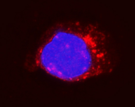
The catalog includes IHC images of human paraffin tissue with M04201 and A04201-1 (catalog IHC captions), plus an IF image of A431 cells with A04201-1 (catalog IF caption).

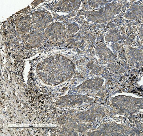
Real IHC data IHC analysis of Integrin beta 5/ITGB5 using anti-Integrin beta 5/ITGB5 antibody (M04201). Integrin beta 5/ITGB5 was detected in paraffin-embedded section of human lung cancer tissue. Heat mediated antigen retrieval was performed in EDTA buffer (pH8.0, epitope retrieval solution). The tissue section was blocked with 10% goat serum. The tissue section was then incubated with 2μg/ml mouse anti-Integrin beta 5/ITGB5 Antibody (M04201) overnight at 4°C. Biotinylated goat anti-mouse IgG was used as secondary antibody and incubated for 30 minutes at 37°C. The tissue section was developed using Strepavidin-Biotin-Complex (SABC) (Catalog # SA1021) with DAB as the chromogen.
Anti-Integrin beta 5/ITGB5 Antibody ® (monoclonal, 9E2)
Cat # M04201
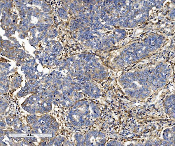
Real IHC data IHC analysis of ITGB5 using anti-ITGB5 antibody (A04201-1). ITGB5 was detected in paraffin-embedded section of human rectal cancer tissue. Heat mediated antigen retrieval was performed in EDTA buffer (pH8.0, epitope retrieval solution). The tissue section was blocked with 10% goat serum. The tissue section was then incubated with 1μg/ml rabbit anti-ITGB5 Antibody (A04201-1) overnight at 4°C. Biotinylated goat anti-rabbit IgG was used as secondary antibody and incubated for 30 minutes at 37°C. The tissue section was developed using Strepavidin-Biotin-Complex (SABC) (Catalog # SA1022) with DAB as the chromogen.
Anti-Integrin beta 5/ITGB5 Antibody ®
Cat # A04201-1

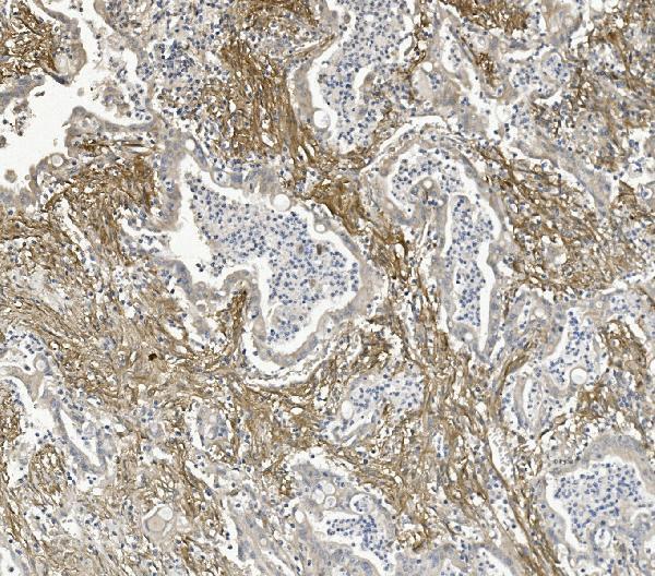
M04201 will render with its human lung cancer paraffin-section IHC image (M04201 IHC caption). A04201-1 will render with its human rectal cancer paraffin-section IHC image; it also has IF data in A431 cells (A04201-1 IHC and IF captions).

Which to pick: For human tissue IHC, M04201 is a mouse monoclonal option and A04201-1 is a rabbit option; each has its own paraffin-section IHC image, and neither caption reports the fixative (catalog applications; M04201 and A04201-1 IHC captions). For IF/ICC, choose A04201-1: both applications are listed and its IF caption shows A431 cells (A04201-1 catalog applications and IF caption). For broader species reactivity, M04201-1 lists human, monkey and rat for IHC, while A04201-1 lists human and mouse; M04201-1 has no IHC image in the supplied catalog (catalog applications, reactivity and image fields).

Each figure is that product's own IHC / IF validation image from its datasheet.

References

  1. UniProt Consortium. UniProt entry P18084 (ITB5_HUMAN, Integrin beta-5).
  2. Human Protein Atlas. ITGB5 tissue IHC expression (reliability: Enhanced).
  3. Human Protein Atlas. ITGB5 subcellular location (ICC-IF): Localized to the plasma membrane and mitochondria..
  4. Human Protein Atlas. ITGB5 antibody validation summary (2 antibodies).
  5. A high-affinity anti-ITGB5 nanobody for hepatocellular carcinoma: Antitumor efficacy and tumor microenvironment reprogramming. The Journal of biological chemistry 2026 — PMC13570297.
  6. ROS-mediated ITGB5 promotes tongue squamous cell carcinoma metastasis through epithelial mesenchymal transition and cell adhesion signal pathway. Journal of cancer research and clinical oncology 2024 — PMC11344732.
  7. ITGB5 is a prognostic factor in colorectal cancer and promotes cancer progression and metastasis through the Wnt signaling pathway. Scientific reports 2025 — PMC11914080.
  8. Integrin-β5, a miR-185-targeted gene, promotes hepatocellular carcinoma tumorigenesis by regulating β-catenin stability. Journal of experimental & clinical cancer research : CR 2018 — PMC5793391.
  9. PubMed PMID:2328726 — UniProt-cited evidence.
  10. PubMed PMID:2371275 — UniProt-cited evidence.
  11. PubMed PMID:2211615 — UniProt-cited evidence.