This website uses cookies to ensure you get the best experience on our website.
- Table of Contents
1 Citations

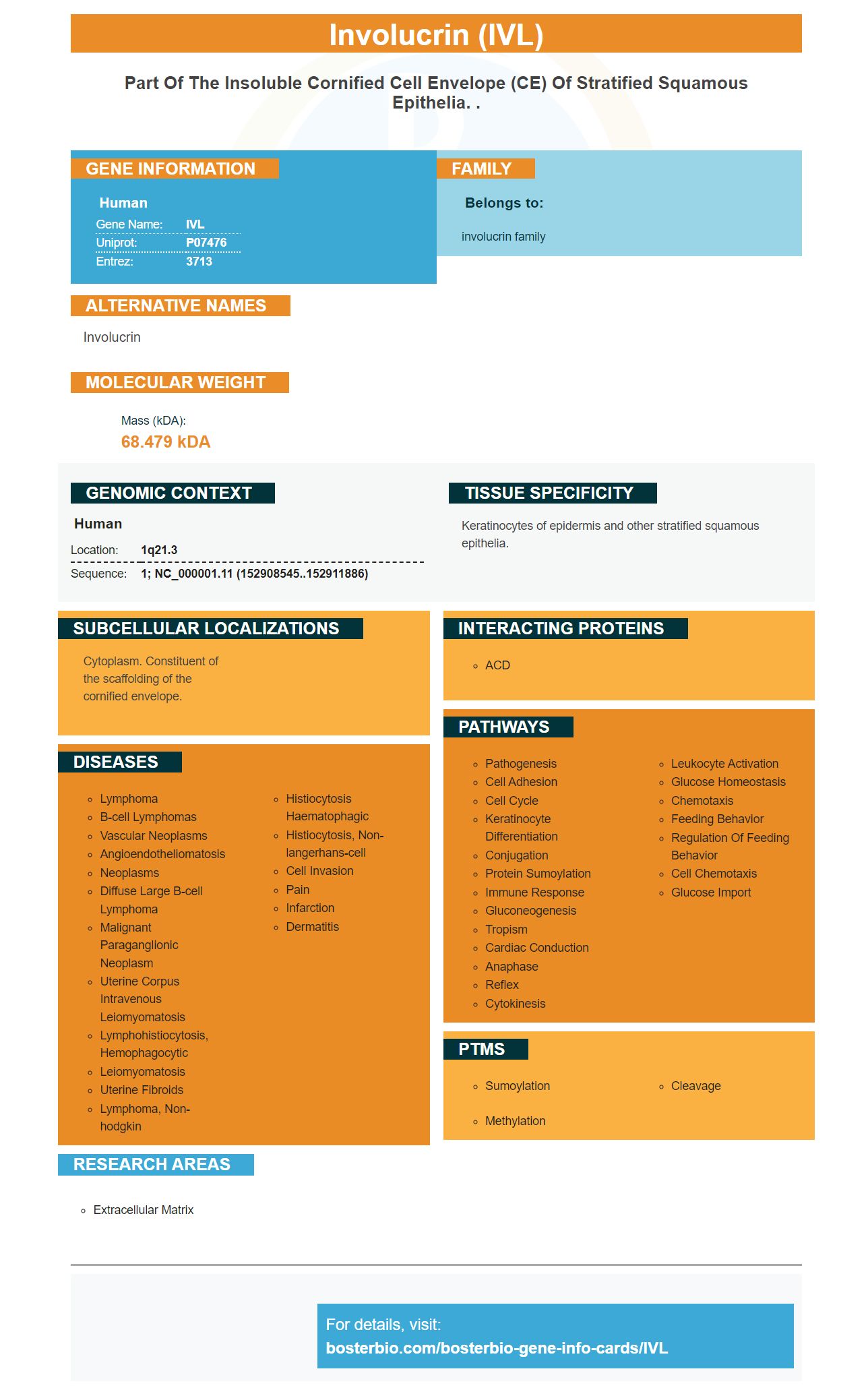
Facts about Involucrin.

| Human | |
|---|---|
| Gene Name: | IVL |
| Uniprot: | P07476 |
| Entrez: | 3713 |

| Belongs to: |
|---|
| involucrin family |

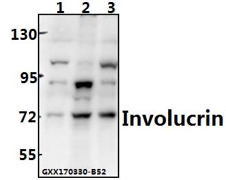
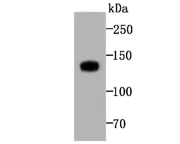
involucrin
Mass (kDA):
68.479 kDA

| Human | |
|---|---|
| Location: | 1q21.3 |
| Sequence: | 1; NC_000001.11 (152908545..152911886) |
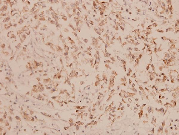
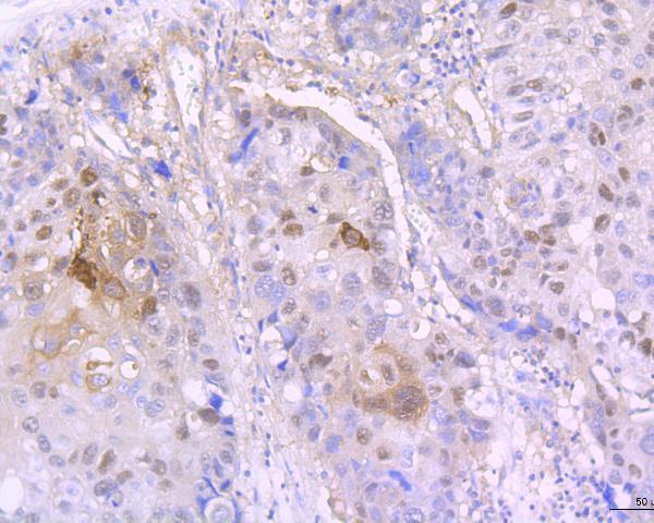
Keratinocytes of epidermis and other stratified squamous epithelia.
Cytoplasm. Constituent of the scaffolding of the cornified envelope.


PMID: 2873896 by Eckert R.L., et al. Structure and evolution of the human involucrin gene.
PMID: 9115270 by Robinson N.A., et al. S100A11, S100A10, annexin I, desmosomal proteins, small proline-rich proteins, plasminogen activator inhibitor-2, and involucrin are components of the cornified envelope of cultured human epidermal keratinocytes.