This website uses cookies to ensure you get the best experience on our website.
- Table of Contents
2 Citations 17 Q&As
2 Citations 16 Q&As
1 Citations
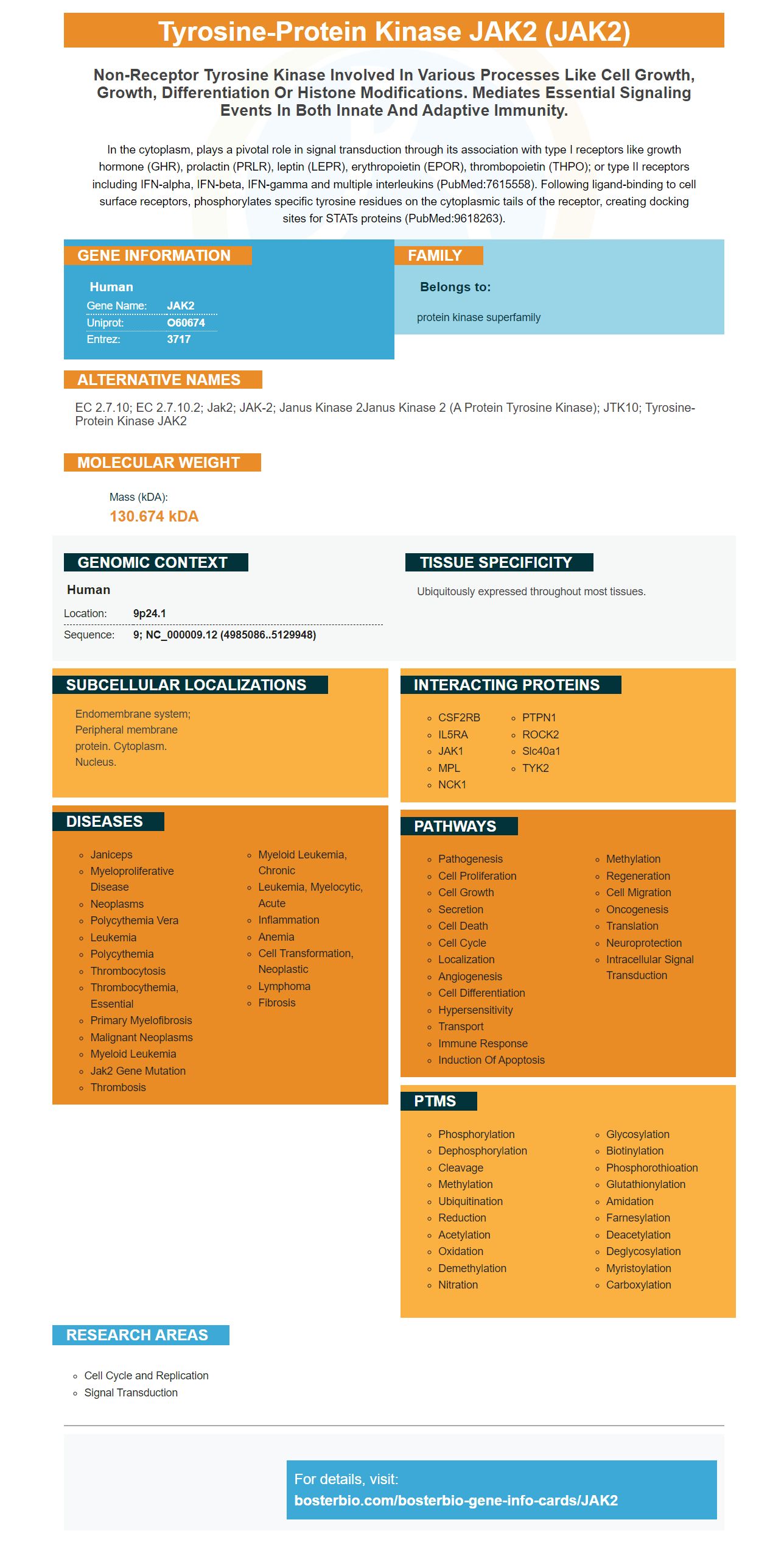
Facts about Tyrosine-protein kinase JAK2.
In the cytoplasm, plays a pivotal role in signal transduction through its association with type I receptors like growth hormone (GHR), prolactin (PRLR), leptin (LEPR), erythropoietin (EPOR), thrombopoietin (THPO); or type II receptors including IFN-alpha, IFN-beta, IFN-gamma and multiple interleukins (PubMed:7615558). Following ligand-binding to cell surface receptors, phosphorylates specific tyrosine residues on the cytoplasmic tails of the receptor, creating docking sites for STATs proteins (PubMed:9618263).
Human | |
---|---|
Gene Name: | JAK2 |
Uniprot: | O60674 |
Entrez: | 3717 |
Belongs to: |
---|
protein kinase superfamily |
EC 2.7.10; EC 2.7.10.2; Jak2; JAK-2; Janus kinase 2Janus kinase 2 (a protein tyrosine kinase); JTK10; tyrosine-protein kinase JAK2
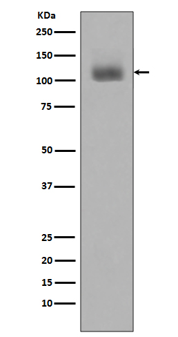
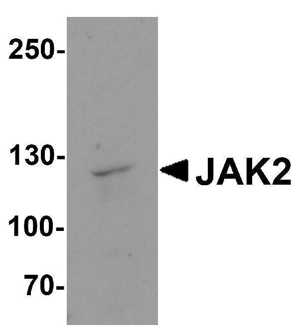
Mass (kDA):
130.674 kDA
Human | |
---|---|
Location: | 9p24.1 |
Sequence: | 9; NC_000009.12 (4985086..5129948) |
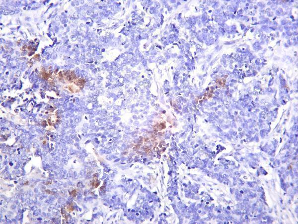
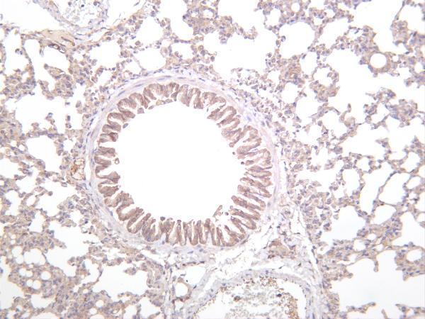
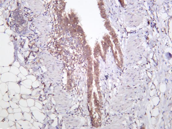
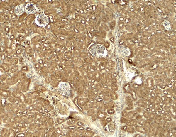
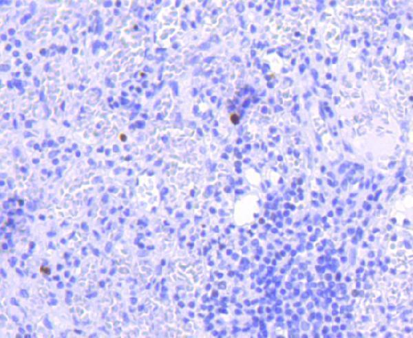
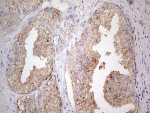
Ubiquitously expressed throughout most tissues.
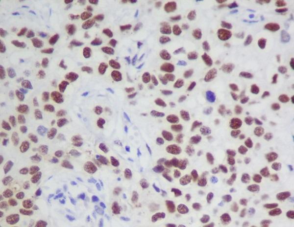
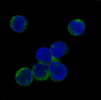
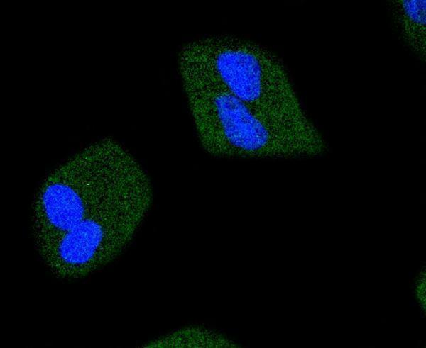
Endomembrane system; Peripheral membrane protein. Cytoplasm. Nucleus.
This article discusses how important BRD4 phosphorylation of tyrosine97/98 is for its stability in cells with rIL6/rIL8. This article also discusses cross-linking VSIG4 to MS4A6D. This article is applicable for all scientists worldwide. Boster has been a major provider of JAK2 marker markers for almost 30 years.
We previously found that VSIG4 directly interacts with MS4A6D on the cell membrane. Five candidate genes were identified that interact with Vsig4 via a mouse PEM. This was done in a bait-dependent way. Transient transfection experiments were performed to determine if VSIG4 & MS4A6D crosslink. These results indicate that the two proteins interact with other molecules on the surface and may also interact to MS4A6D.
MS4A6D is a member in the membrane-spanning subfamily A and interacts VSIG4. VSIG4 blocks macrophages from expressing IL-1b or NLRP3 during inflammatory response. This interaction suggests that VSIG4 plays a significant role in the negative regulation of macrophage-driven intracellularinflammatory responses.
VSIG4 also inhibits transcription MS4A6D. It also inhibits the synthesis 2 transcription factors: Il-1b, and Nlrp3. VSIG4 interferes with the AKT/STAT3/A20 axis. It is not clear how this interaction occurs. These results indicate that VSIG4 inhibits the activation NLRP3/MS4A6D.
It was found that Vsig4A6D expression was present in human brain cell cells via immunohistochemistry, Western blot analysis, and immunohistochemistry. This protein promotes macrophage production of IL-10 in mice. This may counteract inflammatory pathogenesis. It also inhibits spontaneous autoinflammatory disorders that can develop in mice with Vsig4-deficient mice.
EAE and colitis can be caused by mutations of VSIG4 in mouse models. Deficient mice were more likely than wild-type control mice to develop EAE, and were more susceptible a dextran sulfate salt-induced colitis. These mice also had an increase in IL-1b, NLRP3, and other inflammatory markers in vivo. The inhibitor of Vsig4 by CY09 reduces the activity these leukocytes which suggests therapeutic potential.
VSIG4 can also affect IL-2 production. It has been shown that VSIG4 can inhibit neuroinflammation in perihematomas of ICH. Inhibition of the NLRP3 receptor is also associated with a decreased risk of ICH and increased A20 expression. Silencing MS4a6D decreases A20 levels and enhances NLRP3 expression. These mice models are not large enough to provide direct evidence, but they do contain important features that support VSIG4 cross with MS4A6D.
Split-ubiquitin allowed the generation of cDNA libraries in macrophage cells by using the VSIG4 encoding genetic. The Vsig4 encoding gene was cloned to pBT3–SUC as bait constructs. The library of plasmids was used to transform yeast cells bearing the bait constructs. Positive transformants were selected from these libraries and retransformed into reporter strain NMY51.
These findings support the hypothesis that VSIG4 and MS4A6D interact with each other to increase IL-2 production in T cell. Furthermore, these interactions may promote the activation of the GITR receptor, which in turn induces the production of IL-2. MS4A family members have been shown to amplify Ag signals through lateral coassociation. Consequently, these proteins are used by T cells preoccupied with immune defense to increase their sensitivity and ensure they eliminate Ags.
The JAK2 gene regulates growth factors and cytokines production. When activated the protein causes an increase of cellular growth, differentiation, and cell proliferation. To monitor the effects of rIL6 on the cell cycle, we used JAK2 as a marker. We found that rIL6 treatment decreased JAK2 expression, but that rIL8 had no effect upon JAK2 activity.
The JAK2 protein was detected by immunohistochemistry. The JAK2 protein is expressed in various tissues and organs. Nuclear accumulation of pJAK2 was induced by rIL6. This study used a NanoBRET (tm) BRD4/Histone H3.3 Interaction Assay. To conduct this experiment, 293T cells were transfected with active JAK2-V617F. The cells were then treated with the indicated inhibitors. The interaction between BRD4-histone H3.3 was enhanced by the JAK2-V617FF mutants. Pacritinib was applied to reverse this effect.
Transfected cells were treated with small RNAs (siRNA). These siRNAs were compatible for Lipofectamine (RNAiMAX). The sequences can be found in Supplementary Table 3
The JAK2 marker was detected in all rIL6 and -IL8-treated cell lines and human monocytes. JAK2 phosphorylation is increased by rIL6 or rIL8. rIL8 also increases JAK2 expression. This research is especially useful for cancer patients.
This study also showed HSC clonal abnormalities provide fertile ground for malignant mutations. It was also shown that clinical markers can be used to predict subtypes of MPN. The genomic classifications of these cells could also be used to identify patients who are at greatest risk of disease progression, or the expansion of a malignant strain.
Molecular analysis of JAK2 in MPN cells revealed that this gene promotes synthesis of PD-L1, a protein that is necessary for anti-neoplastic T-cell responses. These findings could help in immunotherapeutic approaches to JAK-mutated cells. This research also identifies the metabolic pathways that regulate the JAK gene and promotes clonal evolution.
The inhibitory effect of JQ1 alone was small, but when combined with pacritinib or HT-29 cells, the inhibitory effect was greater. JQ1 alone produced only modest effects on the phenotype of PDC cells, suggesting that blocking the JAK2-JAK2 pathway might help overcome BETi resistance.
The JAK2 gene marker is crucial for the analysis of rIL6- and t-IL8 mediated immune responses in tumors, and other tissues. In addition to the immune response, JAK2-signaling plays an important role in inflammation and lymphocyte biology. If it is not functioning properly, rIL6/t-IL8 treatment may promote the development cancerous cells.
PMID: 9618263 by Saltzman A., et al. Cloning and characterization of human Jak-2 kinase: high mRNA expression in immune cells and muscle tissue.
PMID: 9446644 by Dalal I., et al. Cloning and characterization of the human homolog of mouse Jak2.
*More publications can be found for each product on its corresponding product page