This website uses cookies to ensure you get the best experience on our website.


- Table of Contents
Plan chromogenic JMJD1C IHC on paraffin sections around the reported nuclear tissue pattern (HPA tissue IHC). Compare adrenal glandular cells with ovarian follicle cells (HPA tissue IHC), starting the catalog antibody at 2.5 μg/mL (datasheet).

Expected localisation, validated protocols, controls and antibodies — the at-a-glance facts below, then the full design guide.
| Expected localisation | Nuclear staining in tissue (HPA tissue IHC) | |
| Staining pattern | Widespread nuclear staining across cell types (HPA tissue IHC) | |
| Antigen retrieval | Tris-EDTA pH 9.0 HIER, 95–98 °C, 20 min (rule: nuclear antigen) | |
| Positive control | Adrenal gland+4 more · see all | |
| Negative control | Ovary+1 more · see all |
| Fixation | Keep fixation consistent across sections (standard IHC practice; not target-specific) | |
| Caveat | Staining and RNA show medium consistency (HPA tissue IHC) | |
| Regulation | No established regulator of staining intensity (UniProt) | |
| Isoform / epitope | 3 isoforms; epitope coverage unknown (UniProt; datasheet) |
The catalog antibody protocol uses Tris-EDTA retrieval (page antigen retrieval); the published IHC options below cover testis, thyroid and clinical tissue specimens (PMC5722903; PMC10178269; PMC2965226; PMC8473479).
| Sample | Tissue sections; selected-image fixative not specified (standard IHC workflow) |
| Fixation | Image fixative and duration unreported (datasheet A05189); verify before use. |
| Sectioning | 4–5 µm sections on charged slides (standard) |
| Deparaffinisation | Xylene, graded ethanol series to water (standard) |
| Antigen retrieval | Heat-induced epitope retrieval in Tris-EDTA buffer, pH 9.0, 20 min at 95–98 °C (standard rule: nuclear antigen) |
| Peroxidase block | 3% H2O2, 10 min, room temperature (standard) |
| Blocking | 10% normal serum of the secondary host, 30 min, room temperature (standard) |
| Primary antibody | Rabbit anti-JMJD1C, 2.5 μg/mL (datasheet A05189) |
| Primary incubation | Overnight at 4 °C (standard) |
| Detection | HRP-polymer secondary, DAB chromogen 5–10 min (standard) |
| Counterstain | Hematoxylin, blue, dehydrate and mount (standard) |
| Expected result | JMJD1C-positive staining in glandular cells of adrenal gland (HPA tissue IHC: Medium). HPA tissue profile: Ubiquitous nuclear expression. No signal in the no-primary control. |
JMJD1C is a nuclear protein with no transmembrane segment (UniProt Q15652: subcellular location and topology). In paraffin-section IHC, expect predominantly nuclear staining across many cell types, including adrenal glandular cells and cortical neurons (HPA: ubiquitous nuclear expression; Medium staining in those cells). Treat this as a guide to interpretation: HPA rates the tissue pattern Approved, with medium agreement between staining and RNA data, pending external verification (HPA: tissue IHC reliability).
| Predominantly nuclear staining in glandular or neuronal cells, at about the Medium level shown in the HPA examples. | This fits the expected IHC pattern (HPA: ubiquitous nuclear expression; Medium in adrenal glandular and cerebral cortical neuronal cells). Compare nuclei within the same section before judging intensity; Medium is an HPA category, not a universal scoring threshold (HPA: tissue IHC levels). |
| Strong, widespread cytoplasmic staining dominates while nuclei remain weak or blank. | Recheck specificity and detection background: a dominant cytoplasmic IHC pattern conflicts with the reported nuclear tissue profile (HPA: ubiquitous nuclear expression). A small cytosolic component is possible in IF/ICC (HPA: additional cytosol location, approved), so compartment alone does not prove an artefact. |
| A conspicuous signal appears in ovarian follicle cells or soft-tissue chondrocytes. | HPA reports JMJD1C as Not detected in these sampled cells (HPA: ovary follicle cells; soft-tissue chondrocytes). Check cross-reactivity or endogenous detection activity using appropriate controls (general IHC practice). These examples do not establish that every such cell is always negative (HPA: sampled tissue IHC pattern). |
| Brown deposit spreads across stroma, lumens, or tissue edges without clear nuclear boundaries. | Treat this as background until controls support a cellular signal (general chromogenic IHC practice). Reassess blocking, washes, primary-antibody concentration, and the detection-only control (general IHC practice); diffuse deposit does not match the reported nuclear pattern (HPA: tissue IHC profile). |
| No nuclear signal appears in a section expected to contain HPA Medium-positive cells. | Check that the relevant cells are present, then assess retrieval, antibody performance, and detection controls (general IHC practice). HPA's examples provide reference patterns, not a guarantee for every specimen (HPA: Medium in adrenal glandular cells and cortical neurons; tissue reliability pending external verification). |
| How strong is the tissue-pattern evidence? | HPA labels its JMJD1C tissue IHC profile Approved, with medium agreement between antibody staining and RNA expression and external verification pending (HPA: reliability). Use the listed cells as practical comparators, while treating an unexpected result as a reason to run controls rather than as proof of altered biology (general IHC practice). |
| Which cell populations offer useful comparisons? | Adrenal glandular and cerebral cortical neuronal cells are Medium; hippocampal neurons and breast glandular cells are Low (HPA: tissue IHC levels). Ovarian follicle cells are Not detected in the supplied profile (HPA: tissue IHC). Score the named cell population and its nuclei, rather than assigning one intensity to an entire organ (general IHC practice). |
| Could isoforms or epitope choice change interpretation? | Three JMJD1C isoforms are listed (UniProt Q15652: isoforms 1–3). The supplied record gives no antibody epitope or isoform-specific IHC pattern, so staining cannot be assigned to a particular isoform from these data (UniProt Q15652: isoform record; HPA: supplied antibody-validation summary). |
| What does antibody validation establish? | HPA056175 is Approved for IHC and ICC; HPA066195 is Approved for ICC, with no IHC status supplied (HPA: antibody validation). Approval supports use of the reported pattern as a reference; it does not resolve an unexpected stain in a new specimen without controls (HPA: tissue reliability; general IHC practice). |
| IF/ICC Q&A: should all signal be nuclear? | Mainly nucleoplasmic staining with an additional cytosolic location is reported in IF/ICC (HPA: approved subcellular locations). That observation helps interpret an IF image, but does not establish a cytosolic IHC pattern or provide an IF protocol (HPA: subcellular ICC-IF; HPA: nuclear tissue IHC profile). |
| Situation | Likely cause | Next action |
|---|---|---|
| The expected nuclei are blank, including in a Medium-positive comparator (HPA: tissue IHC). | The run may have failed at retrieval, primary-antibody binding, or chromogenic detection (general IHC practice); these data do not identify JMJD1C-specific retrieval sensitivity (HPA and UniProt: supplied evidence). | Verify the named cells are present; review the antibody's IHC instructions and compare a suitable positive section and detection controls in the same run (general IHC practice). |
| Only cytoplasm stains strongly in the test and comparator sections (HPA: expected nuclear IHC profile). | Nonspecific binding or detection background is possible (general IHC practice). Additional cytosol was observed by IF/ICC, but its extent in paraffin IHC is not established here (HPA: ICC-IF subcellular location). | Check nuclear counterstain and cellular boundaries, then compare primary-omission and other appropriate controls before scoring the cytoplasmic signal (general IHC practice). |
| Follicle cells or chondrocytes stain unexpectedly (HPA: Not detected in supplied tissue examples). | Cross-reactivity or endogenous chromogenic detection activity may produce signal (general IHC practice); the HPA examples alone cannot assign its cause (HPA: tissue IHC profile). | Run detection-only and primary-omission controls, inspect whether deposit is nuclear, and compare a supported positive cell population (general IHC practice; HPA: nuclear IHC profile). |
| The section has broad haze or precipitate that obscures nuclei (general chromogenic IHC practice). | Inadequate blocking or washing, overly concentrated reagents, or detection precipitate are possible workflow causes (general IHC practice); no JMJD1C-specific cause is supplied (HPA and UniProt: supplied evidence). | Review blocking, reagent preparation, washes, and the detection-only control; score JMJD1C only where individual nuclei can be resolved (general IHC practice; HPA: nuclear profile). |
| Signal varies sharply between fields of the same section (general IHC observation). | Cell composition or uneven processing may contribute (general IHC practice); HPA reports different levels among named cell populations and does not establish a fixation effect for JMJD1C (HPA: tissue IHC levels). | Identify the cell type in each field, compare matched regions, and check section quality and run controls before attributing the difference to JMJD1C expression (general IHC practice). |
| An IF/ICC image shows both nuclear and cytosolic fluorescence (HPA: subcellular ICC-IF profile). | That distribution can fit HPA's mainly nucleoplasmic and additional cytosolic locations (HPA: both locations approved), though fluorescence background remains possible (general IF practice). | Compare the nuclear and cytosolic signal with appropriate fluorescence controls (general IF practice); interpret this image using the separate IF/ICC guide, without treating it as a paraffin-IHC protocol (HPA: ICC-IF and tissue IHC are distinct evidence sets). |


Comprehensive Human Protein Atlas IHC scoring per tissue (reliability: Approved — Medium consistency between antibody staining and RNA expression data. Pending external verification.). Rows are taken directly from the HPA tissue chart — click any row's HPA link to view the source.
| Tissue | Cell type | Level | Evidence | Source |
|---|---|---|---|---|
| Adrenal gland | Glandular cells | Medium | Protein (IHC) | HPA → |
| Appendix | Glandular cells | Medium | Protein (IHC) | HPA → |
| Bronchus | Respiratory epithelial cells | Medium | Protein (IHC) | HPA → |
| Caudate | Neuronal cells | Medium | Protein (IHC) | HPA → |
| Cerebellum | Cells in molecular layer | Medium | Protein (IHC) | HPA → |
Troubleshoot JMJD1C staining in paraffin section IHC by checking retrieval, tissue processing, nuclear localisation and controls; use the IF guidance when comparing modalities.
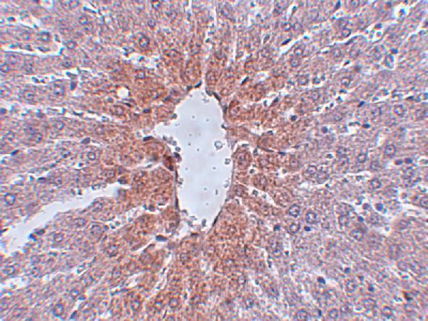
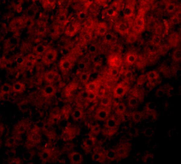
A05189 lists IHC-P and IF applications and human, mouse, and rat reactivity (catalog: A05189 applications/reactivity); its IHC and IF images show rat liver (catalog: A05189 image captions).
A05189 has a rat liver IHC image at 2.5 μg/mL (catalog: A05189 IHC image caption). The same SKU has a rat liver IF image at 20 μg/mL and lists human, mouse, and rat reactivity (catalog: A05189 IF image caption; reactivity).
Which to pick: Choose A05189 for paraffin-section tissue IHC because IHC-P is listed and its own IHC image shows rat liver staining; the fixative is unreported (catalog: A05189 applications; IHC image caption). For IF, choose A05189 based on its rat liver IF image; ICC validation is unreported (catalog: A05189 applications; IF image caption). For work across species, A05189 lists human, mouse, and rat reactivity; clonality is unreported (catalog: A05189 reactivity; clone field).