This website uses cookies to ensure you get the best experience on our website.


- Table of Contents
Plan KCNK1 chromogenic IHC-P around variable cytoplasmic staining, with high levels in brain and glandular epithelial cells (HPA tissue IHC). Start the catalog antibody at 5 μg/mL and use matched staining controls (datasheet: 5 μg/mL; standard IHC practice).

Expected localisation, validated protocols, controls and antibodies — the at-a-glance facts below, then the full design guide.
| Expected localisation | Variable cytoplasmic staining (HPA tissue IHC) | |
| Staining pattern | Variable cytoplasmic staining; high in brain and glandular cells (HPA tissue IHC) | |
| Antigen retrieval | Citrate pH 6.0 HIER, 95–98 °C, 20 min (rule: cytoplasmic / membrane antigen) | |
| Positive control | Kidney+4 more · see all | |
| Negative control | None in HPA (detected in all 45 tissues); use no-primary + isotype controls |
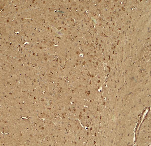
| Fixation | Matched tissue-IHC evidence does not establish the fixation claim. Validate the specimen-specific method before use. (selected-SKU IHC image A04502-1) | |
| Caveat | Antibody staining has low consistency with RNA expression (HPA tissue IHC) | |
| Regulation | Expression varies by tissue (UniProt) | |
| Isoform / epitope | No annotated isoforms; epitope location may matter (UniProt) |
The catalog antibody has an IHC-P protocol (datasheet); the published bladder tissue microarray protocol adds an EDTA retrieval condition (PMC11059691).
| Sample | Tissue sections; selected-image fixative not specified (standard IHC workflow) |
| Fixation | Image fixative and duration unreported (datasheet A04502-1); verify before use. |
| Sectioning | 4–5 µm sections on charged slides (standard) |
| Deparaffinisation | Xylene, graded ethanol series to water (standard) |
| Antigen retrieval | Heat-induced epitope retrieval in citrate buffer, pH 6.0, 20 min at 95–98 °C (standard rule: cytoplasmic / membrane antigen) |
| Peroxidase block | 3% H2O2, 10 min, room temperature (standard) |
| Blocking | 10% normal serum of the secondary host, 30 min, room temperature (standard) |
| Primary antibody | Rabbit anti-KCNK1, 5 μg/mL (datasheet A04502-1) |
| Primary incubation | Overnight at 4 °C (standard) |
| Detection | HRP-polymer secondary, DAB chromogen 5–10 min (standard) |
| Counterstain | Hematoxylin, blue, dehydrate and mount (standard) |
| Expected result | KCNK1-positive staining in cells in tubules of kidney (HPA tissue IHC: High). HPA tissue profile: General cytoplasmic expression at variable levels. High levels in brain and glandular epithelial cells. No signal in the no-primary control. |
KCNK1 is a four-pass membrane channel that can appear at the cell surface and in recycling endosomes (UniProt O00180 topology and subcellular location). In paraffin-section IHC, expect cytoplasmic staining in kidney tubule cells and placental trophoblasts, with variable staining elsewhere (HPA: High in both cell types; general cytoplasmic profile). HPA rates the tissue staining Approved but reports low consistency with RNA expression (HPA: reliability description).
| Strong cytoplasmic stain in kidney tubule cells or placental trophoblasts (HPA: High in both). | This matches the reported IHC pattern. Assess the stained cells and compartment together; intensity alone cannot establish specificity (HPA: general cytoplasmic profile; general IHC practice). |
| Apical membrane accentuation in kidney proximal tubules or bronchial epithelium (UniProt O00180: subcellular location). | These sites are compatible with reported KCNK1 localization. Other kidney tubules may show intracellular staining, so a purely membranous rule would be too narrow (UniProt O00180: kidney localization). |
| Dominant nuclear staining with little cytoplasmic or membrane signal (HPA: cytoplasmic profile; UniProt O00180: subcellular location). | Treat this as discordant with the supplied localization evidence. Review morphology, counterstain and controls before calling the nuclei KCNK1 positive (general IHC practice). |
| Staining in unexpected cells, or a diffuse deposit across tissue compartments (HPA: reported cell-specific pattern). | Cross-reactivity, endogenous detection activity or background may explain it; compare cell types and the no-primary control before assigning target expression (general IHC practice). |
| No signal in kidney tubule cells or placental trophoblasts (HPA: High in both). | Check assay performance before interpreting the section as negative. HPA reports these cells as strong comparators, although its Approved IHC assessment has low RNA consistency (HPA: tissue IHC reliability). |
| Membrane topology and intracellular distribution (UniProt O00180: four transmembrane segments; recycling endosome). | Surface-only scoring could miss intracellular KCNK1. The supplied record does not identify the antibody epitope, so topology alone cannot set an antigen-retrieval condition (UniProt O00180: topology; general IHC practice). |
| Strength of tissue evidence (HPA: IHC Approved; low consistency with RNA expression). | Use the HPA pattern as a comparison, with a specificity caveat. A matching stain supports interpretation but does not independently validate an antibody in a new assay (HPA: reliability description; general IHC practice). |
| Q: What should IF/ICC show? (HPA: subcellular ICC-IF; UniProt O00180: subcellular location). | A: HPA calls vesicles the approved main location; UniProt also reports membranes and recycling endosomes. Interpret IF/ICC on its separate guide page, since these images do not define an IHC-P protocol (HPA: subcellular ICC-IF; UniProt O00180: subcellular location). |
| Situation | Likely cause | Next action |
|---|---|---|
| Kidney tubules and placental trophoblasts are unstained (HPA: High in both). | An IHC run problem is possible; an unstained comparator does not by itself establish absent KCNK1 (general IHC practice). | Check section integrity and the run's positive and negative controls, then review antigen retrieval and antibody conditions for this assay (general IHC practice). |
| The whole section has diffuse chromogen (general IHC practice). | Inadequate blocking or washing, or endogenous detection activity, can obscure cell-associated staining (general IHC practice). | Inspect the no-primary control; adjust blocking, washing or endogenous enzyme quenching as indicated by that control (general IHC practice). |
| Nuclei dominate while cytoplasm is faint (HPA: cytoplasmic profile). | The pattern conflicts with reported localization and may reflect background or an interpretation error (HPA: tissue IHC profile; general IHC practice). | Compare with counterstain and no-primary control; require a plausible cell-associated pattern before scoring positive (general IHC practice). |
| Staining appears mainly in cells outside the reported pattern (HPA: tissue IHC cell types). | Cross-reactivity or endogenous detection activity is possible; HPA's Approved rating does not resolve a new assay's specificity (HPA: reliability; general IHC practice). | Check negative controls and compare a reported positive tissue on the same run before assigning those cells KCNK1 expression (HPA: High kidney tubules and trophoblasts; general IHC practice). |
| Apical-only scoring makes kidney staining seem inconsistent (UniProt O00180: kidney localization). | KCNK1 is reported at proximal-tubule apical membrane and in subapical or cytoplasmic compartments elsewhere in kidney (UniProt O00180: kidney localization). | Record compartment and tubule morphology separately; compare the result with HPA's High tubule-cell staining without assuming every tubule has one pattern (HPA: kidney; UniProt O00180: kidney localization). |
| Brain stain is weaker than kidney stain (HPA: Medium in cerebellar granular layer; High in kidney tubules). | Those reported IHC levels differ, and HPA warns of low consistency between antibody staining and RNA expression (HPA: tissue IHC and reliability description). | Compare like cell populations and the run controls; do not treat relative brightness across organs as a stand-alone specificity test (general IHC practice). |


Comprehensive Human Protein Atlas IHC scoring per tissue (reliability: Approved — Low consistency between antibody staining and RNA expression data.). Rows are taken directly from the HPA tissue chart — click any row's HPA link to view the source.
| Tissue | Cell type | Level | Evidence | Source |
|---|---|---|---|---|
| Kidney | Cells in tubules | High | Protein (IHC) | HPA → |
| Placenta | Trophoblastic cells | High | Protein (IHC) | HPA → |
| Adrenal gland | Glandular cells | Medium | Protein (IHC) | HPA → |
| Appendix | Glandular cells | Medium | Protein (IHC) | HPA → |
| Breast | Myoepithelial cells | Medium | Protein (IHC) | HPA → |
| Tissue | Cell type | Level | Evidence | Source |
|---|---|---|---|---|
| None in HPA: KCNK1 is detected in all 45 scored tissues. Use a no-primary (secondary-only) and an isotype control instead. | ||||
Troubleshoot KCNK1 staining in paraffin sections by checking retrieval, compartment, cell type, controls, and how the signal is scored.
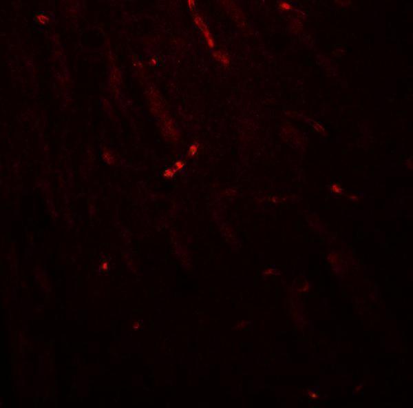
A04502-1 has IHC and IF images from mouse brain tissue (catalog image captions) and listed reactivity with human, mouse and rat (catalog reactivity).
A04502-1 is listed for IHC-P and IF (catalog applications). Its IHC and IF images both show mouse brain tissue (catalog image captions); the images do not establish performance in human or rat tissue (catalog image captions).
Which to pick: Choose A04502-1 for paraffin-section IHC: IHC-P is listed (catalog applications), and its own IHC caption shows mouse brain tissue stained at 5 μg/mL (A04502-1 IHC image caption); the fixative is unreported (A04502-1 IHC image caption). For IF, A04502-1 has a mouse brain tissue image at 20 μg/mL (A04502-1 IF image caption); ICC validation is unreported (catalog applications and image captions). For cross-species studies, A04502-1 lists human, mouse and rat reactivity (catalog reactivity), while its displayed tissue images are mouse only (catalog image captions); clonality is unreported (catalog clone field).