This website uses cookies to ensure you get the best experience on our website.


- Table of Contents
Plan KIF2C staining in paraffin sections using the expected nuclear testis and cytoplasmic lymphoid patterns (HPA tissue IHC). This guide covers tissue controls, fixation consistency, detection, and interpretation of low-level staining.

Expected localisation, validated protocols, controls and antibodies — the at-a-glance facts below, then the full design guide.
| Expected localisation | Nuclear in testis; cytoplasmic in lymphoid tissues and intestines (HPA tissue IHC) | |
| Staining pattern | Pachytene spermatocyte nuclei; lymphoid cytoplasm (HPA tissue IHC) | |
| Antigen retrieval | EDTA pH 8.0 HIER, heat-mediated (datasheet PB9230) | |
| Positive control | Testis+2 more · see all | |
| Negative control | Adrenal gland+4 more · see all |
| Fixation | Matched tissue-IHC evidence does not establish the fixation claim. Validate the specimen-specific method before use. (selected-SKU IHC image PB9230) | |
| Caveat | Low germinal-center staining may be missed (HPA tissue IHC) | |
| Regulation | High in testis and thymus; low in gut (UniProt) | |
| Isoform / epitope | 2 isoforms; isoform 2 is testis-specific; epitope coverage is unknown (UniProt) |
The catalog antibody’s IHC-P protocol is accompanied by published KIF2C IHC methods for Wilms tumor, tissue sections, and liver cancer (PMC10505208; PMC8828917; PMC9171145).
| Sample | Tissue sections; selected-image fixative not specified (standard IHC workflow) |
| Fixation | Image fixative and duration unreported (datasheet PB9230); verify before use. |
| Sectioning | 4–5 µm sections on charged slides (standard) |
| Deparaffinisation | Xylene, graded ethanol series to water (standard) |
| Antigen retrieval | Heat retrieval: EDTA pH 8.0 (datasheet PB9230); 20 min, 95–100 °C (standard) |
| Peroxidase block | 3% H2O2, 10 min, room temperature (standard) |
| Blocking | 10% goat serum (datasheet PB9230) |
| Primary antibody | Rabbit anti-KIF2C, 0.5-1μg/ml (datasheet PB9230) |
| Primary incubation | Overnight at 4 °C (datasheet PB9230) |
| Detection | HRP-conjugated secondary, DAB chromogen (datasheet PB9230) |
| Counterstain | Hematoxylin, blue, dehydrate and mount (standard) |
| Expected result | KIF2C-positive staining in pachytene spermatocytes of testis (HPA tissue IHC: High). HPA tissue profile: Nuclear expression in testis and cytoplasmic expression in lymphoid tissues and intestines. No signal in the no-primary control. |
KIF2C is a nonmembrane protein associated with the cytoskeleton, nucleus, centromere and kinetochore (UniProt Q99661: localization and topology). In IHC, expect strong nuclear staining in testicular pachytene spermatocytes and moderate staining in thymic cortical cells; HPA also describes cytoplasmic expression in lymphoid tissues and intestines (HPA: tissue IHC, Enhanced reliability). Interpret the stained cell type alongside its compartment, because the reported pattern varies by tissue (HPA: tissue IHC).
| Strong nuclear staining in pachytene spermatocytes, with weaker or absent staining in surrounding cells (HPA: testis, High). | This is the clearest supplied positive tissue pattern. Evaluate the named cells, rather than requiring uniform staining across the section (HPA: testis, High; general IHC practice). |
| Moderate staining in thymic cortical cells or appendix endocrine cells (HPA: thymus and appendix, Medium). | These are additional observed positives. Cytoplasmic signal can be compatible with the HPA lymphoid tissue profile; a testis-like nuclear pattern is not required in every tissue (HPA: tissue IHC profile). |
| Predominantly membrane-outline or extracellular staining, without staining in an expected cellular compartment. | Treat this as a possible artefact and review controls: KIF2C has no transmembrane segment or signal peptide, and its reported locations are intracellular (UniProt Q99661: topology, processing and localization). |
| Strong staining in cells listed as undetected, or broad colour deposition across unrelated cell types (HPA: negative tissue IHC entries). | Cross-reactivity or endogenous detection activity is possible, not proven. Check the exact cell type and a no-primary control before assigning the signal to KIF2C (HPA: tissue IHC; general IHC practice). |
| No signal in pachytene spermatocytes in a testis section (HPA: testis, High). | The run lacks its strongest supplied positive reference. Check tissue preservation, retrieval, primary antibody conditions and detection controls before calling an unknown sample negative (general IHC practice). |
| Tissue and cell selection (HPA: tissue IHC). | Testicular pachytene spermatocytes are High; thymic cortical and appendix endocrine cells are Medium. Bone marrow hematopoietic cells are Low, so a weak result there is less decisive (HPA: tissue IHC). |
| IHC antibody evidence (HPA: antibody validation). | HPA079172 and CAB080091 have Enhanced IHC validation; HPA006219 has Approved ICC status but no IHC status in the supplied record. Match the antibody's validation to the assay (HPA: antibody validation; general IHC practice). |
| Intracellular target and processing (UniProt Q99661: topology and processing). | KIF2C has no transmembrane segment or signal peptide; the reported chain spans residues 2–725. These facts support intracellular interpretation but do not identify the antibody epitope or predict antigen retrieval response (UniProt Q99661; general IHC practice). |
| Isoform distribution (UniProt Q99661: isoforms and tissue specificity). | UniProt lists two isoforms and describes isoform 2 as testis-specific. The supplied evidence does not show which isoforms the IHC antibodies recognize, so testis staining cannot be assigned to one isoform (UniProt Q99661; HPA: antibody validation). |
| IF/ICC Q&A: What localization is reported? | HPA reports approved centrosome and midbody localization; nucleoplasm and cytosol are uncertain. Its ICC images include Rh30, SiHa and U2OS. These IF observations do not define an IHC-P protocol (HPA: subcellular ICC-IF). |
| Antigen retrieval (general IHC practice). | Follow the IHC-validated antibody's stated retrieval conditions and assess them with a positive control. The supplied UniProt and HPA records give no KIF2C-specific fixation or retrieval sensitivity (general IHC practice; UniProt Q99661; HPA: tissue IHC). |
| Situation | Likely cause | Next action |
|---|---|---|
| Testis positive control is blank (HPA: pachytene spermatocytes, High). | A failed staining run or unsuitable antibody conditions are possible; the slide alone cannot identify which step failed (general IHC practice). | Check control tissue, the IHC-validated antibody's retrieval and dilution instructions, and detection reagents; repeat with a known-positive section (general IHC practice). |
| Weak signal in bone marrow hematopoietic cells (HPA: bone marrow, Low). | Low expression is compatible with the HPA observation; intensity alone does not establish an assay failure (HPA: tissue IHC). | Compare with testicular pachytene spermatocytes on the same run before interpreting a weak unknown sample (HPA: testis, High; general IHC practice). |
| Diffuse chromogen obscures cellular boundaries. | Nonspecific antibody binding, endogenous enzyme activity or excess detection signal may contribute (general IHC practice). | Review no-primary controls, blocking and detection steps; adjust antibody conditions according to the IHC-validated antibody's instructions (general IHC practice). |
| Strong staining appears in an HPA-undetected cell type (HPA: tissue IHC negative entries). | Cross-reactivity or endogenous detection activity is possible; an HPA negative entry alone cannot diagnose the mechanism (HPA: tissue IHC; general IHC practice). | Confirm the cell identity, inspect a no-primary control and compare an independent IHC-validated antibody where available (HPA: antibody validation; general IHC practice). |
| Signal outlines membranes or extracellular material while expected cells remain unstained. | This conflicts with the reported intracellular locations and lack of a transmembrane segment or signal peptide (UniProt Q99661: localization, topology and processing). | Check controls and chromogen deposition, then reassess the compartment in a known-positive testis section (HPA: testis, High; general IHC practice). |
| IHC appears to disagree with ICC-IF localization. | HPA reports nuclear testis and cytoplasmic lymphoid or intestinal tissue patterns; its ICC centrosome and midbody findings come from a different assay (HPA: tissue IHC and subcellular ICC-IF). | Interpret each result against its own tissue, cell type and assay validation; use IHC-validated antibodies for the paraffin section comparison (HPA: antibody validation; general IHC practice). |


Comprehensive Human Protein Atlas IHC scoring per tissue (reliability: Enhanced — High consistency between antibody staining and RNA expression data.). Rows are taken directly from the HPA tissue chart — click any row's HPA link to view the source.
| Tissue | Cell type | Level | Evidence | Source |
|---|---|---|---|---|
| Testis | Pachytene spermatocytes | High | Protein (IHC) | HPA → |
| Appendix | Endocrine cells | Medium | Protein (IHC) | HPA → |
| Thymus | Cortical cells | Medium | Protein (IHC) | HPA → |
| Tissue | Cell type | Level | Evidence | Source |
|---|---|---|---|---|
| Adrenal gland | Glandular cells | Not detected | Protein (IHC) | HPA → |
| Breast | Adipocytes | Not detected | Protein (IHC) | HPA → |
| Bronchus | Respiratory epithelial cells | Not detected | Protein (IHC) | HPA → |
| Caudate | Glial cells | Not detected | Protein (IHC) | HPA → |
| Cerebellum | Cells in granular layer | Not detected | Protein (IHC) | HPA → |
Troubleshoot KIF2C staining in paraffin sections by checking retrieval, cellular compartment, tissue context and controls before interpreting chromogenic signal.
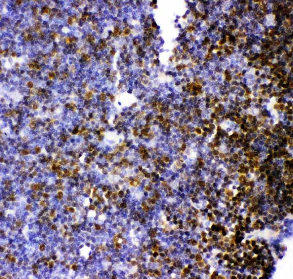
PB9230 has paraffin-section IHC images from rat, mouse, and human tissues and an IF image from U20S cells (PB9230 IHC/IF captions).
PB9230 is the only card that will render (cards: PB9230); its IHC images show rat thymus, rat testis, mouse testis, and human testis (PB9230 IHC captions). It lists IF/ICC applications and human, mouse, and rat reactivity (PB9230 catalog: applications/reactivity); its IF image shows U20S cells (PB9230 IF caption).
Which to pick: Choose PB9230 for tissue IHC: its captions show paraffin sections, but do not report the fixative (PB9230 IHC captions). For IF/ICC, PB9230 has an IF image of U20S cells (PB9230 IF caption); M03202-1 lists human ICC/IF but has no figure in the payload (M03202-1 catalog: applications/reactivity/image alts). For cross-species work, PB9230 lists human, mouse, and rat reactivity, while M03202-1 lists human reactivity only (PB9230 and M03202-1 catalogs: reactivity).