This website uses cookies to ensure you get the best experience on our website.


- Table of Contents
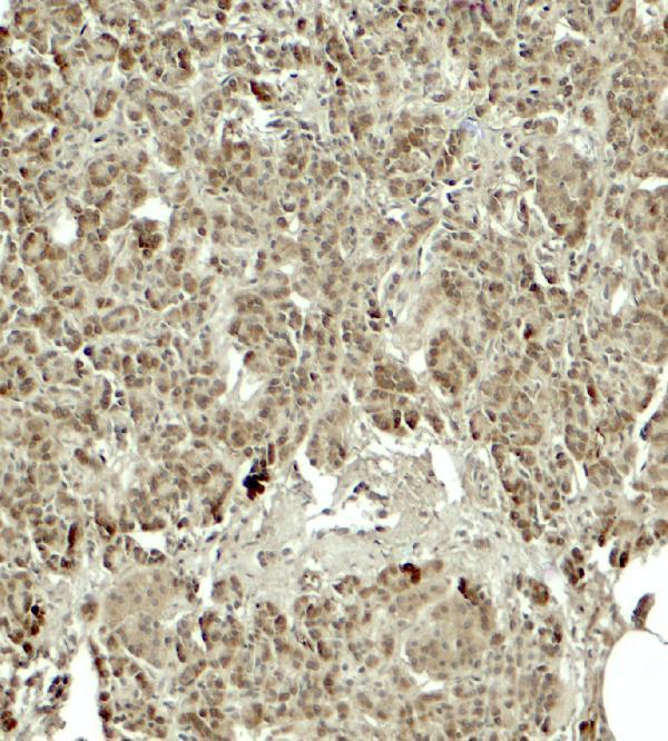
Use exocrine pancreatic cells as a positive control for paraffin-section KIRREL2 IHC (HPA tissue IHC), starting the catalog antibody at 5 μg/mL (datasheet: IHC-P). Compare the observed cytoplasmic staining (HPA tissue IHC) with the annotated cell-contact membrane location (UniProt).

Expected localisation, validated protocols, controls and antibodies — the at-a-glance facts below, then the full design guide.
| Expected localisation | Cytoplasmic in exocrine pancreas (HPA tissue IHC); cell-contact membrane annotated (UniProt) | |
| Staining pattern | Exocrine pancreatic cells show cytoplasmic staining (HPA tissue IHC) | |
| Antigen retrieval | Citrate pH 6.0 HIER, 95–98 °C, 20 min (rule: cytoplasmic / membrane antigen) | |
| Positive control | Pancreas | |
| Negative control | Adipose tissue+4 more · see all |
| Fixation | Keep fixation consistent across sections (standard IHC practice; not target-specific); Matched tissue-IHC evidence does not establish the fixation claim. Validate the specimen-specific method before use. (selected-SKU IHC image A07812) | |
| Caveat | Tissue localization conflicts with limited external data (HPA tissue IHC) | |
| Regulation | Pancreas-enriched RNA expression (HPA tissue RNA) | |
| Isoform / epitope | 4 isoforms; map epitopes to extracellular or cytoplasmic regions (UniProt) |
The catalog antibody’s IHC-P protocol is accompanied by one published KIRREL2 protocol using FFPE tissue sections (PMC9288368: methods).
| Sample | Tissue sections; selected-image fixative not specified (standard IHC workflow) |
| Fixation | Image fixative and duration unreported (datasheet A07812); verify before use. |
| Sectioning | 4–5 µm sections on charged slides (standard) |
| Deparaffinisation | Xylene, graded ethanol series to water (standard) |
| Antigen retrieval | Heat-induced epitope retrieval in citrate buffer, pH 6.0, 20 min at 95–98 °C (standard rule: cytoplasmic / membrane antigen) |
| Peroxidase block | 3% H2O2, 10 min, room temperature (standard) |
| Blocking | 10% normal serum of the secondary host, 30 min, room temperature (standard) |
| Primary antibody | Rabbit anti-KIRREL2, 5 μg/mL (datasheet A07812) |
| Primary incubation | Overnight at 4 °C (standard) |
| Detection | HRP-polymer secondary, DAB chromogen 5–10 min (standard) |
| Counterstain | Hematoxylin, blue, dehydrate and mount (standard) |
| Expected result | KIRREL2-positive staining in exocrine glandular cells of pancreas (HPA tissue IHC: High). HPA tissue profile: Cytoplasmic expression in exocrine pancreas. No signal in the no-primary control. |
For paraffin IHC, expect strong cytoplasmic staining in pancreatic exocrine glandular cells (HPA: High in exocrine pancreas; IHC reliability Enhanced). Interpret compartment carefully: UniProt places KIRREL2 at cell contacts on the membrane and reports high expression in islet beta cells (UniProt Q6UWL6 localization and tissue specificity). HPA reports medium staining–RNA consistency and limited external data that contradict its protein localization (HPA reliability description).
| Strong cytoplasmic staining in pancreatic exocrine glandular cells, with readable tissue structure. | This matches the documented tissue IHC pattern (HPA: High in exocrine glandular cells). Record whether staining is broadly cytoplasmic or concentrated at cell borders: UniProt places KIRREL2 along cell contacts, but HPA's IHC image summary describes cytoplasmic expression (UniProt Q6UWL6 localization; HPA tissue IHC profile). The compartment discrepancy limits a definitive localization call. |
| Predominantly nuclear staining, or staining in a compartment unsupported by either source. | Treat this as a possible staining artefact and inspect the controls before calling it KIRREL2 (general IHC practice). UniProt reports a membrane protein at cell contacts, whereas HPA describes cytoplasmic exocrine staining; neither supports a predominantly nuclear pattern (UniProt Q6UWL6 topology and localization; HPA tissue IHC profile). |
| Strong staining in cells scored as not detected, with little or no staining in pancreatic exocrine cells. | Consider antibody cross-reactivity or endogenous chromogenic detection activity, then check matched controls (general IHC practice). HPA scores adipocytes in adipose tissue and adrenal glandular cells as Not detected, while pancreatic exocrine glandular cells are High (HPA tissue IHC). A positive result in one unexpected cell type alone does not establish KIRREL2 expression. |
| Diffuse colour across cells and surrounding tissue, obscuring cell boundaries. | Interpret the field as background until detection and washing controls are satisfactory (general IHC practice). Diffuse staining cannot reliably establish the cell-specific exocrine pattern reported by HPA or resolve its disagreement with UniProt localization (HPA tissue IHC profile; UniProt Q6UWL6 localization). Score only staining distinguishable from the local background. |
| No interpretable signal in pancreatic exocrine glandular cells. | A negative result in the strongest supplied IHC reference tissue calls for a run and reagent check before biological interpretation (HPA: High in pancreatic exocrine glandular cells; general IHC practice). Check tissue integrity, retrieval, primary antibody, detection, and counterstain using the chosen assay's instructions (general IHC practice). This result alone cannot establish target absence. |
| Tissue and compartment agreement | HPA identifies exocrine cytoplasmic staining, while UniProt identifies beta-cell enrichment and cell-contact membrane localization (HPA tissue IHC; UniProt Q6UWL6). Document which pattern the slide shows. HPA's reliability note reports medium staining–RNA consistency and contradictory limited external localization data (HPA reliability description), so avoid declaring either compartment settled by this record. |
| Antibody validation | HPA071587 has Enhanced IHC validation; HPA074326 has no IHC status listed, though both have Approved ICC status (HPA antibodies). Enhanced denotes a pattern reproduced by independent antibodies or orthogonal data (HPA validation summary). Record the antibody identifier when comparing slides, because validation status is antibody specific (HPA antibodies). |
| Epitope location and retrieval | KIRREL2 has an extracellular region at residues 21–510, a transmembrane segment at 511–531, and a cytoplasmic region at 532–708 (UniProt Q6UWL6 topology). These boundaries help interpret an epitope if the antibody's immunogen is known; the supplied record does not locate that immunogen or establish a KIRREL2-specific retrieval condition (UniProt Q6UWL6 topology; supplied HPA antibody data). |
| Alternative forms and processing | UniProt lists four isoforms, a signal peptide at residues 1–20, and a mature chain at 21–708 (UniProt Q6UWL6). Antibodies directed at different sequences may therefore need separate interpretation (general antibody practice), but the supplied record does not identify an antibody epitope or show isoform-specific IHC staining (supplied HPA antibody data). |
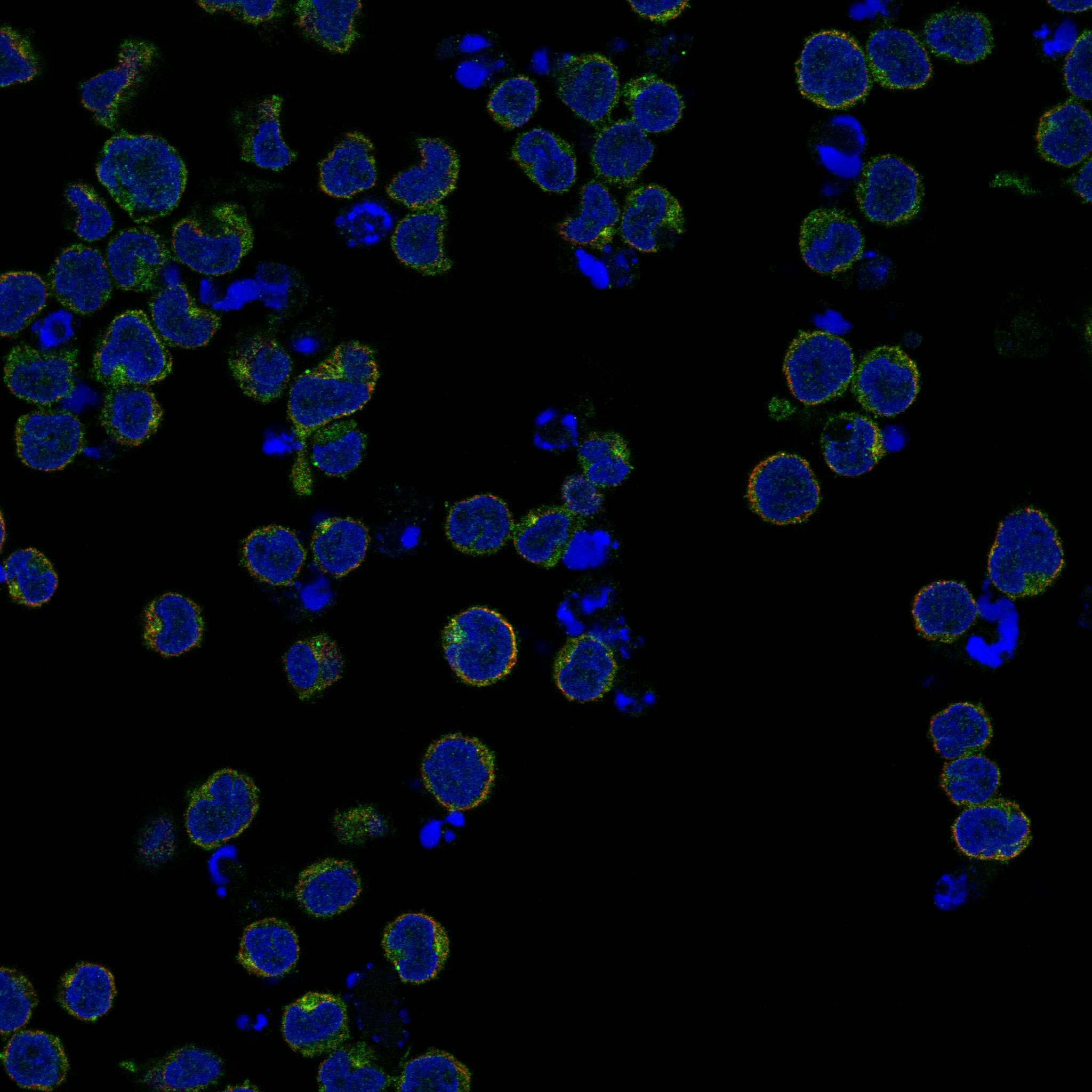
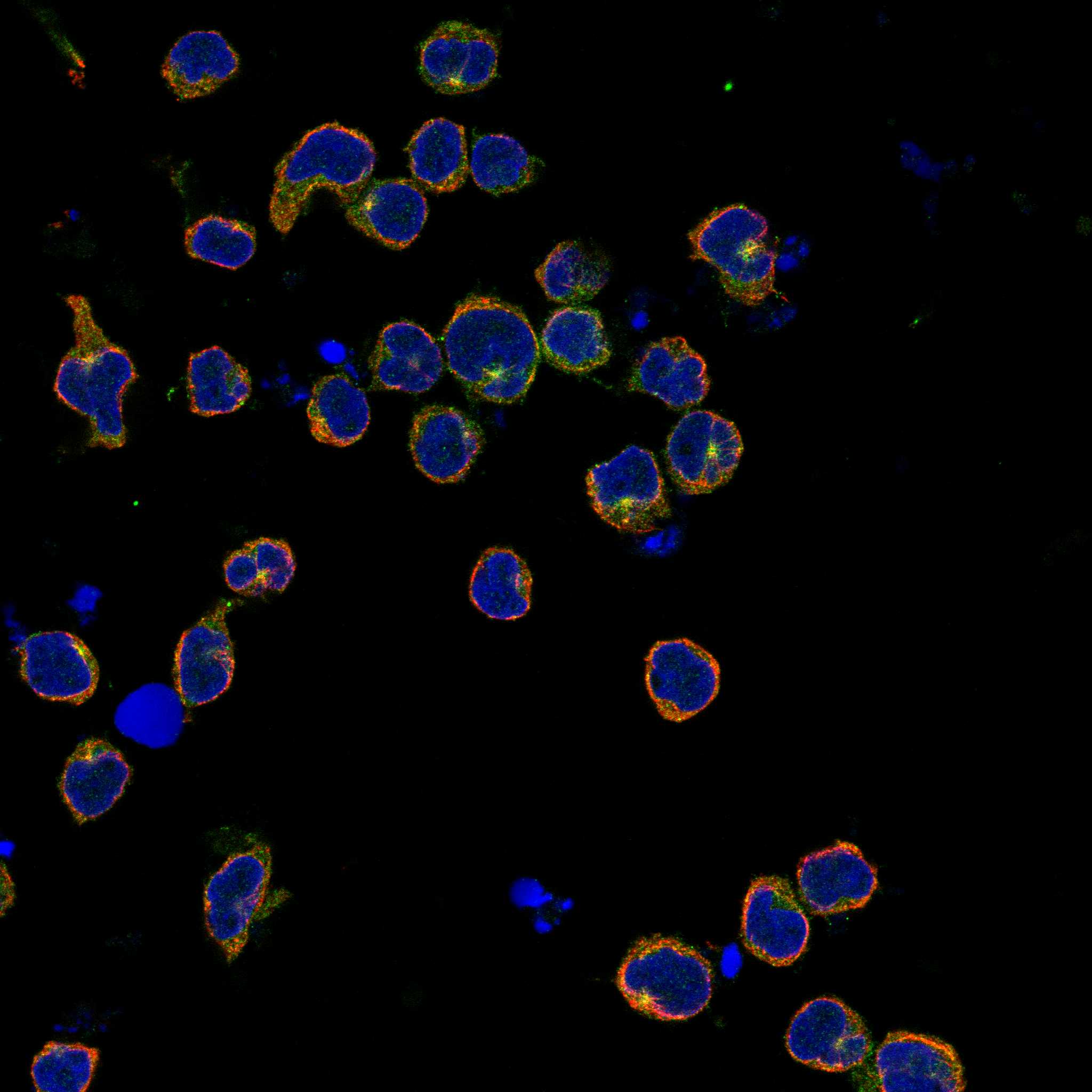
| IF/ICC Q&A | What should an IF/ICC image show? HPA reports mainly centriolar satellite localization in REH and U2OS images, with both listed antibodies Approved for ICC (HPA subcellular; HPA antibodies). That observation belongs to cell-based IF interpretation; it does not establish a paraffin IHC compartment or replace the tissue pattern above (HPA subcellular; HPA tissue IHC). |
| Situation | Likely cause | Next action |
|---|---|---|
| Pancreatic exocrine cells show no signal. | The positive tissue result has not been reproduced; the cause is unresolved (HPA: High in exocrine glandular cells). | Confirm the tissue and cell identification, then review retrieval, primary antibody, detection reagents, and a concurrent positive control under the chosen IHC procedure (general IHC practice). Do not infer a target-specific fixation effect: none is documented here (supplied HPA and UniProt records). |
| Signal appears mainly nuclear. | This compartment conflicts with both reported patterns (HPA: cytoplasmic exocrine profile; UniProt Q6UWL6: membrane at cell contacts). | Inspect a primary-antibody omission control and compare nuclear colour with background in adjacent cells (general IHC practice). Reassess tissue morphology and antibody specificity before assigning a KIRREL2 score (general IHC practice). |
| HPA Not detected cells stain as strongly as pancreas. | Cross-reactivity or endogenous detection activity is possible (general IHC practice); HPA reports Not detected staining in adipose adipocytes and adrenal glandular cells (HPA tissue IHC). | Compare a matched primary-antibody omission control, check the detection system's endogenous-activity control, and repeat with an independently validated IHC antibody if available (general IHC practice; HPA: HPA071587 Enhanced IHC). |
| Colour is diffuse and masks cell boundaries. | Background or overdeveloped chromogen may prevent cell-level interpretation (general IHC practice). | Check wash steps, detection development, counterstain balance, and the omission control under the chosen procedure (general IHC practice). Score only cell-associated signal that remains distinguishable from nearby background (general IHC practice). |
| Exocrine cytoplasm stains, but cell borders do not stand out. | The sources disagree on compartment: HPA reports cytoplasmic IHC, while UniProt reports localization along cell contacts (HPA tissue IHC; UniProt Q6UWL6 localization). | Report the observed cytoplasmic pattern and antibody identifier without calling absent border accentuation a failed assay (HPA tissue IHC; HPA antibodies). Use morphology and appropriate controls for the interpretation (general IHC practice). |
| An ICC/IF image appears punctate near the centrosomal region. | HPA reports mainly centriolar satellite localization in its cell-based images (HPA subcellular). | Interpret the image on the separate IF/ICC guide page and identify the cell line and antibody used (HPA subcellular; HPA antibodies). Keep its compartment call separate from the exocrine paraffin IHC readout (HPA subcellular; HPA tissue IHC). |


Comprehensive Human Protein Atlas IHC scoring per tissue (reliability: Enhanced — Medium consistency between antibody staining and RNA expression data. Protein localization is contradicted by limited external data.). Rows are taken directly from the HPA tissue chart — click any row's HPA link to view the source.
| Tissue | Cell type | Level | Evidence | Source |
|---|---|---|---|---|
| Pancreas | Exocrine glandular cells | High | Protein (IHC) | HPA → |
| Tissue | Cell type | Level | Evidence | Source |
|---|---|---|---|---|
| Adipose tissue | Adipocytes | Not detected | Protein (IHC) | HPA → |
| Adrenal gland | Glandular cells | Not detected | Protein (IHC) | HPA → |
| Appendix | Glandular cells | Not detected | Protein (IHC) | HPA → |
| Bone marrow | Hematopoietic cells | Not detected | Protein (IHC) | HPA → |
| Breast | Adipocytes | Not detected | Protein (IHC) | HPA → |
Troubleshoot KIRREL2 staining by comparing signal intensity, cell type and subcellular pattern with the available tissue evidence.
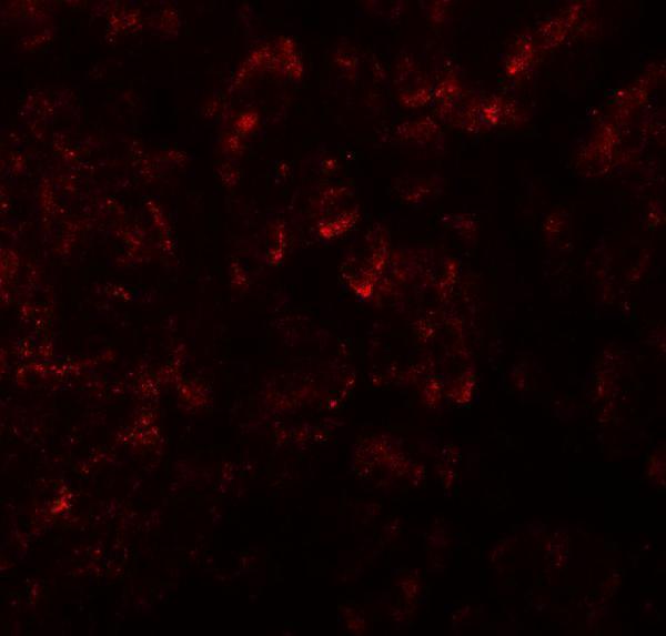
A07812 is listed for IHC-P and IF (catalog: applications), with images of human pancreas tissue for both methods (catalog: image captions). Its listed reactivity covers human, mouse, and rat (catalog: reactivity).
A07812 will render with its own IHC image of human pancreas tissue at 5 μg/mL (catalog: A07812 IHC image caption). Its IF image also shows human pancreas tissue, at 20 μg/mL (catalog: A07812 IF image caption).
Which to pick: Choose A07812 for paraffin-section tissue IHC because IHC-P is listed (catalog: applications) and its own IHC image shows human pancreas tissue (catalog: A07812 IHC image caption); the fixative is unreported (catalog: A07812 IHC image caption). For IF, A07812 has a human pancreas image (catalog: A07812 IF image caption); ICC is not listed (catalog: applications). For cross-species experiments, A07812 lists human, mouse, and rat reactivity (catalog: reactivity), while its IHC and IF images show human tissue only (catalog: image captions).