This website uses cookies to ensure you get the best experience on our website.


- Table of Contents
Plan KISS1 IHC-P using placental syncytiotrophoblasts as a high-staining reference (HPA tissue IHC). The guide covers formaldehyde-fixed paraffin sections (datasheet M00689) and interpretation of a secreted protein whose RNA and protein locations may differ (HPA tissue IHC).

Expected localisation, validated protocols, controls and antibodies — the at-a-glance facts below, then the full design guide.
| Expected localisation | Cytoplasmic in placental trophoblasts (HPA tissue IHC) | |
| Staining pattern | Strong cytoplasmic staining in placental syncytiotrophoblasts (HPA tissue IHC) | |
| Antigen retrieval | Citrate pH 6 HIER, heat-mediated (datasheet M00689) | |
| Positive control | Placenta+1 more · see all | |
| Negative control | Adipose tissue+4 more · see all |
| Fixation | Formaldehyde-fixed paraffin sections (selected-SKU IHC image M00689); Matched tissue-IHC evidence does not establish the fixation claim. Validate the specimen-specific method before use. (selected-SKU IHC image M00689) | |
| Caveat | Secreted protein; RNA and protein locations can differ (HPA tissue IHC) | |
| Regulation | Higher in first-trimester than term trophoblasts (UniProt) | |
| Isoform / epitope | No annotated isoforms; secreted chain begins at residue 20 (UniProt) |
The catalog antibody’s IHC-P protocol is followed by published chromogenic KISS1 IHC protocols for osteosarcoma, canine reproductive tissues, and human and rat carotid body and superior cervical ganglion (PMC3896032; PMC13082194; PMC3284150).
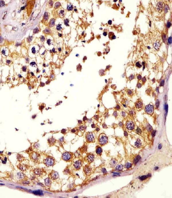
| Sample | Formaldehyde-fixed, paraffin-embedded human testis tissue (datasheet M00689) |
| Fixation | Image formalin-fixed; duration unreported (datasheet M00689); verify before use. |
| Sectioning | 4–5 µm sections on charged slides (standard) |
| Deparaffinisation | Xylene, graded ethanol series to water (standard) |
| Antigen retrieval | Heat retrieval: Citrate pH 6 (datasheet M00689); 20 min, 95–100 °C (standard) |
| Peroxidase block | 3% H2O2, 10 min, room temperature (standard) |
| Blocking | 10% normal serum of the secondary host, 30 min, room temperature (standard) |
| Primary antibody | Rabbit anti-KISS1, 1:25 (datasheet M00689) |
| Primary incubation | 1 hours at 37°C (datasheet M00689) |
| Detection | HRP-polymer secondary, DAB chromogen 5–10 min (standard) |
| Counterstain | Hematoxylin, blue, dehydrate and mount (standard) |
| Expected result | KISS1-positive staining in syncytiotrophoblasts - cell body of placenta (HPA tissue IHC: High). HPA tissue profile: Selective cytoplasmic expression in placental trophoblasts. No signal in the no-primary control. |
In paraffin IHC, expect selective cytoplasmic KISS1 staining in placental trophoblasts, strongest in syncytiotrophoblast cell bodies (HPA tissue IHC: Enhanced; high in syncytiotrophoblasts). KISS1 is a secreted protein with no transmembrane segment, so a sharply defined membrane pattern is unexpected (UniProt Q15726: secreted; no transmembrane segment). HPA notes that tissue RNA and protein locations can differ for secreted proteins (HPA tissue IHC).
| Strong cytoplasmic staining in placental syncytiotrophoblast cell bodies. | This matches the principal positive pattern: high staining in these cells (HPA tissue IHC: placenta, syncytiotrophoblasts, High). Score the named cells and their cytoplasm, since HPA describes selective trophoblast expression rather than uniform staining across the section (HPA tissue IHC: tissue profile). |
| Moderate staining in round or early spermatids in testis. | This is a supported secondary positive pattern at medium level (HPA tissue IHC: testis, round or early spermatids, Medium). Its lower reported level means it should not be treated as equivalent to the placental high-positive reference (HPA tissue IHC: positive tissues). |
| Predominantly nuclear staining or a crisp membrane rim in placental cells. | This differs from the reported cytoplasmic IHC pattern (HPA tissue IHC: selective cytoplasmic expression). A membrane rim also conflicts with the lack of a transmembrane segment (UniProt Q15726: topology). Treat the pattern as suspect and compare it with staining and detection controls (general IHC practice). |
| Strong staining in an HPA-listed negative cell population. | Examples include adipocytes in adipose tissue and glandular cells in adrenal gland, both reported as not detected (HPA tissue IHC: negative tissues). Cross-reactivity or endogenous detection activity is possible; inspect control sections before assigning the signal to KISS1 (general IHC practice). |
| Diffuse staining across tissue, or no staining in placental syncytiotrophoblasts. | Diffuse staining obscures the selective cytoplasmic pattern (HPA tissue IHC: tissue profile); review background controls (general IHC practice). Absence in the high-positive placental cells makes the run inconclusive until section quality, antibody conditions, and detection are checked (HPA tissue IHC: placenta, High; general IHC practice). |
| Placental cell identity and gestational context | HPA localizes high protein staining to syncytiotrophoblast cell bodies (HPA tissue IHC: placenta, High). UniProt reports higher KISS1 expression in first-trimester trophoblasts than at term, with expression in villous trophoblast (UniProt Q15726: tissue specificity). Record the sampled context when comparing sections. |
| Secretion and processing | KISS1 is secreted and has a signal peptide at residues 1–19 (UniProt Q15726: subcellular location; processing). HPA cautions that RNA and protein tissue locations can differ for secreted proteins (HPA tissue IHC: reliability description). Interpret protein staining by its observed cell pattern, without assuming RNA and protein coincide. |
| IHC evidence and controls | HPA assigns the tissue IHC profile Enhanced reliability, citing consistency between staining and RNA data (HPA tissue IHC: reliability). Both listed antibodies, HPA035542 and CAB017775, have Enhanced IHC status (HPA antibodies). Placenta is the high-positive reference; HPA-listed negative cell populations help judge background (HPA tissue IHC: positive and negative tissues). |
| What does IF/ICC show? | HPA reports KISS1 mainly in vesicles by ICC-IF, with vesicles an approved main location (HPA subcellular ICC-IF). This supports a punctate intracellular IF interpretation, while the paraffin IHC reference remains selective cytoplasmic staining in placental trophoblasts (HPA tissue IHC: tissue profile). |
| Antigen retrieval and epitope | The supplied UniProt and HPA records give no antibody epitope or KISS1-specific retrieval result (UniProt Q15726; HPA antibodies). If optimizing retrieval, assess it against the same positive and negative controls under the chosen IHC workflow (general IHC practice); do not infer target-specific fixation sensitivity. |
| Situation | Likely cause | Next action |
|---|---|---|
| Placental syncytiotrophoblasts show no signal. | The expected high-positive cells are absent from the stained result (HPA tissue IHC: placenta, High); a failed staining step or unsuitable section is possible (general IHC practice). | Confirm the named cells are present, then check antibody application, retrieval conditions, and detection with a positive control (general IHC practice). Do not call the tissue KISS1-negative from this run. |
| The entire section has diffuse color. | Background can hide HPA's selective cytoplasmic pattern (HPA tissue IHC: tissue profile). Incomplete blocking, washing, or endogenous detection activity can contribute (general IHC practice). | Review omission and detection controls; adjust blocking, washing, or detection conditions as indicated by those controls (general IHC practice). Reassess cell-specific staining. |
| Signal is mainly nuclear or outlines membranes. | That distribution differs from cytoplasmic placental IHC staining (HPA tissue IHC: tissue profile); KISS1 has no transmembrane segment (UniProt Q15726: topology). | Check compartment assignment against counterstained morphology and controls, then reassess antibody and detection background (general IHC practice). |
| Adipocytes in adipose tissue stain strongly. | Those cells are reported as not detected (HPA tissue IHC: adipose tissue). Cross-reactivity or endogenous detection activity is possible (general IHC practice). | Compare antibody-omission and detection controls, and judge the signal alongside the placental positive pattern (general IHC practice; HPA tissue IHC: placenta, High). |
| Testis staining is weaker than placenta. | HPA reports medium staining in round or early spermatids and high staining in placental syncytiotrophoblasts (HPA tissue IHC: positive tissues). | Identify the spermatid population before scoring; compare tissues using their reported cell types and levels (HPA tissue IHC: positive tissues). |
| Punctate IF signal appears different from the IHC pattern. | HPA's approved ICC-IF location is mainly vesicles, while its placental tissue IHC description is cytoplasmic (HPA subcellular ICC-IF; HPA tissue IHC: tissue profile). | Interpret each application against its own HPA observation; use the separate IF/ICC guide for IF workflow decisions (HPA subcellular ICC-IF; HPA tissue IHC). |


![HPA ICC-IF BJ [Human fibroblast] — KISS1 (HPA035542)](https://images.proteinatlas.org/35542/1742_H7_3_cr5804964873f54_blue_red_green.jpg)
![HPA ICC-IF BJ [Human fibroblast] — KISS1 (HPA035542)](https://images.proteinatlas.org/35542/1742_H7_18_cr58049652a0d0f_blue_red_green.jpg)
Comprehensive Human Protein Atlas IHC scoring per tissue (reliability: Enhanced — High consistency between antibody staining and RNA expression data. Secreted protein, tissue location of RNA and protein is expected to differ.). Rows are taken directly from the HPA tissue chart — click any row's HPA link to view the source.
| Tissue | Cell type | Level | Evidence | Source |
|---|---|---|---|---|
| Placenta | Syncytiotrophoblasts - cell body | High | Protein (IHC) | HPA → |
| Testis | Round or early spermatids | Medium | Protein (IHC) | HPA → |
| Tissue | Cell type | Level | Evidence | Source |
|---|---|---|---|---|
| Adipose tissue | Adipocytes | Not detected | Protein (IHC) | HPA → |
| Adrenal gland | Glandular cells | Not detected | Protein (IHC) | HPA → |
| Appendix | Glandular cells | Not detected | Protein (IHC) | HPA → |
| Bone marrow | Hematopoietic cells | Not detected | Protein (IHC) | HPA → |
| Breast | Adipocytes | Not detected | Protein (IHC) | HPA → |
Use the catalog antibody’s IHC-P evidence alongside KISS1 processing and tissue localisation to troubleshoot chromogenic staining in paraffin sections.
Anti-KISS1 antibodies have real paraffin-section IHC data in human testis (M00689 image caption) and IF data in human HeLa cells (M00689-1 image caption).
M00689 lists human IHC-P reactivity and shows staining in a human testis paraffin section (M00689 catalog; M00689 image caption). M00689-1 lists human IF reactivity and shows cytoplasmic staining in HeLa cells (M00689-1 catalog; M00689-1 image caption).
Which to pick: Choose M00689 for tissue IHC: it is rabbit polyclonal, lists IHC-P, and has a paraffin-section image; the caption gives conflicting fixative descriptions, so the exact fixative is unresolved (M00689 catalog; M00689 image caption). Choose M00689-1 for IF/ICC: it is rabbit polyclonal, lists IF, and has a fixed, permeabilized HeLa-cell image (M00689-1 catalog; M00689-1 image caption). Neither SKU lists nonhuman reactivity, so neither has catalog support for a cross-species choice (M00689 catalog; M00689-1 catalog).