KISS1R / KiSS-1 receptor · IHC design guide

Design Immunohistochemistry for KISS1R

Plan KISS1R chromogenic IHC on paraffin sections using 1–2 μg/ml catalog antibody and placenta trophoblasts as a positive control (datasheet A01364-2; HPA tissue IHC). Interpret cytoplasmic tissue staining alongside the annotated cell-membrane location and HPA's low consistency between staining and RNA expression (HPA tissue IHC; UniProt).

Evidence assembled Oct 2026 · For research use; verify linked source records and product datasheet before use
Immunohistochemistry protocol sheet for KISS1R (IHC for KISS1R): expected localisation Cytoplasmic in tissue (HPA tissue IHC); cell membrane (UniProt), antibody A01364-2, validated IHC image, and IHC protocol steps
Printable KISS1R IHC protocol sheet — expected localisation Cytoplasmic in tissue (HPA tissue IHC); cell membrane (UniProt), antibody A01364-2, controls and protocol steps. Open the full KISS1R IHC guide →

KISS1R Immunohistochemistry Experimental Design Guide

Expected localisation, validated protocols, controls and antibodies — the at-a-glance facts below, then the full design guide.

Must know before staining
Expected localisation Cytoplasmic in tissue (HPA tissue IHC); cell membrane (UniProt)
Staining pattern Cytoplasmic staining in several tissues, including hypothalamus (HPA tissue IHC)
Antigen retrieval EDTA pH 8.0 HIER, heat-mediated (datasheet A01364-2)
Positive control ⓘ Adrenal gland+4 more · see all
Negative control ⓘ Adipose tissue+4 more · see all
Important caveats
Reasons your staining may differ from the expected pattern.
Fixation Keep fixation consistent across sections (standard IHC practice; not target-specific). Selected-image fixative and duration unreported (datasheet A01364-2); verify before use.
Caveat Antibody staining has low consistency with RNA expression (HPA tissue IHC)
Regulation Higher in early than term placenta (UniProt)
Isoform / epitope 0 annotated isoforms; extracellular vs cytoplasmic epitope matters (UniProt)
Section 1

Recommended KISS1R IHC & IF Protocols

The catalog antibody’s IHC-P protocol uses EDTA pH 8.0 retrieval (datasheet: A01364-2); three published KISS1R IHC protocols provide tissue-specific alternatives (PMC3284150; PMC6433948; PMC8871750).

Recommended immunohistochemistry (IHC-P) protocol parameters
SampleParaffin-embedded human gall bladder adenosquamous carcinoma tissue; fixative not specified (datasheet A01364-2)
FixationImage fixative and duration unreported (datasheet A01364-2); verify before use.
Sectioning4–5 µm sections on charged slides (standard)
DeparaffinisationXylene, graded ethanol series to water (standard)
Antigen retrievalHeat retrieval: EDTA pH 8.0 (datasheet A01364-2); 20 min, 95–100 °C (standard)
Peroxidase block3% H2O2, 10 min, room temperature (standard)
Blocking10% goat serum (datasheet A01364-2)
Primary antibodyRabbit anti-KISS1R, 1-2 μg/ml (datasheet A01364-2)
Primary incubationOvernight at 4 °C (datasheet A01364-2)
DetectionStreptavidin-biotin complex (SABC), DAB chromogen (datasheet A01364-2)
CounterstainHematoxylin, blue, dehydrate and mount (standard)
Expected resultKISS1R-positive staining in glandular cells of adrenal gland (HPA tissue IHC: High). HPA tissue profile: Cytoplasmic expression in several tissues, including hypothalamus. No signal in the no-primary control.
💡Decision noteStart with heat-mediated EDTA pH 8.0 for the catalog antibody (datasheet: A01364-2); evaluate citrate pH 6.0 if adapting a published protocol (PMC3284150; PMC6433948).
Section 2

What Is the Expected KISS1R Staining Pattern?

KISS1R is a 7-transmembrane receptor assigned to the cell membrane (UniProt Q969F8 topology). In paraffin-section IHC, assess staining in identified cells: HPA reports cytoplasmic expression across several tissues, including hypothalamus, and high staining in placental trophoblastic cells and hippocampal neurons (HPA tissue IHC). Treat the pattern as provisional because HPA rates its IHC antibody Approved and reports low consistency between staining and RNA expression (HPA tissue IHC; HPA antibodies).

What am I looking at on my slide?
Membrane-associated staining, with or without cytoplasmic staining, appears in identified placental trophoblastic cells.This fits the receptor’s membrane assignment and HPA’s high trophoblast staining; cytoplasmic IHC staining is also reported (UniProt Q969F8; HPA tissue IHC). Judge the cell pattern against controls because HPA reports low staining–RNA consistency (HPA tissue IHC).
A strong nuclear-only signal appears while cell borders and cytoplasm are unstained.Nuclear-only staining conflicts with the annotated cell-membrane location and HPA’s cytoplasmic IHC profile (UniProt Q969F8; HPA tissue IHC). Investigate artefact or nonspecific detection before assigning it to KISS1R (general IHC practice).
Strong signal appears in adipocytes instead of the identified cells in a positive tissue.HPA reports KISS1R as not detected in adipocytes and high in placental trophoblastic cells (HPA tissue IHC). Check cross-reactivity and endogenous detection activity before interpreting the adipocyte signal as target staining (general IHC practice).
Color is diffuse across cells, stroma and empty slide areas.That distribution prevents a reliable cell-level call (general IHC practice). Compare a no-primary control, inspect blocking and washes, and check detection reagents for background; HPA’s tissue profile does not validate diffuse staining (HPA tissue IHC; general IHC practice).
No signal appears in a placental trophoblast section.HPA reports high staining in trophoblastic cells, so an absent result warrants a run check (HPA tissue IHC). Verify tissue identity, antibody application and detection with appropriate controls before calling the sample negative (general IHC practice).
💡Expected KISS1R appearanceCall a positive when staining is attributable to identified, HPA-high cells such as placental trophoblasts, with membrane-associated and/or cytoplasmic signal; isolated nuclear staining or diffuse color outside cells is suspect (UniProt Q969F8; HPA tissue IHC; general IHC practice).
How each factor affects the staining
Membrane assignment versus section appearanceUniProt assigns KISS1R to the cell membrane with 7 transmembrane segments; HPA describes cytoplasmic tissue IHC staining (UniProt Q969F8 topology; HPA tissue IHC). Record both compartment and cell identity instead of requiring a continuous membrane outline in every positive section (general IHC practice).
Choice of comparison tissue and cellHPA reports high staining in placental trophoblasts, hippocampal neurons and testicular Leydig cells, while adipocytes are not detected (HPA tissue IHC). Those observations offer comparison patterns, subject to HPA’s reported low staining–RNA consistency (HPA tissue IHC).
Antibody evidenceThe HPA tissue IHC antibody HPA007156 is rated Approved, with low consistency between staining and RNA expression reported for the tissue profile (HPA antibodies; HPA tissue IHC). Agreement with the reported pattern supports interpretation but does not by itself establish specificity (general IHC practice).
Does IF/ICC require the same appearance?HPA ICC-IF places KISS1R mainly at the plasma membrane (supported) and additionally in vesicles (approved); its ICC antibody HPA071913 is Supported (HPA subcellular; HPA antibodies). Use the separate IF/ICC guide for that application; these observations do not supply an IHC-P protocol.
Why is my staining missing, weak or wrong?
SituationLikely causeNext action
Placental trophoblasts show no staining.A run or detection problem is possible because HPA reports high trophoblast staining (HPA tissue IHC; general IHC practice).Confirm tissue and cell identity; check antibody application, detection reagents and a concurrently stained positive control (general IHC practice).
Nuclear staining dominates the section.A nuclear-only pattern conflicts with the membrane annotation and HPA cytoplasmic IHC description (UniProt Q969F8; HPA tissue IHC).Review the no-primary control, counterstain and detection background; withhold a KISS1R-positive call until the cellular pattern is resolved (general IHC practice).
Adipocytes stain strongly.HPA lists adipocytes as not detected; cross-reactivity or endogenous detection activity may contribute (HPA tissue IHC; general IHC practice).Compare a no-primary control and review the blocking and detection steps before scoring adipocyte signal (general IHC practice).
Chromogen covers tissue and empty slide areas.Diffuse deposition can obscure cell-specific staining (general IHC practice).Inspect the no-primary control, washes and detection reagents; repeat interpretation only when cell boundaries can be assessed (general IHC practice).
A positive tissue shows cytoplasmic staining without a crisp membrane rim.HPA describes cytoplasmic tissue IHC expression despite UniProt’s membrane assignment (HPA tissue IHC; UniProt Q969F8).Document compartment, cell type and controls; do not reject the result solely for lacking a continuous rim, given HPA’s low staining–RNA consistency (HPA tissue IHC; general IHC practice).
A low or negative comparison tissue appears weakly positive.HPA levels are observations for specified cells, and weak color may reflect background (HPA tissue IHC; general IHC practice).Compare the same cell type with its HPA entry and the no-primary control; score only distinguishable cellular staining (HPA tissue IHC; general IHC practice).

Sample controls for KISS1R IHC & IF

🧪Run placenta first and assess trophoblastic cells, which HPA scores High (HPA: placenta, trophoblastic cells, High). Use adipose tissue as the negative tissue (HPA: adipocytes, Not detected); on the placenta slide, adjacent non-trophoblastic cells should provide a comparison for background staining, without assuming they are KISS1R-negative.
Positive control tissue: Adrenal gland (Glandular cells, HPA High)
Negative control tissue: Adipose tissue (HPA Not detected)
ICC-IF cell lines (HPA subcellular resource): HPA ICC-IF images show KISS1R in Hep-G2, SH-SY5Y, U2OS, with annotated localisation: Plasma membrane (supported) (HPA subcellular).
Technical controls: Include a no-primary, secondary-only control, a rabbit IgG isotype control matched to the primary antibody’s clonality where known, and a confirmed KISS1R knockout sample as a biological negative (selected-SKU caption: rabbit primary antibody). For placental chromogenic sections, check endogenous peroxidase and biotin background when using the reported biotin-based DAB detection (selected-SKU caption: biotinylated secondary, SABC and DAB).
⚠️Feasibility: The selected-SKU paraffin-section caption reports heat retrieval in EDTA at pH 8.0, but does not establish that this retrieval is required for other specimens (selected-SKU caption: EDTA retrieval). Its fixative is unreported, and the supplied evidence reports no KISS1R-specific fixation window or fixation effect (selected-SKU caption: fixative not stated). ICC-IF images exist for Hep-G2, SH-SY5Y and U2OS, with mainly plasma-membrane and additional vesicular localization, but the evidence does not establish that IF or frozen sections are easier than paraffin IHC (HPA: subcellular localization and ICC-IF images).

HPA tissue IHC evidence for KISS1R

Comprehensive Human Protein Atlas IHC scoring per tissue (reliability: Approved — Low consistency between antibody staining and RNA expression data.). Rows are taken directly from the HPA tissue chart — click any row's HPA link to view the source.

Positive expression · recommended positive controls

TissueCell typeLevelEvidenceSource
Adrenal gland Glandular cells High Protein (IHC) HPA →
Hippocampus Neuronal cells High Protein (IHC) HPA →
Ovary Follicle cells High Protein (IHC) HPA →
Parathyroid gland Glandular cells High Protein (IHC) HPA →
Placenta Trophoblastic cells High Protein (IHC) HPA →

Undetected expression · recommended negative controls

TissueCell typeLevelEvidenceSource
Adipose tissue Adipocytes Not detected Protein (IHC) HPA →
Bone marrow Hematopoietic cells Not detected Protein (IHC) HPA →
Cerebellum Cells in granular layer Not detected Protein (IHC) HPA →
Cervix Glandular cells Not detected Protein (IHC) HPA →
Esophagus Squamous epithelial cells Not detected Protein (IHC) HPA →
Section 3

Advanced KISS1R IHC Tips

Troubleshoot KISS1R staining in paraffin sections using the selected antibody’s tissue image, receptor topology, and tissue and subcellular expression evidence.

Which retrieval conditions should I try first for KISS1R in paraffin sections?
Start with heat mediated retrieval in EDTA at pH 8.0 (datasheet A01364-2). That condition produced the selected antibody’s paraffin section image, although the caption does not report the fixative (datasheet A01364-2). If staining is weak, compare retrieval intensity or duration on adjacent sections while keeping antibody concentration, detection chemistry, and imaging conditions constant (standard IHC practice). Include a known staining control in each run; placenta trophoblastic cells are a candidate, but tissue staining has low consistency with RNA expression (HPA: High in trophoblastic cells; HPA: Approved, low consistency). Excessive retrieval can damage morphology and make compartment assignment unreliable (standard IHC practice).
How should I troubleshoot weak staining when fixation history is uncertain?
The selected tissue image specifies a paraffin section but does not state its fixative, so KISS1R sensitivity to a particular fixation method or duration is unknown (datasheet A01364-2). Record the available processing history and compare adjacent sections from the same block before attributing weak staining to fixation (standard IHC practice). Check morphology and retrieval performance alongside a control section processed in the same run (standard IHC practice). Keep the catalog antibody at 2 μg/ml with overnight incubation at 4°C during that comparison, matching the selected image conditions (datasheet A01364-2). Avoid inferring fixation tolerance from tissue staining patterns or receptor topology (HPA: tissue IHC; UniProt Q969F8 topology).
Should KISS1R staining be membranous or cytoplasmic?
Expect a membrane associated pattern because KISS1R is a 7 transmembrane receptor assigned to the cell membrane (UniProt Q969F8 topology; UniProt Q969F8 subcellular). Plasma membrane localisation is supported in cell imaging, with an additional vesicular location reported (HPA: subcellular). Tissue IHC nevertheless describes cytoplasmic staining in several tissues, including hypothalamus, and reports low consistency with RNA expression (HPA: tissue IHC). Score membrane and cytoplasmic signal separately, documenting whether it outlines cells or appears as discrete intracellular puncta (standard IHC practice). Diffuse staining alone cannot establish receptor localisation; compare cell type, tissue controls, morphology, and detection controls before assigning specificity (standard IHC practice).
How can epitope position change my interpretation of KISS1R staining?
Confirm the catalog antibody’s epitope information before interpreting compartment specific staining; no epitope position is supplied here (datasheet A01364-2). KISS1R has extracellular residues 1–46 and a cytoplasmic tail at 329–398, separated by 7 transmembrane segments (UniProt Q969F8 topology). Its annotated glycosylation sites are at residues 10, 18, and 28, which makes epitope mapping relevant when assessing an extracellular region (UniProt Q969F8 glycosylation). The supplied record lists 0 annotated isoforms; that does not identify the antibody’s binding site (UniProt Q969F8 isoforms; datasheet A01364-2). Compare staining across retrieval conditions with the same detection settings and control tissue before calling a missing pattern biologically absent (standard IHC practice).
How should I evaluate KISS1R by multiplex immunofluorescence?
Treat IF as a separate validation exercise: the selected catalog image documents chromogenic staining of a paraffin section, not an IF result (datasheet A01364-2). Multiplex KISS1R with a marker for the expected cell type, such as a trophoblastic cell marker when examining placenta, and check each channel separately (HPA: High in placental trophoblastic cells; standard IF practice). Choose fluorophores after examining unstained tissue for autofluorescence and include single stain controls to assess bleed through (standard IF practice). If the antibody recognises a cytoplasmic epitope, assess permeabilisation; an extracellular epitope may be accessible without it, but this antibody’s epitope is unspecified (UniProt Q969F8 topology; datasheet A01364-2).
What should I check when KISS1R DAB staining is widespread?
First compare the suspect section with a no primary control and inspect whether colour follows tissue edges, damaged areas, or the expected cell compartment (standard IHC practice). The selected image used 10% goat serum block, biotinylated goat anti rabbit secondary, streptavidin biotin detection, and DAB (datasheet A01364-2). Check endogenous peroxidase blocking and, for this biotin based detection scheme, evaluate endogenous biotin as possible sources of background (standard IHC practice; datasheet A01364-2). Compare antibody titration around the documented 2 μg/ml condition while keeping development and section handling consistent (datasheet A01364-2; standard IHC practice). Interpret diffuse cytoplasmic colour cautiously because tissue staining and RNA show low consistency (HPA: tissue IHC).
How should I quantify KISS1R staining across tissue sections? ⚠ ANSWER MARKED FOR VERIFICATION
Define the cell population and compartment before scoring, then record % positive cells and intensity or calculate an H-score from intensity categories (standard IHC practice). Report membrane and cytoplasmic scores separately because plasma membrane and vesicular locations are reported, while tissue IHC also describes cytoplasmic staining (HPA: subcellular; HPA: tissue IHC). Normalise positive counts to the number of eligible cells, or positive area to analysed tissue area in mm², rather than comparing raw counts across unequal fields (standard IHC practice). Use matched section thickness, exposure or scan settings, and DAB development for comparisons (standard IHC practice). State the cell type and region analysed, since HPA reports distinct levels across cell populations (HPA: tissue IHC).
How can I distinguish genuine KISS1R staining from artefact?
A credible result should recur in the expected cells and show interpretable membrane or vesicular distribution; KISS1R is membrane associated, while tissue IHC also reports cytoplasmic staining (UniProt Q969F8 subcellular; HPA: subcellular; HPA: tissue IHC). Placental trophoblastic cells provide a candidate positive comparison, but the tissue IHC dataset warns of low consistency with RNA expression (HPA: High in trophoblastic cells; HPA: Approved, low consistency). Examine no primary and detection controls for endogenous enzyme colour, and exclude necrotic regions and section edge staining from scoring (standard IHC practice). Diffuse signal in an unexpected cell type or compartment needs independent corroboration before a biological claim (standard IHC practice). Record the control outcomes and localisation alongside the score (standard IHC practice).
Boster reagents

Best KISS1R / KiSS-1 receptor IHC Antibodies

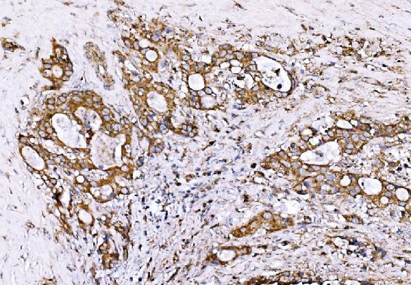
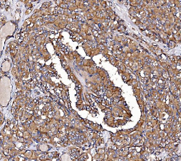
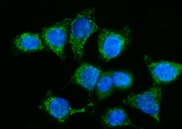
The IHC-validated antibody A01364-2 has images from paraffin-embedded human tumor and mouse colon sections (IHC captions), plus IF data from CACO-2 cells (IF caption).

Real IHC data IHC analysis of GPR54/KISS1R using anti-GPR54/KISS1R antibody (A01364-2). GPR54/KISS1R was detected in a paraffin-embedded section of human gall bladder adenosquamous carcinoma tissue. Heat mediated antigen retrieval was performed in EDTA buffer (pH 8.0, epitope retrieval solution). The tissue section was blocked with 10% goat serum. The tissue section was then incubated with 2 μg/ml rabbit anti-GPR54/KISS1R Antibody (A01364-2) overnight at 4°C. Biotinylated goat anti-rabbit IgG was used as secondary antibody and incubated for 30 minutes at 37°C. The tissue section was developed using Strepavidin-Biotin-Complex (SABC) (Catalog # SA1022) with DAB as the chromogen.
Anti-GPR54/KISS1R Antibody ®
Cat # A01364-2

A01364-2 has IHC images from paraffin-embedded human gall bladder adenosquamous carcinoma, thyroid papillary carcinoma, liver cancer, and mouse colon sections (IHC captions). It also has an IF/ICC image from CACO-2 cells (IF caption); the catalog lists human, mouse, and rat reactivity (catalog applications and reactivity).

Which to pick: Choose A01364-2 for paraffin-section IHC: its captions document staining at 2 μg/ml after EDTA retrieval at pH 8.0 (IHC captions). For IF/ICC, the same SKU has a CACO-2 image at 5 μg/ml (IF caption). It is the listed cross-species option for human, mouse, and rat (catalog reactivity), although clonality and the fixative used for the paraffin sections are unreported (catalog payload; IHC captions).

Each figure is that product's own IHC / IF validation image from its datasheet.

References

  1. UniProt Consortium. UniProt entry Q969F8 (KISSR_HUMAN, KiSS-1 receptor).
  2. Human Protein Atlas. KISS1R tissue IHC expression (reliability: Approved).
  3. Human Protein Atlas. KISS1R subcellular location (ICC-IF): Mainly localized to the plasma membrane. In addition localized to vesicles..
  4. Human Protein Atlas. KISS1R antibody validation summary (2 antibodies).
  5. KISS1 and KISS1R expression in the human and rat carotid body and superior cervical ganglion. European journal of histochemistry : EJH 2011 — PMC3284150.
  6. Kisspeptin/kisspeptin receptor system in pseudopregnant rabbit corpora lutea: presence and function. Scientific reports 2019 — PMC6433948.
  7. Differential Expression of Kisspeptin System and Kisspeptin Receptor Trafficking during Spermatozoa Transit in the Epididymis. Genes 2022 — PMC8871750.
  8. Kisspeptin as a Precision Biomarker in Personalized Pharmacy: Implications for Individualized Monitoring of Early Pregnancy Viability. Pharmacy (Basel, Switzerland) 2026 — PMC13306833.
  9. PubMed PMID:11385580 — UniProt-cited evidence.
  10. PubMed PMID:11414709 — UniProt-cited evidence.
  11. PubMed PMID:11387329 — UniProt-cited evidence.