This website uses cookies to ensure you get the best experience on our website.


- Table of Contents
Plan chromogenic KITLG IHC in paraffin sections using hematopoietic cells in bone marrow as a medium-staining reference (HPA tissue IHC). Interpret cytoplasmic and nuclear tissue staining alongside KITLG’s membrane-bound and soluble forms, and check whether the antibody targets an extracellular or cytoplasmic epitope (HPA tissue IHC; UniProt).

Expected localisation, validated protocols, controls and antibodies — the at-a-glance facts below, then the full design guide.
| Expected localisation | Cytoplasmic and nuclear tissue staining (HPA tissue IHC) | |
| Staining pattern | Leukocytes, marrow and red pulp cells: cytoplasmic/nuclear (HPA tissue IHC) | |
| Antigen retrieval | Citrate pH 6 HIER, heat-mediated (datasheet A01254) | |
| Positive control | Bone marrow | |
| Negative control | Adipose tissue+4 more · see all |
| Fixation | Formaldehyde-fixed mouse tissue was used (selected-SKU IHC image A01254); keep fixation consistent (standard IHC practice; not target-specific) | |
| Caveat | Soluble KITLG can complicate membrane-based interpretation (UniProt) | |
| Regulation | Expression regulation not established (UniProt) | |
| Isoform / epitope | 3 isoforms; epitope location may affect form detection (UniProt) |
Compare the catalog antibody's citrate pH 6 IHC-P protocol (datasheet A01254) with published KITLG staining protocols for PSCC tissue (PMC13457360) and buffalo ovary (PMC10066048).
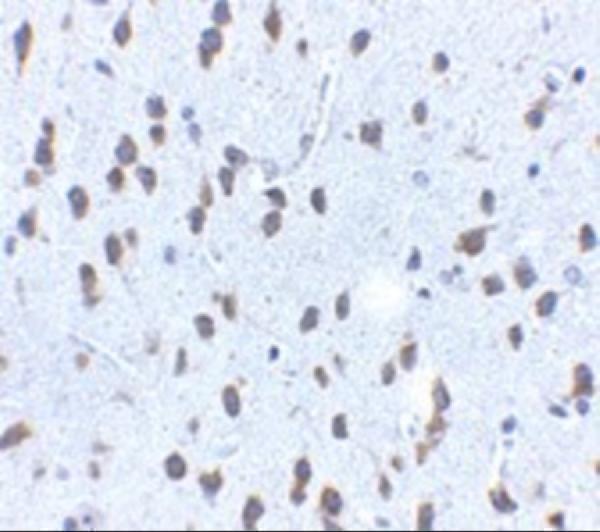
| Sample | Formaldehyde-fixed, paraffin-embedded mouse brain tissue (datasheet A01254) |
| Fixation | Image formalin-fixed; duration unreported (datasheet A01254); verify before use. |
| Sectioning | 4–5 µm sections on charged slides (standard) |
| Deparaffinisation | Xylene, graded ethanol series to water (standard) |
| Antigen retrieval | Heat retrieval: Citrate pH 6 (datasheet A01254); 20 min, 95–100 °C (standard) |
| Peroxidase block | 3% H2O2, 10 min, room temperature (standard) |
| Blocking | 10% serum (datasheet A01254) |
| Primary antibody | Rabbit anti-KITLG, 2.5 μg/mL (datasheet A01254) |
| Primary incubation | Overnight at 4 °C (standard) |
| Detection | HRP-polymer secondary, DAB chromogen 5–10 min (standard) |
| Counterstain | Hematoxylin, blue, dehydrate and mount (standard) |
| Expected result | KITLG-positive staining in hematopoietic cells of bone marrow (HPA tissue IHC: Medium). HPA tissue profile: Cytoplasmic and nuclear expression mainly in peripheral leukocytes, hematopoietic cells and cells in splenic red pulp. No signal in the no-primary control. |
KITLG can occupy the cell membrane, cytoplasm, cell projections and extracellular space; its membrane form has an extracellular region and a cytoplasmic tail (UniProt P21583 topology). In paraffin IHC, expect staining mainly in peripheral leukocytes, hematopoietic cells and splenic red-pulp cells, with cytoplasmic and some nuclear signal reported by HPA (HPA: tissue IHC profile). HPA rates the tissue staining Approved, while noting staining in some cells or structures it has not annotated (HPA: reliability description).
| Bone-marrow hematopoietic cells show moderate cytoplasmic staining. | This matches HPA's clearest listed positive tissue and cell population (HPA: bone marrow, hematopoietic cells, Medium). Assess the named cells rather than assigning one score to the entire section; a mixture of stained and unstained cells is more informative than an overall brown appearance (general IHC practice). |
| Splenic red-pulp cells stain weakly; peripheral leukocytes or hematopoietic cells show cytoplasmic signal, sometimes with nuclear signal. | These findings fit the reported cell distribution, including low staining in splenic red pulp and cytoplasmic and nuclear expression in the broader HPA profile (HPA: spleen, Low; HPA: tissue IHC profile). Nuclear staining alone is therefore not grounds to reject a section; judge its cell distribution and controls (HPA: tissue IHC profile; general IHC practice). |
| Signal is exclusively nuclear across unrelated cell types, with no convincing staining in the expected populations. | Treat this compartment pattern as a possible artefact and review controls. HPA reports nuclear expression, but mainly in its stated cell populations; UniProt lists membrane, cytoplasmic, projection and secreted locations (HPA: tissue IHC profile; UniProt P21583 subcellular location). Neither source supports calling widespread nuclear-only staining a KITLG-positive result. |
| Adipocytes or respiratory epithelial cells stain strongly while expected hematopoietic cells remain negative. | Suspect antibody cross-reactivity or endogenous detection activity before interpreting these cells as KITLG positive. HPA lists adipocytes in adipose tissue and respiratory epithelial cells in bronchus as Not detected (HPA: tissue IHC). A negative HPA observation is a comparison point, not proof that every specimen must be negative (general IHC practice). |
| Brown colour spreads diffusely over cells and surrounding tissue without a discernible cellular pattern. | This is background until it can be separated from cell-associated staining by detection controls and slide review (general IHC practice). KITLG also has a secreted form, so extracellular colour alone cannot identify the producing cell or establish specific ligand staining (UniProt P21583 processing and subcellular location). |
| Membrane form and epitope location | KITLG residues 26–214 are extracellular, 215–237 span the membrane and 238–273 are cytoplasmic (UniProt P21583 topology). The antibody's epitope is unspecified here, so the record cannot predict whether a given stain will favour membrane, cytoplasmic or released material. |
| Processing and isoforms | UniProt lists a membrane chain at residues 26–273, a soluble chain at 26–190 and 3 isoforms (UniProt P21583 processing and isoforms). Shedding can complicate attribution of extracellular signal; without an epitope map, do not claim that this antibody detects all forms equally. |
| Antigen retrieval | The supplied sources give no KITLG-specific retrieval condition or retrieval response. Choose and document a paraffin IHC retrieval condition with a known-positive section, then compare staining and background under that condition (general IHC practice); do not infer fixation sensitivity from HPA staining levels. |
| Antibody evidence | HPA070395 is Approved for IHC and Uncertain for ICC (HPA: antibody validation). HPA's tissue reliability is also Approved but notes staining in unannotated cells or structures (HPA: reliability description). These ratings support cautious pattern comparison, not an assumption that every stained structure is specific. |
| IF/ICC Q: where should signal appear? | A: HPA reports vesicles as its approved ICC-IF location (HPA: subcellular summary). This is an IF interpretation clue, not an IHC-P protocol or an upgrade of HPA070395's Uncertain ICC status (HPA: antibody validation). |
| Situation | Likely cause | Next action |
|---|---|---|
| No stain appears in bone-marrow hematopoietic cells. | The expected positive population may be absent from the sampled field, or the IHC detection workflow may have failed; HPA records Medium staining in these cells (HPA: bone marrow, Medium; general IHC practice). | Confirm hematopoietic cells are present, then inspect the positive-control slide and primary-omission control. Review retrieval and detection conditions using the same run before interpreting the specimen as KITLG negative (general IHC practice). |
| Only faint staining appears in splenic red pulp. | A faint result may match the reported Low level there; it does not by itself show a failed assay (HPA: spleen, Low). | Compare cell-associated signal with local background and the bone-marrow positive reference. Score the red-pulp cells themselves and record the weak intensity rather than increasing the call to a strong positive (HPA: spleen, Low; HPA: bone marrow, Medium; general IHC practice). |
| Strong colour appears in HPA-listed negative cell types. | Cross-reactivity or endogenous chromogen-generating activity is possible; HPA lists adipose-tissue adipocytes and bronchial respiratory epithelium as Not detected (HPA: tissue IHC negatives; general IHC practice). | Check a primary-omission control and review blocking and detection steps. Reassess whether the colour is cell-specific before assigning a KITLG-positive score (general IHC practice). |
| The whole section has diffuse brown background. | Nonspecific detection or inadequate blocking can obscure cell boundaries (general IHC practice). KITLG's soluble form also makes an extracellular deposit ambiguous without cell-associated evidence (UniProt P21583 processing). | Compare the primary-omission control, adjust blocking or detection conditions as appropriate, and require a readable cellular distribution before scoring (general IHC practice). |
| Staining is confined to nuclei throughout the section. | HPA does report nuclear expression mainly in peripheral leukocytes and hematopoietic or splenic red-pulp cells, but widespread nuclear-only staining does not match that qualified profile (HPA: tissue IHC profile). | Identify the stained cell types, compare cytoplasmic signal and controls, and flag a widespread nuclear-only pattern as a possible artefact rather than discarding all nuclear staining (HPA: tissue IHC profile; general IHC practice). |
| IF/ICC vesicles are visible, but IHC appears weak or different. | HPA describes vesicles in ICC-IF, while its IHC tissue profile describes cytoplasmic and nuclear expression; HPA070395 has different validation ratings for the two applications (HPA: subcellular summary; HPA: tissue IHC profile; HPA: antibody validation). | Interpret each preparation against its own HPA observation and controls. Do not transfer an IF/ICC appearance or protocol into the paraffin IHC decision (HPA: subcellular summary; HPA: tissue IHC profile; general IHC practice). |


![HPA ICC-IF BJ [Human fibroblast] — KITLG (HPA070395)](https://images.proteinatlas.org/70395/1534_H2_2_blue_red_green.jpg)
![HPA ICC-IF BJ [Human fibroblast] — KITLG (HPA070395)](https://images.proteinatlas.org/70395/1534_H2_3_blue_red_green.jpg)
Comprehensive Human Protein Atlas IHC scoring per tissue (reliability: Approved — Antibody staining in cells/structures not annotated, view images.). Rows are taken directly from the HPA tissue chart — click any row's HPA link to view the source.
| Tissue | Cell type | Level | Evidence | Source |
|---|---|---|---|---|
| Bone marrow | Hematopoietic cells | Medium | Protein (IHC) | HPA → |
| Tissue | Cell type | Level | Evidence | Source |
|---|---|---|---|---|
| Adipose tissue | Adipocytes | Not detected | Protein (IHC) | HPA → |
| Adrenal gland | Glandular cells | Not detected | Protein (IHC) | HPA → |
| Appendix | Glandular cells | Not detected | Protein (IHC) | HPA → |
| Breast | Adipocytes | Not detected | Protein (IHC) | HPA → |
| Bronchus | Respiratory epithelial cells | Not detected | Protein (IHC) | HPA → |
Troubleshoot chromogenic KITLG staining in paraffin sections by checking retrieval, compartment, cell identity and controls before interpreting signal intensity.
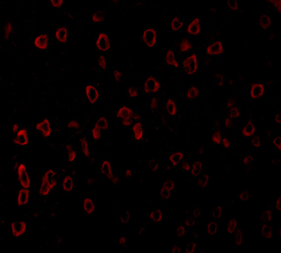
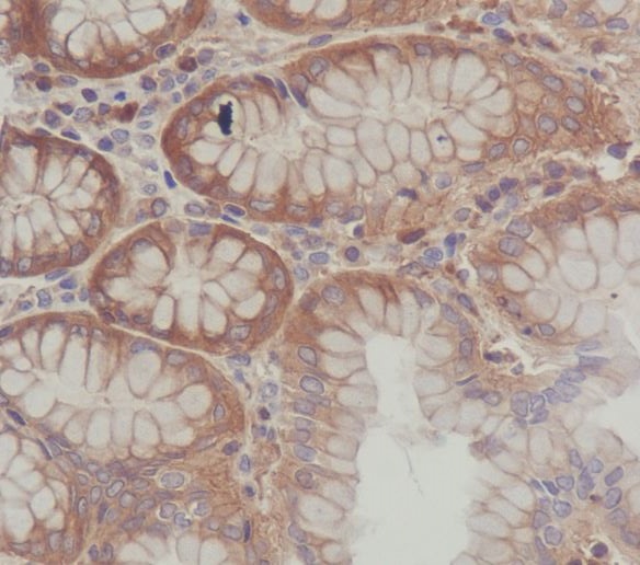
The catalog shows KITLG IHC images from mouse brain and human stomach paraffin sections, plus an IF image from human brain (A01254 and M01254 image captions).
A01254 has IHC data from formaldehyde-fixed mouse brain paraffin sections and IF data from paraformaldehyde-fixed human brain (A01254 image captions). M01254 has IHC data from human stomach paraffin sections; its fixative is unreported (M01254 IHC image caption).
Which to pick: For mouse tissue IHC, A01254 has a documented formaldehyde-fixed paraffin-section protocol at 2.5 μg/mL; for human stomach paraffin sections, M01254 has an IHC image, but its fixative is unreported (A01254 and M01254 IHC image captions). For IF, A01254 has human brain data at 20 μg/mL; M01254 lists ICC/IF without an IF image in the payload (A01254 IF image caption; M01254 application list). For work spanning human, mouse and rat, A01254 lists reactivity with all three, though its supplied IHC image validates mouse tissue only (A01254 reactivity list and IHC image caption).