This website uses cookies to ensure you get the best experience on our website.


- Table of Contents
Plan KLF17 IHC-P using testis, where elongated or late spermatids stain strongly (HPA tissue IHC). Start the catalog antibody at 5 μg/mL (datasheet: 5 μg/mL), and assess nuclear localisation as a molecular expectation (UniProt).

Expected localisation, validated protocols, controls and antibodies — the at-a-glance facts below, then the full design guide.
| Expected localisation | Nuclear location expected (UniProt); tissue compartment unreported (HPA tissue IHC) | |
| Staining pattern | High in elongated or late spermatids; subcellular pattern unreported (HPA tissue IHC) | |
| Antigen retrieval | Tris-EDTA pH 9.0 HIER, 95–98 °C, 20 min (rule: nuclear antigen) | |
| Positive control | Testis | |
| Negative control | Adipose tissue+4 more · see all |
| Fixation | Matched tissue-IHC evidence does not establish the fixation claim. Validate the specimen-specific method before use. (selected-SKU IHC image A09147) | |
| Caveat | Human IHC performance is untested for A09147 (datasheet) | |
| Regulation | Testis-enriched RNA (HPA tissue RNA) | |
| Isoform / epitope | No annotated isoforms or processing; epitope map unresolved (UniProt) |
The catalog antibody’s IHC protocol is complemented by published KLF17 staining methods for human lung and gastric tissue (PMC5503568; PMC8573523).
| Sample | Tissue sections; selected-image fixative not specified (standard IHC workflow) |
| Fixation | Image fixative and duration unreported (datasheet A09147); verify before use. |
| Sectioning | 4–5 µm sections on charged slides (standard) |
| Deparaffinisation | Xylene, graded ethanol series to water (standard) |
| Antigen retrieval | Heat-induced epitope retrieval in Tris-EDTA buffer, pH 9.0, 20 min at 95–98 °C (standard rule: nuclear antigen) |
| Peroxidase block | 3% H2O2, 10 min, room temperature (standard) |
| Blocking | 10% normal serum of the secondary host, 30 min, room temperature (standard) |
| Primary antibody | Rabbit anti-KLF17, 5 μg/mL (datasheet A09147) |
| Primary incubation | Overnight at 4 °C (standard) |
| Detection | HRP-polymer secondary, DAB chromogen 5–10 min (standard) |
| Counterstain | Hematoxylin, blue, dehydrate and mount (standard) |
| Expected result | KLF17-positive staining in elongated or late spermatids of testis (HPA tissue IHC: High). HPA tissue profile: Expression in testis. No signal in the no-primary control. |
KLF17 is a nuclear transcription factor with no transmembrane segment (UniProt Q5JT82). In paraffin-section IHC, expect staining in elongated or late spermatids in testis, the cell population scored High by HPA (HPA tissue IHC). HPA rates its IHC evidence Enhanced, while describing only medium consistency with RNA data and pending external verification (HPA tissue IHC).
| Distinct chromogenic signal over nuclei of elongated or late spermatids in testis. | This matches the reported High testis cell population (HPA tissue IHC) and nuclear location (UniProt Q5JT82). Judge the signal against a counterstain and the distribution of the same cell population across the section (general IHC practice). |
| Predominantly cytoplasmic or surface staining, with little nuclear signal in the expected cells. | That distribution conflicts with the UniProt nuclear assignment and absence of a transmembrane segment (UniProt Q5JT82). Treat it as suspect; inspect controls and morphology before assigning it to KLF17 (general IHC practice). |
| Strong signal in cells other than elongated or late spermatids, such as adrenal glandular cells. | HPA scores adrenal glandular cells Not detected (HPA tissue IHC). Investigate antibody cross-reactivity or detection-system activity; a discrepant stain alone cannot identify which is responsible (general IHC practice). |
| Diffuse brown deposit across tissue, including spaces outside cells. | A widespread deposit does not show the expected cell and nuclear pattern (HPA tissue IHC; UniProt Q5JT82). Review blocking, washes, detection controls and section quality as general chromogenic IHC checks (general IHC practice). |
| No signal in a testis section containing elongated or late spermatids. | This disagrees with the HPA High result for those cells (HPA tissue IHC). First verify that the expected cells are present, then check reagent performance and staining conditions; one negative section does not settle target absence (general IHC practice). |
| Cell population and strength | HPA reports High staining in elongated or late spermatids in testis, not a numerical intensity cutoff (HPA tissue IHC). Score the named cells, and record how many are present, rather than treating an entire testis section as uniformly positive (general IHC practice). |
| Localization and topology | UniProt assigns KLF17 to the nucleus and lists no transmembrane segment (UniProt Q5JT82). Nuclear staining is therefore the expected compartment; neither record supplies a separate tissue-level map of cytoplasmic staining. |
| Evidence strength | HPA lists the tissue IHC result as Enhanced but also states medium consistency with RNA expression and pending external verification (HPA tissue IHC). Use its pattern as a reference for assessment, with those limits stated. |
| Processing and isoforms | UniProt lists a single 1–389 chain, no signal peptide or propeptide, and zero annotated isoforms or glycosylation sites (UniProt Q5JT82). The supplied record gives no epitope map, so it cannot predict how any particular IHC antibody binds. |
| Fixation and retrieval | Target-specific fixation effects are not established by the supplied assay evidence. Verify with a matched IHC source before attributing a result to fixation. |
| IF/ICC Q&A | Q: Is an ICC-IF pattern documented here? A: HPA lists no KLF17 ICC-IF images or main location (HPA subcellular). Nuclear localization comes from UniProt, so an IF result needs its own controls and cannot be called HPA-confirmed (UniProt Q5JT82; general IF practice). |
| Situation | Likely cause | Next action |
|---|---|---|
| Testis section stains weakly or remains blank. | The expected elongated or late spermatids may be absent from the area assessed, or the IHC run may have failed; HPA reports High staining specifically in those cells (HPA tissue IHC). | Confirm cell identity on the counterstain, then compare a documented positive section and run controls. Review antibody and detection steps before changing general retrieval conditions (general IHC practice). |
| Signal sits mainly in cytoplasm instead of nuclei. | The location differs from UniProt's nuclear assignment (UniProt Q5JT82). Background, detection deposits or cell-identification errors are possible explanations, not established KLF17 biology (general IHC interpretation). | Check morphology and a no-primary control; repeat with the IHC-validated antibody and assess whether signal overlaps nuclei in the expected cells (general IHC practice). |
| Adrenal glandular cells or adipocytes stain strongly. | Those named cell populations are scored Not detected in the supplied HPA tissue IHC data (HPA tissue IHC). Cross-reactivity or endogenous detection activity may explain a discrepancy (general IHC practice). | Check the matching cell type in controls, inspect a no-primary control, and review detection-system blocking. Report the discrepancy if it persists rather than assigning target specificity from stain alone (general IHC practice). |
| Diffuse chromogen obscures cell boundaries. | The deposit prevents assessment of the cell-restricted, nuclear reference pattern (HPA tissue IHC; UniProt Q5JT82). Excess detection signal or insufficient washing may contribute (general IHC practice). | Review no-primary controls, washes and detection exposure; adjust these general IHC steps while retaining a positive control for comparison (general IHC practice). |
| A section appears positive only at its edge or in damaged areas. | An edge-restricted deposit does not reproduce the HPA cell population or UniProt compartment assignment (HPA tissue IHC; UniProt Q5JT82). Section artifacts can complicate interpretation (general IHC practice). | Score intact interior tissue, compare serial sections and examine a no-primary control before calling the case positive (general IHC practice). |
| An IF image is offered as confirmation of the IHC result. | HPA supplies no KLF17 ICC-IF image or main-location call in this payload (HPA subcellular). UniProt's nuclear assignment does not validate that particular IF image (UniProt Q5JT82). | Evaluate the IF image with its own nuclear counterstain and controls, and report it separately from the paraffin-section IHC pattern (general IF practice). |


Comprehensive Human Protein Atlas IHC scoring per tissue (reliability: Enhanced — Medium consistency between antibody staining and RNA expression data. Pending external verification.). Rows are taken directly from the HPA tissue chart — click any row's HPA link to view the source.
| Tissue | Cell type | Level | Evidence | Source |
|---|---|---|---|---|
| Testis | Elongated or late spermatids | High | Protein (IHC) | HPA → |
| Tissue | Cell type | Level | Evidence | Source |
|---|---|---|---|---|
| Adipose tissue | Adipocytes | Not detected | Protein (IHC) | HPA → |
| Adrenal gland | Glandular cells | Not detected | Protein (IHC) | HPA → |
| Appendix | Glandular cells | Not detected | Protein (IHC) | HPA → |
| Bone marrow | Hematopoietic cells | Not detected | Protein (IHC) | HPA → |
| Breast | Adipocytes | Not detected | Protein (IHC) | HPA → |
Troubleshoot KLF17 staining by assessing retrieval, nuclear localisation, cell identity and controls in chromogenic paraffin section IHC (UniProt Q5JT82; HPA tissue IHC).
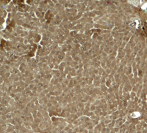
A09147 has a mouse-liver IHC image (image caption: mouse liver) and lists human, mouse and rat reactivity (catalog: reactivity); no IF image is supplied (catalog: IF images).
A09147 will render with an IHC image from mouse liver at 5 μg/mL (image caption: mouse liver, 5 μg/mL). Its listed applications include IHC-P, while the catalog supplies no IF image (catalog: applications; IF images).
Which to pick: Choose A09147 for paraffin-section IHC: IHC-P is listed, and its own image shows mouse-liver IHC at 5 μg/mL (catalog: applications; image caption: mouse liver, 5 μg/mL); the fixative is unreported (image caption). No listed SKU has IF/ICC validation or an IF dilution (catalog: applications; IF dilution). A09147 lists human, mouse and rat reactivity, but the supplied IHC image documents mouse tissue only (catalog: reactivity; image caption: mouse liver).