This website uses cookies to ensure you get the best experience on our website.


- Table of Contents
Plan KLF4 chromogenic IHC around nuclear staining, especially in squamous epithelium (HPA tissue IHC). This guide covers fixation consistency, cell-specific controls, and interpretation of five annotated isoforms (UniProt).

Expected localisation, validated protocols, controls and antibodies — the at-a-glance facts below, then the full design guide.
| Expected localisation | Nuclear staining in several tissues (HPA tissue IHC) | |
| Staining pattern | Nuclear staining, abundant in squamous epithelium (HPA tissue IHC) | |
| Antigen retrieval | Citrate pH 6 HIER, heat-mediated (datasheet A00120) | |
| Positive control | Appendix+4 more · see all | |
| Negative control | Adipose tissue+4 more · see all |
| Fixation | Matched tissue-IHC evidence does not establish the fixation claim. Validate the specimen-specific method before use. (selected-SKU IHC image A00120) | |
| Caveat | Staining varies by cell type across tissues (HPA tissue IHC) | |
| Regulation | BTRC promotes KLF4 degradation (UniProt) | |
| Isoform / epitope | Five isoforms; verify antibody epitope coverage (UniProt) |
Compare the catalog antibody’s IHC-P protocol (datasheet: A00120) with three published KLF4 IHC protocols (PMC4100128; PMC12173638; PMC3198687).
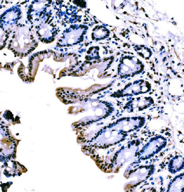
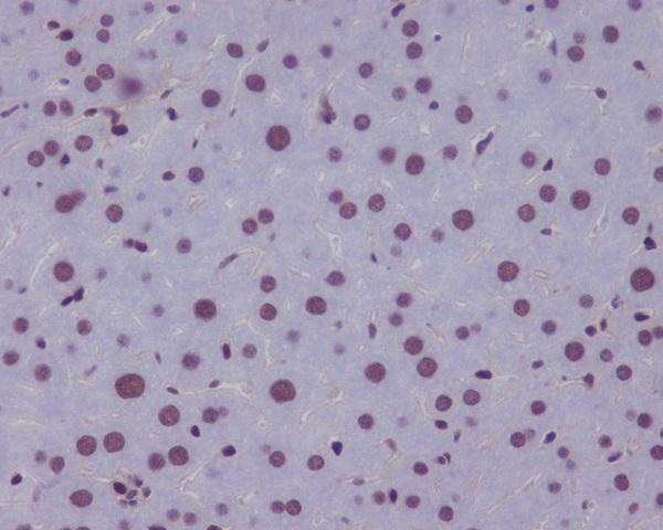
| Sample | Paraffin-embedded rat small intestine tissue; fixative not specified (datasheet A00120) |
| Fixation | Image fixative and duration unreported (datasheet A00120); verify before use. |
| Sectioning | 4–5 µm sections on charged slides (standard) |
| Deparaffinisation | Xylene, graded ethanol series to water (standard) |
| Antigen retrieval | Heat retrieval: Citrate pH 6, 20 min (datasheet A00120) |
| Peroxidase block | 3% H2O2, 10 min, room temperature (standard) |
| Blocking | 10% goat serum (datasheet A00120) |
| Primary antibody | Rabbit anti-KLF4, 2μg/ml (datasheet A00120) |
| Primary incubation | Overnight at 4 °C (datasheet A00120) |
| Detection | Streptavidin-biotin complex (SABC), DAB chromogen (datasheet A00120) |
| Counterstain | Hematoxylin, blue, dehydrate and mount (standard) |
| Expected result | KLF4-positive staining in endocrine cells of appendix (HPA tissue IHC: High). HPA tissue profile: Nuclear expression in several different tissues, highly abundant in squamous epithelium. No signal in the no-primary control. |
KLF4 should stain predominantly in nuclei, especially in squamous epithelial cells of esophagus and oral mucosa and respiratory epithelial cells of bronchus (HPA tissue IHC). UniProt also lists cytoplasmic localization and no transmembrane segment (UniProt O43474). HPA rates the tissue staining profile Enhanced, with medium consistency between antibody staining and RNA expression; assess the named cell populations rather than treating every cell in a tissue as positive (HPA tissue IHC).
| Distinct nuclear chromogen in esophageal or oral squamous epithelium, with little surrounding background (HPA tissue IHC). | This matches HPA's high staining in those cells and its predominantly nuclear tissue profile (HPA tissue IHC). Record the fraction and intensity of stained nuclei within the epithelial population (standard IHC practice). |
| Predominantly membranous staining, or chromogen outside recognizable cells, in a presumed positive section. | A membrane dominant pattern conflicts with KLF4's listed nuclear and cytoplasmic locations and lack of a transmembrane segment (UniProt O43474). Review morphology and the no primary control before assigning the signal to KLF4 (standard IHC practice). |
| Strong staining in adipocytes where HPA reports KLF4 as not detected (HPA tissue IHC). | Treat this as discordant, not proof that the tissue is wholly negative: HPA's call applies to adipocytes (HPA tissue IHC). Cross reactivity or endogenous detection activity is possible; compare a no primary control and a separate positive section (standard IHC practice). |
| Diffuse brown haze across epithelium, stroma and empty spaces, without crisp nuclear boundaries. | The distribution cannot be scored as HPA's cell localized nuclear pattern (HPA tissue IHC). Nonspecific antibody binding or detection background is possible; inspect a no primary control and reassess blocking, washes and chromogen development (standard IHC practice). |
| No nuclear signal in squamous epithelial cells of an esophageal positive control (HPA tissue IHC). | The assay has not demonstrated its expected positive pattern (HPA tissue IHC). Check section integrity, antibody and detection steps, and the antibody's documented IHC conditions before interpreting a study sample as negative (standard IHC practice). |
| Which cells anchor the positive control? | HPA reports high KLF4 in esophageal and oral squamous epithelium, bronchial respiratory epithelium, and endocrine cells of appendix, colon, duodenum and rectum (HPA tissue IHC). |
| Which cells are useful low or negative comparators? | HPA lists adipocytes as not detected in adipose tissue and breast, while kidney tubular cells and lung macrophages are low (HPA tissue IHC). Compare the specified cells, not entire organs. |
| How strong is the tissue evidence? | HPA calls the tissue profile Enhanced but reports medium consistency with RNA expression (HPA tissue IHC). Its listed antibody HPA002926 has Enhanced IHC validation (HPA antibodies). |
| Does isoform information change scoring? | UniProt lists five KLF4 isoforms (UniProt O43474). Their relative staining and recognition by a particular IHC antibody are not supplied; do not assign a staining difference to an isoform. |
| Is a surface or secreted pattern expected? | UniProt lists no transmembrane segment, signal peptide or propeptide, and places KLF4 in nucleus and cytoplasm (UniProt O43474). Surface staining needs independent verification. |
| What should IF/ICC show? | HPA reports nucleoplasm as the main location, with additional cytosol and microtubules in ICC-IF (HPA subcellular). Use that as a localization cross-check; it does not define an IHC chromogen pattern. |
| Situation | Likely cause | Next action |
|---|---|---|
| The positive control stains weakly despite recognizable squamous epithelium. | The observed result falls below HPA's high call for those cells (HPA tissue IHC); the failed step cannot be identified from that comparison alone. | Check the antibody's documented IHC dilution and retrieval conditions, reagent performance and detection controls (standard IHC practice). No KLF4-specific retrieval condition is supplied. |
| Only cytoplasmic staining appears in a high expressing epithelial population. | Cytoplasm is a listed KLF4 location, but HPA describes tissue expression as nuclear (UniProt O43474; HPA tissue IHC). | Score nuclear and cytoplasmic signals separately; compare a positive section and no primary control before accepting a cytoplasm only result (standard IHC practice). |
| A nominally negative tissue shows scattered positive cells. | HPA's not detected entries identify particular cell types; they do not certify every cell in the tissue as KLF4 negative (HPA tissue IHC). | Identify the stained cells morphologically and compare them with HPA's cell specific entry before calling the section a false positive (standard IHC practice; HPA tissue IHC). |
| Background appears in the no primary control. | Signal without primary antibody indicates a detection or tissue background contribution (standard IHC practice). | Review the detection system's recommended endogenous activity block and chromogen development, then repeat the no primary control (standard IHC practice). |
| Background appears only when primary antibody is included. | Nonspecific primary binding is possible when the no primary control is clean (standard IHC practice). | Recheck the antibody's IHC validation and documented dilution; optimize blocking and washes while retaining a known positive section (standard IHC practice). |
| A study sample is negative while its positive control stains as expected. | The assay has demonstrated detectable KLF4 in the HPA positive cell population, but that does not establish expression in every study sample (HPA tissue IHC; standard IHC practice). | Report the negative result for the evaluated cell population and preserve the control result; do not infer a fixation-specific KLF4 effect from these data (standard IHC practice). |

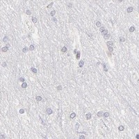
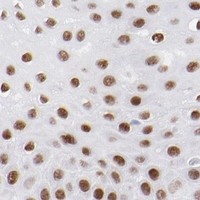
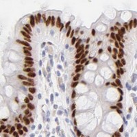
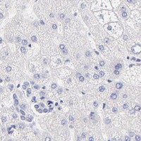


Comprehensive Human Protein Atlas IHC scoring per tissue (reliability: Enhanced — Medium consistency between antibody staining and RNA expression data.). Rows are taken directly from the HPA tissue chart — click any row's HPA link to view the source.
| Tissue | Cell type | Level | Evidence | Source |
|---|---|---|---|---|
| Appendix | Endocrine cells | High | Protein (IHC) | HPA → |
| Bronchus | Respiratory epithelial cells | High | Protein (IHC) | HPA → |
| Colon | Endocrine cells | High | Protein (IHC) | HPA → |
| Duodenum | Endocrine cells | High | Protein (IHC) | HPA → |
| Esophagus | Squamous epithelial cells | High | Protein (IHC) | HPA → |
| Tissue | Cell type | Level | Evidence | Source |
|---|---|---|---|---|
| Adipose tissue | Adipocytes | Not detected | Protein (IHC) | HPA → |
| Adrenal gland | Glandular cells | Not detected | Protein (IHC) | HPA → |
| Breast | Adipocytes | Not detected | Protein (IHC) | HPA → |
| Caudate | Glial cells | Not detected | Protein (IHC) | HPA → |
| Cerebellum | Cells in granular layer | Not detected | Protein (IHC) | HPA → |
Troubleshoot KLF4 staining in paraffin sections by checking retrieval, compartment, cell identity and controls before comparing chromogenic signal across samples.
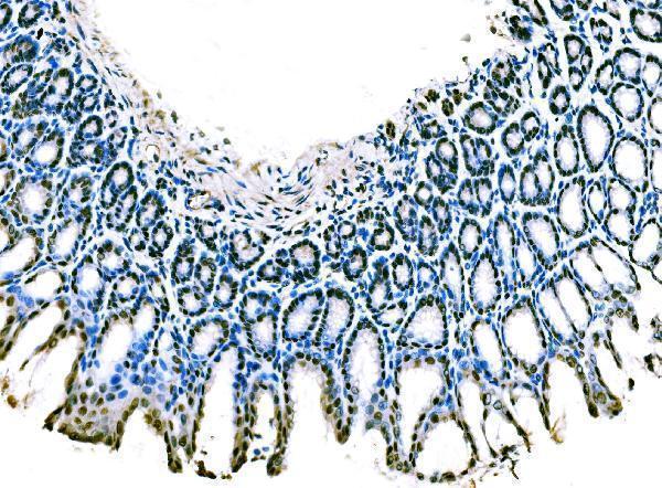
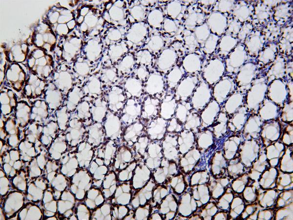
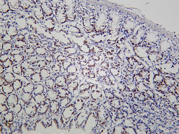
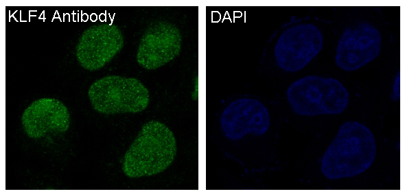
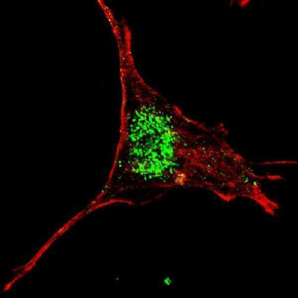
Validated anti-KLF4 antibodies have real IHC images from rat, mouse and human tissues, plus an IF image from SY5Y cells (catalog image captions); catalog reactivity covers human, mouse and rat (catalog reactivity).
A00120 is pictured on rat small intestine, A00120-3 on mouse stomach, M00120-3 on mouse colon, and M00120 on mouse liver (each SKU’s IHC image caption). A00120-2 is pictured by IF in SY5Y cells (A00120-2 IF image caption).
Which to pick: For paraffin-section IHC, choose A00120-3: its mouse stomach image used EDTA retrieval at pH 8.0 and 2 μg/ml primary antibody; the fixative is unreported (A00120-3 IHC image caption). For IF/ICC, choose M00120 because both applications are listed and it has an IF image in Hela cells (M00120 applications and IF image caption). For tissue IHC across species, consider monoclonal M00120-3: its images show mouse, rat and human colon, but report no retrieval method or fixative (M00120-3 catalog description and IHC image captions).