KLK3 / Prostate-specific antigen · IHC design guide

Design Immunohistochemistry for KLK3

Plan chromogenic KLK3 IHC in paraffin sections using prostate glandular cells as a positive control (HPA tissue IHC). The documented workflow uses 2 µg/ml primary antibody (datasheet A01505-3); account for KLK3 secretion when interpreting staining (UniProt).

Evidence assembled Oct 2026 · For research use; verify linked source records and product datasheet before use
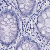
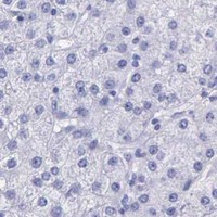
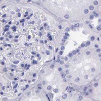
Immunohistochemistry protocol sheet for KLK3 (IHC for KLK3): expected localisation Cytoplasmic in prostate glandular cells (HPA tissue IHC), antibody A01505-3, validated IHC image, and IHC protocol steps
Printable KLK3 IHC protocol sheet — expected localisation Cytoplasmic in prostate glandular cells (HPA tissue IHC), antibody A01505-3, controls and protocol steps. Open the full KLK3 IHC guide →

KLK3 Immunohistochemistry Experimental Design Guide

Expected localisation, validated protocols, controls and antibodies — the at-a-glance facts below, then the full design guide.

Must know before staining
Expected localisation Cytoplasmic in prostate glandular cells (HPA tissue IHC)
Staining pattern High cytoplasmic staining in prostate glandular cells (HPA tissue IHC)
Antigen retrieval EDTA pH 8.0 HIER, heat-mediated (datasheet A01505-3)
Positive control ⓘ Prostate
Negative control ⓘ Adipose tissue+4 more · see all
Important caveats
Reasons your staining may differ from the expected pattern.
Fixation Keep fixation consistent across sections (standard IHC practice; not target-specific)
Caveat Secreted KLK3 may stain away from its RNA source (HPA tissue IHC)
Regulation Prostate-enriched expression (HPA tissue IHC)
Isoform / epitope 5 isoforms; mature chain starts at residue 25—check epitope (UniProt)
Section 1

Recommended KLK3 IHC & IF Protocols

The catalog antibody’s IHC-P protocol (datasheet A01505-3) is accompanied by published IHC examples with usable protocol details (PMC5405195; PMC11520440).

Recommended immunohistochemistry (IHC-P) protocol parameters
SampleParaffin-embedded human prostatic cancer tissue; fixative not specified (datasheet A01505-3)
FixationImage fixative and duration unreported (datasheet A01505-3); verify before use.
Sectioning4–5 µm sections on charged slides (standard)
DeparaffinisationXylene, graded ethanol series to water (standard)
Antigen retrievalHeat retrieval: EDTA pH 8.0 (datasheet A01505-3); 20 min, 95–100 °C (standard)
Peroxidase block3% H2O2, 10 min, room temperature (standard)
Blocking10% goat serum (datasheet A01505-3)
Primary antibodyRabbit anti-KLK3, 2-5μg/ml (datasheet A01505-3)
Primary incubationOvernight at 4 °C (datasheet A01505-3)
DetectionStreptavidin-biotin complex (SABC), DAB chromogen (datasheet A01505-3)
CounterstainHematoxylin, blue, dehydrate and mount (standard)
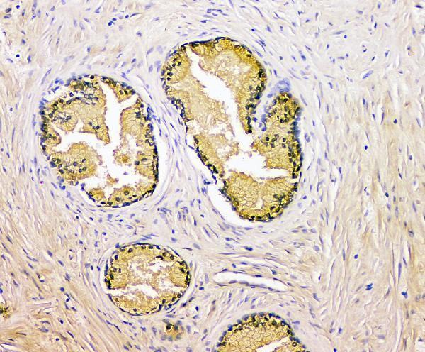
Expected resultKLK3-positive staining in glandular cells of prostate (HPA tissue IHC: High). HPA tissue profile: Selective cytoplasmic expression in prostate. No signal in the no-primary control.
💡Decision noteStart with heat-mediated EDTA pH 8.0 retrieval for the catalog antibody (datasheet A01505-3); neither included article specifies a retrieval method (PMC5405195; PMC11520440).
Section 2

What Is the Expected KLK3 Staining Pattern?

In paraffin-section IHC, expect selective cytoplasmic KLK3 staining in prostate glandular cells (HPA: High in prostate glandular cells; selective cytoplasmic expression). KLK3 is secreted and has no transmembrane segment, so a membrane-restricted pattern is unexpected (UniProt P07288: secreted; no transmembrane segment). HPA rates the tissue pattern Enhanced, while cautioning that its protein signal may include proteins from more than one gene (HPA: reliability Enhanced; specificity caution).

What am I looking at on my slide?
Strong cytoplasmic stain in prostate glandular cells, with a selective distribution (HPA: High in prostate glandular cells; selective cytoplasmic expression).This matches the reported tissue IHC pattern. Score the stained glandular cells and their cytoplasmic intensity; do not treat the HPA reliability rating as proof of antibody specificity in your preparation (HPA: reliability Enhanced; specificity caution).
Predominantly nuclear or sharply membrane-restricted stain in prostate glandular cells (HPA: selective cytoplasmic expression).This conflicts with the reported cytoplasmic pattern and secreted, non-transmembrane topology. Investigate staining artefact or antibody specificity before scoring it as KLK3 (HPA: selective cytoplasmic expression; UniProt P07288: secreted; no transmembrane segment).
Strong stain in adipocytes or in adrenal or appendix glandular cells (HPA: Not detected in these listed cell types).Those cells are reported as negative in the supplied HPA tissue IHC. Check cross-reactivity and endogenous detection activity; staining alone cannot identify which caused the signal (HPA: Not detected in listed cell types; general IHC practice).
Diffuse color across tissue and blank spaces, obscuring cell boundaries (general IHC practice).This is background, not a scoreable cytoplasmic pattern. Review blocking, detection-reagent background, washes and chromogen exposure against a control lacking primary antibody (general IHC practice).
No glandular-cell signal in a known-positive prostate section (HPA: High in prostate glandular cells).The run has not reproduced the reference pattern. Check section integrity, antibody dilution, retrieval and detection with run controls before interpreting an unknown sample as negative (HPA: High in prostate glandular cells; general IHC practice).
💡Expected KLK3 appearanceA positive IHC result is high, selective cytoplasmic staining of prostate glandular cells; dominant nuclear or membrane-restricted stain, or strong stain in HPA-listed negative cells, is suspect (HPA: tissue IHC pattern and listed negatives; UniProt P07288: no transmembrane segment).
How each factor affects the staining
Tissue and cell selection (HPA: prostate glandular cells High; listed negative cell types Not detected).A prostate section provides the clearest reference pattern. Interpret negative controls at the cell-type level: HPA reports, for example, adrenal and appendix glandular cells as Not detected, rather than declaring every cell in those tissues negative (HPA: tissue IHC).
Secretion and processing (UniProt P07288: secreted; signal peptide 1–17; propeptide 18–24; mature chain 25–261).The expected cellular signal is cytoplasmic, but tissue location need not track the cells making the RNA for a secreted protein. Avoid assigning every extracellular deposit or luminal signal to a producing cell (HPA: secreted-protein location caution; UniProt P07288: processing).
Antibody-validation scope (HPA: CAB000070 IHC Enhanced; HPA000764 IHC Supported).These are different IHC validation statuses. HPA also cautions that the tissue signal may target protein from more than one gene; use morphology and controls when interpreting unexpected staining (HPA: antibody statuses; tissue reliability description).
Isoforms and glycosylation (UniProt P07288: 5 isoforms; one glycosylation site at residue 69).The record establishes molecular variation, but gives no epitope map or evidence that it changes staining by a particular antibody. Do not infer isoform selectivity or a glycosylation-related retrieval effect from this record (UniProt P07288: isoforms; glycosylation).
Antigen retrieval and fixation sensitivity (general IHC practice; supplied HPA and UniProt records).Use the antibody's validated IHC-P conditions and run controls when assessing retrieval. Target-specific effects of fixation or retrieval on KLK3 signal are unreported in the supplied evidence; HPA's tissue pattern does not establish them (general IHC practice; HPA: tissue IHC scope).
IF/ICC: what pattern can be claimed here? (HPA: subcellular summary Secreted; no ICC-IF image lines).Secreted is the supplied subcellular summary, but HPA provides no ICC-IF cell-line images or assigned main intracellular location. This IHC pattern cannot establish an IF/ICC compartment or serve as an IF/ICC protocol (HPA: subcellular record).
Why is my staining missing, weak or wrong?
SituationLikely causeNext action
Prostate glandular cells are unstained (HPA: High in prostate glandular cells).A failed stain or unsuitable assay conditions remain possible; the supplied sources give no KLK3-specific fixation explanation (general IHC practice; HPA and UniProt: no fixation-effect evidence).Confirm tissue and run controls, then check the validated IHC-P dilution, retrieval and detection steps before calling the sample negative (general IHC practice).
Staining is mainly nuclear or confined to cell outlines (HPA: selective cytoplasmic expression).The pattern conflicts with HPA tissue IHC and KLK3's lack of a transmembrane segment; artefact or nonspecific binding is possible (HPA: tissue IHC; UniProt P07288: topology; general IHC practice).Compare with the prostate cytoplasmic reference and a control lacking primary antibody; review morphology before scoring (HPA: tissue IHC; general IHC practice).
Adrenal or appendix glandular cells stain strongly (HPA: Not detected in those cell types).Cross-reactivity or endogenous detection activity may contribute; the unexpected stain alone does not distinguish them (HPA: listed negatives; general IHC practice).Inspect a control lacking primary antibody and the detection controls, then reassess antibody-dependent staining in the same cell types (general IHC practice).
Color covers most of the section and tissue-free areas (general IHC practice).Diffuse detection background or excess chromogen can obscure the selective cytoplasmic pattern (general IHC practice; HPA: selective cytoplasmic expression).Check reagent-only background, blocking, wash steps and chromogen development; score cells only once their boundaries can be resolved (general IHC practice).
Extracellular or luminal color is the only apparent signal (general IHC practice).KLK3 is secreted, so protein location can differ from RNA location; the supplied HPA pattern still identifies cytoplasmic glandular staining (UniProt P07288: secreted; HPA: secreted-protein caution; tissue IHC).Record the extracellular finding separately and seek the expected glandular cytoplasmic pattern in the positive control before calling the section positive (HPA: tissue IHC; general IHC practice).
Results differ between IHC antibodies (HPA: CAB000070 Enhanced; HPA000764 Supported).Their supplied validation statuses differ, and HPA cautions that antibody staining may include proteins from more than one gene; the record does not identify an epitope-specific cause (HPA: antibody statuses; specificity caution).Compare staining in matched prostate glandular cells and HPA-listed negative cell types, with the same run controls, before resolving a discordant result (HPA: tissue IHC; general IHC practice).

Sample controls for KLK3 IHC & IF

🧪Run prostate first: its glandular cells should stain strongly (HPA: High in prostate glandular cells). Use adipose tissue as a negative tissue (HPA: Not detected in adipocytes); on the prostate slide, assess adjacent nonglandular cells for absent staining while distinguishing any luminal signal from cellular staining (UniProt P07288: secreted).
Positive control tissue: Prostate (Glandular cells, HPA High)
Negative control tissue: Adipose tissue (HPA Not detected)
ICC-IF cell lines (HPA subcellular resource): HPA carries no ICC-IF cell line for KLK3; derive a cell-line control from the positive tissue's cell type (Glandular cells) and confirm it by RNA or western blot first.
Technical controls: Include a no-primary, secondary-only control; a concentration-matched rabbit IgG isotype control (caption: rabbit primary antibody); and, if available, a KLK3 knockout or validated peptide-block control. For chromogenic detection, control endogenous peroxidase and biotin signal, particularly with the caption’s streptavidin–biotin complex and DAB method (caption: SABC with DAB).
⚠️Feasibility: A target-specific fixation window or fixation effect is unreported, and the selected A01505-3 tissue-IHC caption does not state a fixative (caption: fixative not stated). Heat retrieval in EDTA at pH 8.0 is a documented starting condition; its necessity has not been established by the supplied evidence (caption: heat retrieval in EDTA, pH 8.0). The evidence does not establish whether frozen sections or IF would be easier; KLK3 secretion can make luminal or extracellular signal a localisation concern (UniProt P07288: secreted).

HPA tissue IHC evidence for KLK3

Comprehensive Human Protein Atlas IHC scoring per tissue (reliability: Enhanced — High consistency between antibody staining and RNA expression data. Caution, targets protein from more than one gene. Secreted protein, tissue location of RNA and protein is expected to differ.). Rows are taken directly from the HPA tissue chart — click any row's HPA link to view the source.

Positive expression · recommended positive controls

TissueCell typeLevelEvidenceSource
Prostate Glandular cells High Protein (IHC) HPA →

Undetected expression · recommended negative controls

TissueCell typeLevelEvidenceSource
Adipose tissue Adipocytes Not detected Protein (IHC) HPA →
Adrenal gland Glandular cells Not detected Protein (IHC) HPA →
Appendix Glandular cells Not detected Protein (IHC) HPA →
Bone marrow Hematopoietic cells Not detected Protein (IHC) HPA →
Breast Adipocytes Not detected Protein (IHC) HPA →
Section 3

Advanced KLK3 IHC Tips

Troubleshoot KLK3 staining in paraffin sections by checking retrieval, glandular cell localisation, detection background and scoring against matched controls.

How should I retrieve KLK3 antigen when paraffin-section staining is weak?
Start with heat-mediated EDTA retrieval at pH 8.0 for paraffin sections (datasheet A01505-3). The selected image shows KLK3 detection in human prostatic cancer tissue after this retrieval, followed by 2 µg/ml primary antibody overnight at 4°C (caption A01505-3). If staining is weak, compare retrieval heating conditions on adjacent sections while keeping antibody concentration and detection constant (standard IHC practice). Include a prostate glandular-cell positive control because selective cytoplasmic expression is reported there (HPA tissue IHC). Excessively harsh retrieval can damage section morphology, so assess staining together with preserved gland architecture (standard IHC practice).
Could fixation explain weak or uneven KLK3 staining?
The selected KLK3 image uses a paraffin-embedded human prostatic cancer section, but its fixative is unreported (caption A01505-3). Target-specific sensitivity to fixation is therefore unknown from the supplied evidence (caption A01505-3). For a troubleshooting run, compare sections with documented, consistent fixation and processing histories, then apply the same EDTA retrieval at pH 8.0 (standard IHC practice; datasheet A01505-3). Examine morphology and staining in a prostate glandular-cell control before attributing a negative specimen to biology (HPA tissue IHC; standard IHC practice). Record fixation differences alongside results so an apparent intensity shift is not mistaken for altered KLK3 expression (standard IHC practice).
Where should convincing KLK3 signal appear in a prostate section?
Look primarily for cytoplasmic staining in prostate glandular cells, the selective tissue pattern reported for KLK3 (HPA tissue IHC). KLK3 is secreted and has no transmembrane segment, so a crisp membrane-only pattern lacks support from its annotated topology (UniProt P07288 topology; UniProt P07288 subcellular location). Secreted protein may also appear outside producing cells; assess such staining against adjacent glandular cells and section morphology rather than treating it as cell-autonomous expression (UniProt P07288 subcellular location; standard IHC interpretation). Compare the pattern with a matched negative control and the documented prostate positive control (HPA tissue IHC; standard IHC practice). Predominantly nuclear staining warrants a background review (UniProt P07288 subcellular location; standard IHC practice).
Could KLK3 processing or isoforms change what this antibody detects?
KLK3 has 5 annotated isoforms, and the reference precursor contains a signal peptide at residues 1–17 and a propeptide at 18–24 (UniProt P07288 isoforms; UniProt P07288 processing). Its annotated mature chain spans residues 25–261, with a glycosylation site at residue 69 (UniProt P07288 processing; UniProt P07288 glycosylation). The supplied caption does not identify the catalog antibody's epitope or establish which isoforms it recognizes (caption A01505-3). Accordingly, do not interpret weaker IHC as a specific processing or isoform change without epitope information and an independent assay (standard IHC interpretation). Keep retrieval and detection matched when comparing specimens (standard IHC practice).
How can I assess KLK3 by IF alongside a glandular-cell marker?
For a separate IF/ICC workflow, multiplex KLK3 with a validated marker that identifies prostate glandular cells, where KLK3 staining is reported (HPA tissue IHC; standard IF practice). Choose a fluorophore whose emission separates from the specimen's autofluorescence, and inspect unstained and single-colour controls before judging colocalisation (standard IF practice). KLK3 is secreted, lacks a transmembrane segment, and shows cytoplasmic tissue staining, so use permeabilisation when assessing intracellular antigen (UniProt P07288 topology; HPA tissue IHC; standard IF practice). Optimise permeabilisation against retained morphology and signal (standard IF practice). The supplied paraffin-section caption documents chromogenic IHC, so it does not establish IF performance for this antibody (caption A01505-3).
How do I distinguish KLK3 staining from chromogenic detection background?
The selected IHC image used 10% goat serum blocking, a biotinylated secondary antibody, streptavidin-biotin detection and DAB (caption A01505-3). For diffuse staining, run a no-primary control and examine whether the detection system stains tissue independently of the KLK3 antibody (standard IHC practice). Apply a peroxidase block for DAB detection and assess endogenous biotin background when using a biotin-based system (standard IHC practice). Recheck blocking, washes and the reported 2 µg/ml primary concentration before changing several conditions together (caption A01505-3; standard IHC practice). A convincing result should retain the selective prostate glandular-cell pattern (HPA tissue IHC).
What should I score when comparing KLK3 IHC across sections? ⚠ ANSWER MARKED FOR VERIFICATION
Define the scoring compartment as viable prostate glandular cells, the reported site of high KLK3 protein expression (HPA tissue IHC). Record the percentage of positive glandular cells and their staining intensity; an H-score combines intensity grades 0–3 with their percentages for a possible range of 0–300 (standard IHC scoring practice). Use the number of evaluable glandular cells as the denominator, and document excluded damaged or necrotic regions (standard IHC practice). Score sections with consistent retrieval, development and imaging conditions because these can affect apparent intensity (standard IHC practice). Review extracellular staining separately from cell-associated scores because KLK3 is secreted (UniProt P07288 subcellular location).
When is an unexpected KLK3-positive area likely to be artefact?
A credible positive pattern includes cytoplasmic staining in prostate glandular cells (HPA tissue IHC). Treat isolated nuclear or membrane-only signal cautiously because KLK3 is annotated as secreted, without a transmembrane segment (UniProt P07288 subcellular location; UniProt P07288 topology). Inspect section edges and necrotic areas for staining that follows tissue damage rather than intact glandular cells (standard IHC interpretation). Compare suspicious areas with a no-primary control, particularly when DAB and a biotin-based detection system are used (caption A01505-3; standard IHC practice). HPA cautions that secreted protein location may differ from RNA location, so discordance alone does not establish artefact (HPA tissue IHC).
Boster reagents

Best KLK3 / Prostate-specific antigen IHC Antibodies

Catalog antibodies cover human-reactive IHC and IF/ICC applications, with IHC images from human prostatic cancer and mouse prostate sections and an ICC image from PC-3M cells (catalog applications/reactivity; image captions).

Real IHC data IHC analysis of Prostate Specific Antigen/KLK3 using anti-Prostate Specific Antigen/KLK3 antibody (A01505-3). Prostate Specific Antigen/KLK3 was detected in paraffin-embedded section of human prostatic cancer tissue. Heat mediated antigen retrieval was performed in EDTA buffer (pH8.0, epitope retrieval solution). The tissue section was blocked with 10% goat serum. The tissue section was then incubated with 2μg/ml rabbit anti-Prostate Specific Antigen/KLK3 Antibody (A01505-3) overnight at 4°C. Biotinylated goat anti-rabbit IgG was used as secondary antibody and incubated for 30 minutes at 37°C. The tissue section was developed using Strepavidin-Biotin-Complex (SABC) (Catalog # SA1022) with DAB as the chromogen.
Anti-Prostate Specific Antigen/KLK3 Antibody ®
Cat # A01505-3
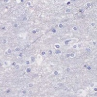
Real IHC data IHC analysis of KLK3 using anti-KLK3 antibody (PB9259). KLK3 was detected in a paraffin-embedded section of human prostatic cancer tissue. Heat mediated antigen retrieval was performed in EDTA buffer (pH 8.0, epitope retrieval solution). The tissue section was blocked with 10% goat serum. The tissue section was then incubated with 2 μg/ml rabbit anti-KLK3 Antibody (PB9259) overnight at 4°C. Biotinylated goat anti-rabbit IgG was used as secondary antibody and incubated for 30 minutes at 37°C. The tissue section was developed using Strepavidin-Biotin-Complex (SABC) (Catalog # SA1022) with DAB as the chromogen.
Anti-Prostate Specific Antigen/KLK3 Antibody ®
Cat # PB9259
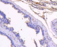
Real IHC data Immunohistochemical analysis of paraffin-embedded mouse prostate tissue using anti-PSA antibody. Counter stained with hematoxylin.
Anti-PSA KLK3 Antibody
Cat # A01505-2

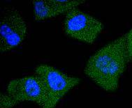
A01505-3 and PB9259 have IHC images of paraffin-embedded human prostatic cancer sections (A01505-3 and PB9259 IHC captions). A01505-2 has an IHC image of paraffin-embedded mouse prostate and an ICC image of PC-3M cells; its listed reactivity is Human (A01505-2 image captions; catalog reactivity).

Which to pick: For human tissue IHC, choose A01505-3 or PB9259: both list Human reactivity and show paraffin-section staining (catalog reactivity; A01505-3 and PB9259 IHC captions). For IF/ICC, choose A01505-2 because those applications are listed and its ICC caption shows stained PC-3M cells (A01505-2 applications; ICC caption). For mouse tissue, A01505-2 has a mouse prostate IHC image, but its listed reactivity is Human; the IHC captions report paraffin embedding without specifying the fixative (A01505-2 IHC caption; catalog reactivity; IHC captions).

Each figure is that product's own IHC / IF validation image from its datasheet.

References

  1. UniProt Consortium. UniProt entry P07288 (KLK3_HUMAN, Prostate-specific antigen).
  2. Human Protein Atlas. KLK3 tissue IHC expression (reliability: Enhanced).
  3. Human Protein Atlas. KLK3 subcellular location (ICC-IF): Secreted.
  4. Human Protein Atlas. KLK3 antibody validation summary (2 antibodies).
  5. Targeting the p300/CBP Axis in Lethal Prostate Cancer. Cancer discovery 2021 — PMC8102310.
  6. c-Myc Antagonises the Transcriptional Activity of the Androgen Receptor in Prostate Cancer Affecting Key Gene Networks. EBioMedicine 2017 — PMC5405195.
  7. GREB1 amplifies androgen receptor output in human prostate cancer and contributes to antiandrogen resistance. eLife 2019 — PMC6336405.
  8. GOLM1 promotes prostate cancer progression via interaction with PSMD1 and enhancing AR-driven transcriptional activation. Journal of cellular and molecular medicine 2024 — PMC11520440.
  9. PubMed PMID:2436946 — UniProt-cited evidence.
  10. PubMed PMID:2467258 — UniProt-cited evidence.
  11. PubMed PMID:2471958 — UniProt-cited evidence.