Click image to see more details
Product Info Summary
| SKU: | A01505-3 |
|---|---|
| Size: | 100 μg/vial |
| Reactive Species: | Human |
| Host: | Rabbit |
| Application: | ELISA, IHC, WB |
Customers Who Bought This Also Bought

Product info
Product Name
Anti-Prostate Specific Antigen/KLK3 Antibody Picoband®
SKU/Catalog Number
A01505-3
Size
100 μg/vial
Form
Lyophilized
Description
Boster Bio Anti-Prostate Specific Antigen/KLK3 Antibody Picoband® catalog # A01505-3. Tested in ELISA, IHC, WB applications. This antibody reacts with Human. The brand Picoband indicates this is a premium antibody that guarantees superior quality, high affinity, and strong signals with minimal background in Western blot applications. Only our best-performing antibodies are designated as Picoband, ensuring unmatched performance.
Storage & Handling
Store at -20˚C for one year from date of receipt. After reconstitution, at 4˚C for one month. It can also be aliquotted and stored frozen at -20˚C for six months. Avoid repeated freeze-thaw cycles.
Cite This Product
Anti-Prostate Specific Antigen/KLK3 Antibody Picoband® (Boster Biological Technology, Pleasanton CA, USA, Catalog # A01505-3)
Host
Rabbit
Contents
Each vial contains 4mg Trehalose, 0.9mg NaCl and 0.2mg Na2HPO4.
Clonality
Polyclonal
Isotype
Rabbit IgG
Immunogen
E.coli-derived human Prostate Specific Antigen/KLK3 recombinant protein (Position: A64-D255).
Cross-reactivity
No cross-reactivity with other proteins.
Reactive Species
A01505-3 is reactive to KLK3 in Human
Observed Molecular Weight
34 kDa
Calculated molecular weight
28.7 kDa
Background of KLK3
Prostate-specific antigen (PSA), also known as gamma-seminoprotein or kallikrein-3 (KLK3), is a glycoprotein enzyme encoded in humans by the KLK3 gene. KLK3 is a member of the kallikrein-related peptidase family and is secreted by the epithelial cells of the prostate gland. This gene is mapped to 19q13.33. KLK3 is present in small quantities in the serum of men with healthy prostates, but is often elevated in the presence of prostate cancer or other prostate disorders. KLK3 is produced for the ejaculate where it liquifies the semen in the seminal coagulum and allows sperm to swim freely. It is also believed to be instrumental in dissolving the cervical mucous cap, allowing the entry of sperm. It is not a unique indicator of prostate cancer, but may also detect prostatitis or benign prostatic hyperplasia.
Antibody Validation
Boster validates all antibodies on WB, IHC, ICC, Immunofluorescence, and ELISA with known positive control and negative samples to ensure specificity and high affinity, including thorough antibody incubations.
Application & Images
Applications
A01505-3 is guaranteed for ELISA, IHC, WB Boster Guarantee
Recommend Dilution
| Application | Dilution | Species |
|---|---|---|
| Western blot | 0.25-0.5μg/ml | Human |
| Immunohistochemistry (Paraffin-embedded Section) | 2-5μg/ml | Human |
| ELISA | 0.1-0.5μg/ml | - |
Tested application
Suggested blocking solution with 5% non-fat milk or BSA; (*)Recommended protein loading: 20-40 µg per lane
Use TE buffer pH 9.0 for antigen retrieval; (*) citrate buffer pH 6.0 is an alternative.
Validation Images & Assay Conditions
Click image to see more details
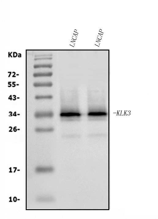
Western blot analysis of Prostate Specific Antigen/KLK3 using anti-Prostate Specific Antigen/KLK3 antibody (A01505-3).
Electrophoresis was performed on a 5-20% SDS-PAGE gel at 70V (Stacking gel) / 90V (Resolving gel) for 2-3 hours. The sample well of each lane was loaded with 30ug of sample under reducing conditions.
Lane 1: human LNCAP whole cell lysates,
Lane 2: human LNCAP whole cell lysates.
After Electrophoresis, proteins were transferred to a Nitrocellulose membrane at 150mA for 50-90 minutes. Blocked the membrane with 5% Non-fat Milk/ TBS for 1.5 hour at RT. The membrane was incubated with rabbit anti-Prostate Specific Antigen/KLK3 antigen affinity purified polyclonal antibody (Catalog # A01505-3) at 0.5 μg/mL overnight at 4°C, then washed with TBS-0.1%Tween 3 times with 5 minutes each and probed with a goat anti-rabbit IgG-HRP secondary antibody at a dilution of 1:5000 for 1.5 hour at RT. The signal is developed using an Enhanced Chemiluminescent detection (ECL) kit (Catalog # EK1002) with Tanon 5200 system. A specific band was detected for Prostate Specific Antigen/KLK3 at approximately 34KD. The expected band size for Prostate Specific Antigen/KLK3 is at 34KD.

Click image to see more details
IHC analysis of Prostate Specific Antigen/KLK3 using anti-Prostate Specific Antigen/KLK3 antibody (A01505-3).
Prostate Specific Antigen/KLK3 was detected in paraffin-embedded section of human prostatic cancer tissue. Heat mediated antigen retrieval was performed in EDTA buffer (pH8.0, epitope retrieval solution). The tissue section was blocked with 10% goat serum. The tissue section was then incubated with 2μg/ml rabbit anti-Prostate Specific Antigen/KLK3 Antibody (A01505-3) overnight at 4°C. Biotinylated goat anti-rabbit IgG was used as secondary antibody and incubated for 30 minutes at 37°C. The tissue section was developed using Strepavidin-Biotin-Complex (SABC) (Catalog # SA1022) with DAB as the chromogen.

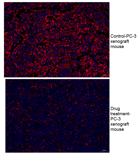
Click image to see more details
IF analysis of Prostate Specific Antigen/KLK3 using anti-Prostate Specific Antigen/KLK3 antibody (A01505-3).
Paraffin-embedded tumor sections were used to detect Prostate Specific Antigen (PSA/KLK3). PC-3 cells were inoculated into nude mouse to establish tumors. Tumor-bearing mice were subsequently treated with docetaxel, while control mouse remained untreated. Heat mediated antigen retrieval was performed in EDTA buffer (pH 8.0, epitope retrieval solution). The tissue section was blocked with 10% goat serum. The tissue section was then incubated with 1:500 rabbit anti-Prostate Specific Antigen/KLK3 Antibody (A01505-3) overnight at 4°C. DyLight®594 Conjugated Goat Anti-Rabbit IgG (H+L) (BA1142) was used as secondary antibody at 1:500 dilution and incubated for 45 minutes at 37°C. The section was counterstained with DAPI. Visualize using a fluorescence microscope and filter sets appropriate for the label used.

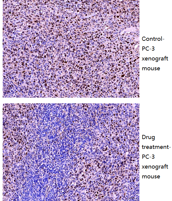
Click image to see more details
IHC analysis of Prostate Specific Antigen/KLK3 using anti-Prostate Specific Antigen/KLK3 antibody (A01505-3).
Paraffin-embedded tumor sections were used to detect Prostate Specific Antigen (PSA/KLK3). PC-3 cells were inoculated into nude mouse to establish tumors. Tumor-bearing mice were subsequently treated with docetaxel, while control mouse remained untreated. Heat mediated antigen retrieval was performed in EDTA buffer (pH8.0, epitope retrieval solution). The tissue section was blocked with 10% goat serum. The tissue section was then incubated with 1:500 rabbit anti-Prostate Specific Antigen/KLK3 Antibody (A01505-3) overnight at 4°C. Two-step IHC Detection Kit (Rabbit IgG) was used as secondary antibody and incubated for 45 minutes at 37°C. The tissue section was developed using Strepavidin-Biotin-Complex (SABC) (Catalog # SA1022) with DAB as the chromogen.
Specific Publications For Anti-Prostate Specific Antigen/KLK3 Antibody Picoband® (A01505-3)
Loading publications
Recommended Resources
Here are featured tools and databases that you might find useful.
- Boster's Pathways Library
- Protein Databases
- Bioscience Research Protocol Resources
- Data Processing & Analysis Software
- Photo Editing Software
- Scientific Literature Resources
- Research Paper Management Tools
- Molecular Biology Software
- Primer Design Tools
- Bioinformatics Tools
- Phylogenetic Tree Analysis
Customer Reviews
Have you used Anti-Prostate Specific Antigen/KLK3 Antibody Picoband®?
Share your experimental results or join a short interview to earn up to $1,000 in product credits or other rewards.
2 Reviews For Anti-Prostate Specific Antigen/KLK3 Antibody Picoband®
IHC using Anti-Ki67 antibody(A01505-3) showed clear nuclear staining with minimal background, revealing markedly reduced Ki67-positive tumor cells in docetaxel-treated PC-3 xenografts compared to control mouse.
Excellent
| SKU | A01505-3 |
|---|---|
| Application | Immunohistochemistry |
| Sample | PC-3 xenograft mouse tissue |
| Sample Processing Description | (1) PC-3 cells were implanted in nude mice to form tumors as the control group; (2) tumor-bearing mice were treated with the drug docetaxel. |
| Other Reagents | Goat serum, DAPI, Anti-fade mounting medium |
| Primary Antibody | Prostate Specific Antigen/KLK3 Antibody Picoband® |
| Primary Incubation | 1:500, overnight at 4 ℃ |
| Secondary Antibody | Two-step IHC Detection Kit (Rabbit IgG) |
| Secondary Incubation | 1:500, 45 min at 37℃ |
| Detection | Imaging system:Leica DM2500 |
| Results Summary | Ki67 is a nuclear protein directly associated with cell proliferation and is widely recognized as a marker of proliferating cells due to its importance in evaluating tumor growth rate and patient prognosis. Higher Ki67 indices indicate more actively proliferating tumor cells, usually correlating with faster growth and increased aggressiveness. After treatment with docetaxel, extensive tumor cell death occurred, and proliferative activity was partially suppressed, resulting in fewer Ki67-positive tumor cells. These results clearly reflect the inhibitory effect of the drug on tumor proliferation. |
Dengxiao Zhang, Gansu Provincial People’s Hospital
Verified customer
Submitted 2026-01-30
Immunofluorescence using KLK3 antibody (A01505-3) showed clear and specific staining in PC-3 xenograft tumors, with markedly reduced PSA expression in tumors treated with docetaxel compared to untreated controls, consistent with expected drug effects.
Excellent
| SKU | A01505-3 |
|---|---|
| Application | Immunofluorescence |
| Sample | PC-3 xenograft mouse tissue |
| Sample Processing Description | (1) PC-3 cells were implanted in nude mice to form tumors as the control group; (2) tumor-bearing mice were treated with the drug docetaxel. |
| Other Reagents | Goat serum, DAPI, Anti-fade mounting medium |
| Primary Antibody | Prostate Specific Antigen/KLK3 Antibody Picoband® |
| Primary Incubation | 1:500, overnight at 4 ℃ |
| Secondary Antibody | DyLight 594–conjugated Goat Anti-Rabbit IgG (H+L)) |
| Secondary Incubation | 1:500, 45 min at 37℃ |
| Detection | Imaging system:Leica DM2500 |
| Results Summary | PSA, or prostate-specific antigen, is a multifaceted tumor marker that can promote the proliferation and migration of prostate cancer cells, driving tumor growth. After treatment with docetaxel, its secretion is expected to decrease, which is confirmed by the experimental results showing markedly reduced PSA expression in the xenograft tumor cells following treatment. |
Dengxiao Zhang, Gansu Provincial People’s Hospital
Verified customer
Submitted 2026-01-26
Customer Q&As
Have a question?
Find answers in Q&As, reviews.
Can't find your answer?
Submit your question
