KRT13 / Keratin, type I cytoskeletal 13 · IHC design guide

Design Immunohistochemistry for KRT13

Plan KRT13 chromogenic IHC around cytoplasmic staining in squamous epithelia and urothelium (HPA tissue IHC). Use the reported tissue staining levels to choose positive controls and assess cell-specific staining (HPA tissue IHC).

Evidence assembled Oct 2026 · For research use; verify linked source records and product datasheet before use
Immunohistochemistry protocol sheet for KRT13 (IHC for KRT13): expected localisation Cytoplasmic in squamous epithelia and urothelium (HPA tissue IHC), antibody M04299, validated IHC image, and IHC protocol steps
Printable KRT13 IHC protocol sheet — expected localisation Cytoplasmic in squamous epithelia and urothelium (HPA tissue IHC), antibody M04299, controls and protocol steps. Open the full KRT13 IHC guide →

KRT13 Immunohistochemistry Experimental Design Guide

Expected localisation, validated protocols, controls and antibodies — the at-a-glance facts below, then the full design guide.

Must know before staining
Expected localisation Cytoplasmic in squamous epithelia and urothelium (HPA tissue IHC)
Staining pattern Cytoplasmic staining in squamous and urothelial cells (HPA tissue IHC)
Antigen retrieval EDTA pH 8.0 HIER, heat-mediated (datasheet M04299)
Positive control ⓘ Cervix+4 more · see all
Negative control ⓘ Adipose tissue+4 more · see all
Important caveats
Reasons your staining may differ from the expected pattern.
Fixation Keep fixation consistent across sections (standard IHC practice; not target-specific)
Caveat Mixed cell populations can alter apparent staining intensity (HPA tissue IHC)
Regulation No specific expression regulator reported (UniProt)
Isoform / epitope 3 isoforms; epitope coverage is unspecified (UniProt)
Section 1

Recommended KRT13 IHC & IF Protocols

The catalog antibody protocol uses heat-mediated EDTA retrieval at pH 8.0 (datasheet M04299). The published option describes chromogenic KRT13 staining of human prostate sections (PMC5053503).

Recommended immunohistochemistry (IHC-P) protocol parameters
SampleParaffin-embedded human breast cancer tissue; fixative not specified (datasheet M04299)
FixationImage fixative and duration unreported (datasheet M04299); verify before use.
Sectioning4–5 µm sections on charged slides (standard)
DeparaffinisationXylene, graded ethanol series to water (standard)
Antigen retrievalHeat retrieval: EDTA pH 8.0 (datasheet M04299); 20 min, 95–100 °C (standard)
Peroxidase block3% H2O2, 10 min, room temperature (standard)
Blocking10% goat serum (datasheet M04299)
Primary antibodyRabbit monoclonal (clone GAC-11) anti-KRT13, 1:50 (datasheet M04299)
Primary incubationOvernight at 4 °C (datasheet M04299)
DetectionHRP-conjugated secondary, DAB chromogen (datasheet M04299)
CounterstainHematoxylin, blue, dehydrate and mount (standard)
Expected resultKRT13-positive staining in squamous epithelial cells of cervix (HPA tissue IHC: High). HPA tissue profile: Selective cytoplasmic expression in squamous epithelia and urothelium. No signal in the no-primary control.
💡Decision noteStart with EDTA at pH 8.0 for the catalog antibody (datasheet M04299); the prostate study reports antigen retrieval without specifying its conditions (PMC5053503).
Section 2

What Is the Expected KRT13 Staining Pattern?

KRT13 is an intermediate filament protein expected to give cytoplasmic staining in squamous epithelial cells and urothelial cells (UniProt P13646 function; HPA tissue IHC). HPA rates its tissue IHC pattern Enhanced, citing high consistency between antibody staining and RNA expression (HPA tissue IHC). KRT13 has no transmembrane segment, so a membrane outline is not the expected pattern (UniProt P13646 topology).

What am I looking at on my slide?
Cytoplasmic staining in cervical, esophageal or oral squamous epithelial cells.This fits HPA's High staining in those cells and its selective cytoplasmic profile (HPA tissue IHC). Judge the signal within the epithelial cells, rather than treating staining anywhere in the section as a positive result (general IHC practice).
A crisp nuclear or cell-surface outline dominates the positive epithelial cells.That compartment conflicts with HPA's cytoplasmic IHC profile and intermediate filament ICC-IF localization (HPA tissue IHC; HPA subcellular). Review the staining pattern and controls before assigning it to KRT13 (general IHC practice).
Strong staining appears chiefly in bronchial respiratory epithelial cells or bone marrow hematopoietic cells.HPA reports KRT13 as Not detected in those cell types (HPA tissue IHC). Cross-reactivity or endogenous detection activity is possible; the staining alone does not distinguish them (general IHC practice).
Color spreads across stroma, empty spaces or much of the section without a cellular pattern.This lacks the selective epithelial, cytoplasmic pattern reported by HPA (HPA tissue IHC). Consider nonspecific background or detection reagent activity and compare with the negative control (general IHC practice).
No staining appears in a well-preserved cervical, esophageal or oral squamous epithelium control.Those are High-staining HPA reference tissues (HPA tissue IHC). A negative run is inconclusive until section quality, antibody incubation and detection performance have been checked (general IHC practice).
💡Expected KRT13 appearanceCall the result positive when squamous epithelial cytoplasm shows clear staining in HPA High tissues such as cervix, esophagus or oral mucosa; staining confined to nuclei, stroma or HPA Not detected cell types is suspect (HPA tissue IHC; HPA subcellular).
How each factor affects the staining
Tissue and cell typeHPA reports High staining in cervical, esophageal and oral squamous cells; Medium in tonsillar and vaginal squamous cells and bladder urothelial cells (HPA tissue IHC). Compare intensity within the named cell type, since a tissue label alone does not identify the stained cells (general IHC practice).
Subcellular patternHPA describes selective cytoplasmic tissue staining and intermediate filament localization by ICC-IF (HPA tissue IHC; HPA subcellular). UniProt describes KRT13 as a structural intermediate filament component that pairs with KRT4 (UniProt P13646 function).
Skin exceptionHPA lists High staining in cells of the skin basal layer, despite summarizing the overall tissue profile as selective staining in squamous epithelia and urothelium (HPA tissue IHC). Record the actual cell layer before calling skin staining discordant.
Isoforms and antibody coverageUniProt lists 3 KRT13 isoforms, but these payloads give no antibody epitope or isoform coverage (UniProt P13646 isoforms; HPA antibodies). Do not infer that a negative stain excludes every isoform.
Antibody validationHPA lists IHC Enhanced for HPA030877 and CAB000133; HPA069771 has no IHC status in the supplied antibody record (HPA antibodies). HPA's validation supports pattern interpretation but does not set an incubation, retrieval method or dilution.
Why is my staining missing, weak or wrong?
SituationLikely causeNext action
A High-reference squamous epithelium gives no signal.The result conflicts with HPA's High cervical, esophageal or oral squamous staining; the cause is unresolved (HPA tissue IHC).Check that the expected cells are present, then review antibody incubation, detection reagents and a run control (general IHC practice). Avoid scoring the specimen negative until the control works.
Nuclear staining is prominent while cytoplasm is weak.A nuclear-dominant pattern conflicts with cytoplasmic IHC and intermediate filament ICC-IF localization (HPA tissue IHC; HPA subcellular).Compare the same cell type in a positive control and review the negative control for nonspecific signal (general IHC practice). Do not score nuclear color alone as KRT13.
A HPA Not detected cell type stains strongly.For bronchial respiratory epithelium or bone marrow hematopoietic cells, that conflicts with HPA's observations (HPA tissue IHC). Cross-reactivity or endogenous detection activity may explain it (general IHC practice).Check cell identity and the negative control; review blocking and detection reagents if the control also stains (general IHC practice).
The whole section has diffuse chromogen.A widespread haze lacks HPA's selective epithelial cytoplasmic distribution (HPA tissue IHC). Nonspecific binding or detection background is possible (general IHC practice).Compare with a negative control, then review wash and blocking steps and whether chromogen development was excessive (general IHC practice). Reassess only after cellular detail is visible.
Bladder or vaginal staining is weaker than cervical staining.HPA reports Medium urothelial and vaginal squamous staining, versus High cervical squamous staining (HPA tissue IHC). That difference alone does not establish a technical failure.Score the relevant cells and use an appropriate positive run control before changing conditions (general IHC practice). Do not require identical intensity across these tissues.
IF/ICC Q&A: should the fluorescence trace a cell membrane?HPA places KRT13 at intermediate filaments in ICC-IF; UniProt reports no transmembrane segment (HPA subcellular; UniProt P13646 topology).Interpret a filamentous intracellular pattern as consistent with the IF evidence. Treat a membrane-only pattern as unresolved and consult the separate IF/ICC guide; no IF protocol is specified here.

Sample controls for KRT13 IHC & IF

🧪Run esophagus first: squamous epithelial cells should stain for KRT13 (HPA: High in esophageal squamous epithelial cells). Use adipose tissue as a negative tissue, with adipocytes unstained (HPA: Not detected in adipocytes); stromal cells on the esophagus slide should provide an internal negative because KRT13 is a mucosal epithelial intermediate filament (UniProt P13646 function).
Positive control tissue: Cervix (Squamous epithelial cells, HPA High)
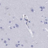
Negative control tissue: Adipose tissue (HPA Not detected)
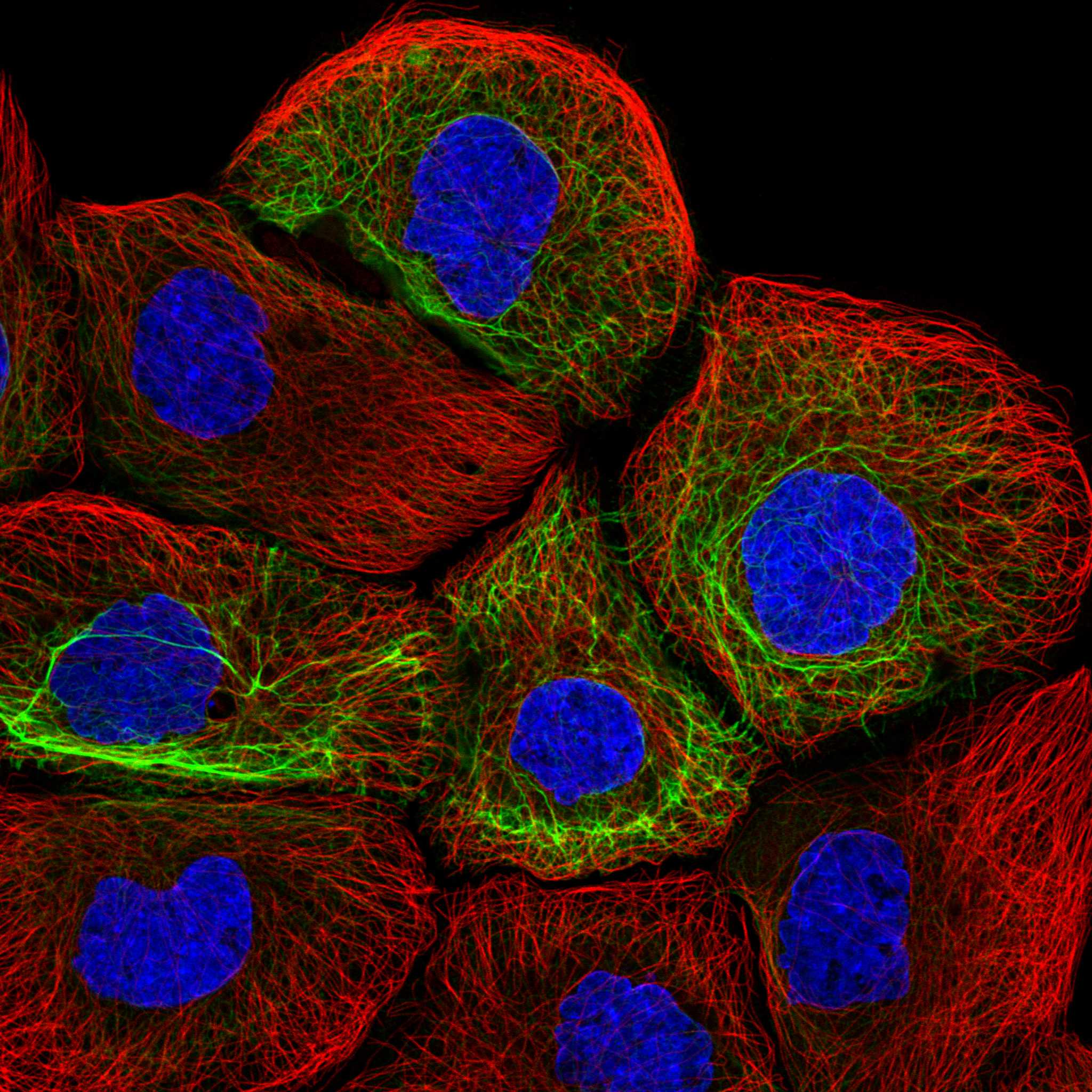
ICC-IF cell lines (HPA subcellular resource): HPA ICC-IF images show KRT13 in A-431, HeLa, U2OS, HaCaT, with annotated localisation: Intermediate filaments (enhanced) (HPA subcellular).
Technical controls: Include a no-primary, secondary-only control and an isotype-matched rabbit IgG control alongside a KRT13 knockout specimen as a biological negative (caption: rabbit anti-KRT13 antibody; standard IHC practice). Quench endogenous peroxidase before HRP–DAB detection and check the esophagus slide for residual background (caption: peroxidase secondary and DAB; standard IHC practice).
⚠️Feasibility: A target-specific fixation window or fixation effect is unreported, and the selected M04299 paraffin-section caption does not state the fixative (M04299 caption). That caption uses heat-mediated retrieval in EDTA at pH 8.0, but it does not establish whether retrieval is required; HPA shows intermediate-filament localization by ICC-IF, without establishing that IF or frozen sections are easier than paraffin IHC (M04299 caption; HPA: subcellular ICC-IF). In esophagus, inspect the squamous epithelial surface for section damage that could make staining difficult to score (standard IHC practice).

HPA tissue IHC evidence for KRT13

Comprehensive Human Protein Atlas IHC scoring per tissue (reliability: Enhanced — High consistency between antibody staining and RNA expression data.). Rows are taken directly from the HPA tissue chart — click any row's HPA link to view the source.

Positive expression · recommended positive controls

TissueCell typeLevelEvidenceSource
Cervix Squamous epithelial cells High Protein (IHC) HPA →
Esophagus Squamous epithelial cells High Protein (IHC) HPA →
Oral mucosa Squamous epithelial cells High Protein (IHC) HPA →
Skin Cells in basal layer High Protein (IHC) HPA →
Tonsil Squamous epithelial cells Medium Protein (IHC) HPA →

Undetected expression · recommended negative controls

TissueCell typeLevelEvidenceSource
Adipose tissue Adipocytes Not detected Protein (IHC) HPA →
Adrenal gland Glandular cells Not detected Protein (IHC) HPA →
Appendix Glandular cells Not detected Protein (IHC) HPA →
Bone marrow Hematopoietic cells Not detected Protein (IHC) HPA →
Breast Adipocytes Not detected Protein (IHC) HPA →
Section 3

Advanced KRT13 IHC Tips

Troubleshoot KRT13 staining in paraffin sections by checking retrieval, epithelial localisation, controls, and scoring before interpreting chromogenic signal.

Which retrieval conditions should I start with for weak KRT13 staining?
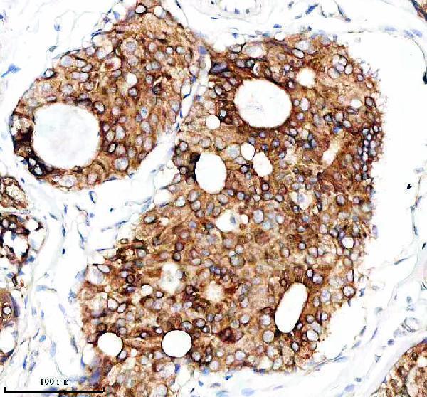
Start with heat-mediated retrieval in EDTA at pH 8.0 for paraffin sections (datasheet M04299). The pictured IHC section used this retrieval before 10% goat-serum blocking and overnight incubation with the catalog antibody at 1:50 and 4°C (datasheet M04299). If staining remains weak, compare a longer retrieval exposure or an alternative buffer on adjacent sections while keeping antibody dilution and detection constant (standard IHC practice). Include a squamous epithelial positive control, such as cervix or esophagus, and a no-primary control in each comparison (HPA tissue IHC; standard IHC practice). Avoid selecting conditions solely for stronger DAB color if epithelial detail or the expected cytoplasmic pattern deteriorates (HPA tissue IHC; standard IHC practice).
Could fixation explain a weak or patchy KRT13 result?
KRT13-specific sensitivity to fixation is unknown from the supplied evidence, and the catalog tissue caption does not state its fixative (datasheet M04299). Record the actual fixative and fixation duration for each specimen, then compare sections processed under matched conditions before changing the antibody dilution (standard IHC practice). Use the documented EDTA pH 8.0 retrieval as the starting condition for this catalog antibody (datasheet M04299). If a matched positive-control section stains while the test section does not, examine tissue preservation and epithelial content before attributing the result to fixation (HPA tissue IHC; standard IHC practice). Do not infer fixation tolerance from KRT13 filament localisation, modified residues, or the tissue expression profile (UniProt P13646; HPA tissue IHC).
What staining pattern should count as KRT13 in a tissue section?
Score a predominantly cytoplasmic pattern in the appropriate epithelial cells, consistent with KRT13 intermediate filaments (HPA tissue IHC; HPA subcellular). Squamous epithelial cells in cervix, esophagus, and oral mucosa are documented high-expression references; urothelial cells in urinary bladder are a documented medium-expression reference (HPA tissue IHC). Compare staining within intact epithelial layers and confirm that the DAB signal follows cell bodies rather than empty spaces or section edges (standard IHC practice). KRT13 forms keratin filaments with KRT4, supporting a cytoplasmic network expectation rather than an isolated nuclear signal (UniProt P13646). Review morphology alongside a counterstained section, since a positive color alone cannot establish the correct cell type (HPA tissue IHC; standard IHC practice).
How should I handle discordant staining when the antibody epitope is unspecified?
KRT13 has 3 reported isoforms, but the supplied antibody caption does not identify its epitope or establish isoform coverage (UniProt P13646; datasheet M04299). Avoid assigning a negative section to loss of one isoform without antibody mapping or an independent assay (UniProt P13646; standard IHC practice). Its intermediate-filament rod spans residues 104–416, while reported modifications include methylarginines at 27 and 35 and phosphoserine at 427 (UniProt P13646). Those features identify possible regions to check when epitope information becomes available; they do not establish that this antibody is modification sensitive (UniProt P13646; datasheet M04299). Compare retrieval conditions and a second independently characterised reagent on matched sections if discordance persists (standard IHC practice).
How can IF help verify a disputed chromogenic KRT13 pattern?
Use IF as a complementary localisation check with a marker identifying the epithelial population under study, and assess whether KRT13 signal overlaps the expected cells (HPA tissue IHC; standard IF practice). Choose a fluorophore channel after inspecting unstained tissue autofluorescence, and include single-stain controls when multiplexing (standard IF practice). KRT13 is an intermediate-filament protein with no annotated transmembrane segment, so intracellular access may require permeabilisation after fixation (HPA subcellular; UniProt P13646; standard IF practice). Optimise permeabilisation because the antibody epitope and its IF performance are not specified by the catalog IHC caption (datasheet M04299; standard IF practice). Treat IF agreement as supporting evidence while retaining the paraffin-section IHC controls for the chromogenic result (standard IHC/IF practice).
What should I change when KRT13 DAB staining is diffuse?
First inspect a no-primary control to distinguish detection-system background from signal dependent on the primary antibody (standard IHC practice). The documented tissue workflow blocked with 10% goat serum and used a peroxidase-conjugated secondary followed by DAB development (datasheet M04299). For chromogenic IHC, check that endogenous peroxidase blocking, washing, and DAB development are appropriate for the specimen; these are general workflow checks, not KRT13-specific findings (standard IHC practice). If background tracks the primary antibody, compare a lower concentration against the documented 1:50 condition while monitoring a positive-control epithelium (datasheet M04299; HPA tissue IHC; standard IHC practice). Do not count diffuse extracellular color as KRT13-positive cells, given the expected cytoplasmic epithelial pattern (HPA tissue IHC; HPA subcellular).
How should I score heterogeneous KRT13 staining across epithelial areas? ⚠ ANSWER MARKED FOR VERIFICATION
Define the epithelial compartment and exclude folds, necrosis, and poorly preserved edges before scoring (standard IHC practice). Record the percentage of positive epithelial cells and staining intensity, then calculate an H-score from those components if intensity differences matter (standard IHC practice). Normalise cell counts to the number of evaluable epithelial cells, or report positive-cell density per mm² of evaluable epithelium when area is the preferred denominator (standard IHC practice). Keep retrieval, antibody dilution, DAB development, and image thresholds consistent across compared sections (standard IHC practice). Report squamous and urothelial compartments separately when both are present, because the documented KRT13 expression profile differs by cell type (HPA tissue IHC).
When is an apparent KRT13-positive focus more likely to be artefact?
A convincing focus follows intact epithelial cells with cytoplasmic staining, consistent with KRT13 expression in squamous epithelia and urothelium (HPA tissue IHC; HPA subcellular). Isolated nuclear color, staining confined to a cut edge, or signal concentrated in necrotic material warrants review before calling the focus positive (HPA subcellular; standard IHC practice). Compare the focus with a no-primary control to assess endogenous enzyme activity or detection-system deposits (standard IHC practice). Breast cancer tissue appears in the catalog IHC caption, but that image alone does not establish which cells in every breast specimen should stain (datasheet M04299). Resolve uncertain foci with morphology, an appropriate epithelial positive control, and repeat staining on an adjacent section when available (HPA tissue IHC; standard IHC practice).
Boster reagents

Best KRT13 / Keratin, type I cytoskeletal 13 IHC Antibodies

M04299 has a human paraffin-section IHC image (M04299 IHC caption) and is listed for human and mouse IHC and IF/ICC (catalog: applications, reactivity).

Real IHC data IHC analysis of KRT13 using anti-KRT13 antibody (M04299). KRT13 was detected in a paraffin-embedded section of human breast cancer tissue. Heat mediated antigen retrieval was performed in EDTA buffer (pH 8.0, epitope retrieval solution). The tissue section was blocked with 10% goat serum. The tissue section was then incubated with 1:50 rabbit anti-KRT13 Antibody (M04299) overnight at 4°C. Peroxidase Conjugated Goat Anti-rabbit IgG was used as secondary antibody and incubated for 30 minutes at 37°C. The tissue section was developed using HRP Conjugated Rabbit IgG Super Vision Assay Kit (Catalog # SV0002) with DAB as the chromogen.
Anti-Cytokeratin 13 Rabbit Monoclonal Antibody
Cat # M04299

M04299 will render with an IHC figure showing staining in a paraffin-embedded human breast cancer section (M04299 IHC caption). The catalog also lists IF/ICC and mouse reactivity, but provides no IF or mouse image (catalog: applications, reactivity, image captions).

Which to pick: For tissue IHC, choose M04299: its rabbit monoclonal antibody has a pictured paraffin-section result using EDTA retrieval at pH 8.0 and a 1:50 primary dilution (catalog: host, clonality; M04299 IHC caption). For IF/ICC or mouse samples, M04299 is the listed option based on its application and reactivity entries; the supplied figure demonstrates neither use (catalog: applications, reactivity, image captions). The IHC caption does not report the tissue fixative (M04299 IHC caption).

Each figure is that product's own IHC / IF validation image from its datasheet.

References

  1. UniProt Consortium. UniProt entry P13646 (K1C13_HUMAN, Keratin, type I cytoskeletal 13).
  2. Human Protein Atlas. KRT13 tissue IHC expression (reliability: Enhanced).
  3. Human Protein Atlas. KRT13 subcellular location (ICC-IF): Localized to the intermediate filaments..
  4. Human Protein Atlas. KRT13 antibody validation summary (3 antibodies).
  5. Exfoliative Cytology and Genetic Analysis for a Non-Invasive Approach to the Diagnosis of White Sponge Nevus: Case Series. Bioengineering (Basel, Switzerland) 2023 — PMC9952746.
  6. Immunohistochemical Expression of CK13 and Molecular Analysis of KRT13 and APC in Odontogenic Ghost Cell Lesions, Adenoid Ameloblastoma, and Conventional Ameloblastoma. Head and neck pathology 2026 — PMC13294416.
  7. Meta-Analysis of COVID-19 BAL Single-Cell RNA Sequencing Reveals Alveolar Epithelial Transitions and Unique Alveolar Epithelial Cell Fates. American journal of respiratory cell and molecular biology 2023 — PMC10704119.
  8. Keratin 13 Is Enriched in Prostate Tubule-Initiating Cells and May Identify Primary Prostate Tumors that Metastasize to the Bone. PloS one 2016 — PMC5053503.
  9. PubMed PMID:2475110 — UniProt-cited evidence.
  10. PubMed PMID:2477803 — UniProt-cited evidence.
  11. PubMed PMID:2483837 — UniProt-cited evidence.