This website uses cookies to ensure you get the best experience on our website.


- Table of Contents
Plan chromogenic KRT9 IHC in paraffin sections using sole epidermis as a positive tissue (HPA tissue IHC). The catalog antibody has a paraffin IHC example (datasheet M03692); assess epidermal staining without assuming a subcellular pattern (HPA tissue IHC).

Expected localisation, validated protocols, controls and antibodies — the at-a-glance facts below, then the full design guide.
| Expected localisation | Sole epidermal cells; compartment unreported (HPA tissue IHC) | |
| Staining pattern | High staining in sole epidermal cells; subcellular pattern unreported (HPA tissue IHC) | |
| Antigen retrieval | EDTA pH 8.0 HIER, heat-mediated (datasheet M03692) | |
| Positive control | Sole of foot | |
| Negative control | Adipose tissue+4 more · see all |
| Fixation | Keep fixation consistent across sections (standard IHC practice; not target-specific); Matched tissue-IHC evidence does not establish the fixation claim. Validate the specimen-specific method before use. (selected-SKU IHC image M03692) | |
| Caveat | Sampling outside sole may miss the restricted tissue signal (HPA tissue IHC) | |
| Regulation | Expression tracks terminal palmoplantar differentiation (UniProt) | |
| Isoform / epitope | No annotated isoforms or cleavage; epitope region unspecified (UniProt) |
The catalog antibody’s IHC-P protocol is accompanied by published KRT9 protocols for mouse footpad epidermis (PMC3923277) and melanoma and nevus tissue (PMC9470607).
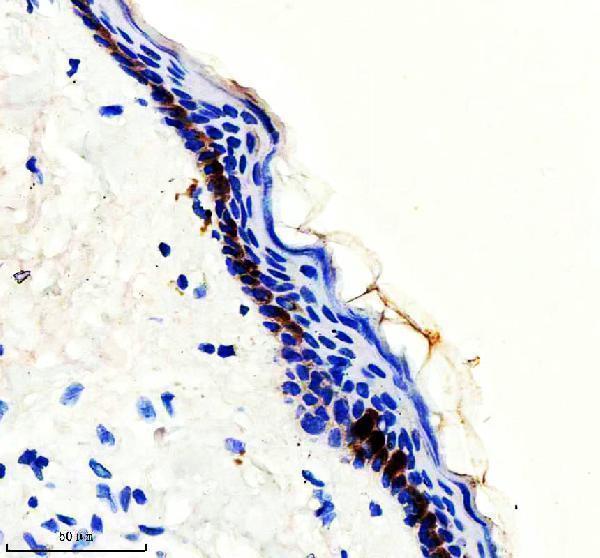
| Sample | Paraffin-embedded human skin cancer tissue; fixative not specified (datasheet M03692) |
| Fixation | Image fixative and duration unreported (datasheet M03692); verify before use. |
| Sectioning | 4–5 µm sections on charged slides (standard) |
| Deparaffinisation | Xylene, graded ethanol series to water (standard) |
| Antigen retrieval | Heat retrieval: EDTA pH 8.0 (datasheet M03692); 20 min, 95–100 °C (standard) |
| Peroxidase block | 3% H2O2, 10 min, room temperature (standard) |
| Blocking | 10% goat serum (datasheet M03692) |
| Primary antibody | Rabbit monoclonal (clone GGH-11) anti-KRT9, 1:50 (datasheet M03692) |
| Primary incubation | Overnight at 4 °C (datasheet M03692) |
| Detection | HRP-conjugated secondary, DAB chromogen (datasheet M03692) |
| Counterstain | Hematoxylin, blue, dehydrate and mount (standard) |
| Expected result | KRT9-positive staining in epidermal cells of sole of foot (HPA tissue IHC: High). HPA tissue profile: Exclusively expressed in sole of foot. No signal in the no-primary control. |
KRT9 is a structural keratin of suprabasal palmoplantar epidermal cells, where cytoplasmic filament staining is expected (UniProt P35527 function). It has no transmembrane segment; UniProt does not annotate a subcellular location (UniProt P35527 topology; UniProt P35527 subcellular record). Sole-of-foot epidermal cells stain at a high level, with HPA reliability rated Supported (HPA tissue IHC).
| Strong staining follows the suprabasal epidermal layers in a sole-of-foot section (HPA tissue IHC; UniProt P35527 function). | This fits the reported high signal in sole epidermal cells and KRT9's filament role in suprabasal palmoplantar keratinocytes (HPA tissue IHC; UniProt P35527 function). Judge the epidermal layer and cytoplasmic pattern together; intensity alone is insufficient (general IHC practice). |
| Signal is mainly nuclear or outlines cell membranes instead of the cytoplasm (UniProt P35527 function; UniProt P35527 topology). | Treat this as a localization mismatch requiring review, since KRT9 forms intermediate filament networks and has no transmembrane segment (UniProt P35527 function; UniProt P35527 topology). Check morphology and the negative control before assigning it to KRT9 (general IHC practice). |
| Adipocytes or respiratory epithelial cells stain conspicuously (HPA tissue IHC). | HPA reports KRT9 as not detected in the sampled adipocytes and bronchial respiratory epithelial cells (HPA tissue IHC). Consider cross-reactivity or endogenous chromogen-generating activity; compare a negative control and verify which cells are stained (general IHC practice). |
| Chromogen spreads across stroma, section edges, and epidermis without a clear cellular pattern (general IHC practice). | Diffuse background does not establish KRT9 expression (general IHC practice). Review reagent background, blocking, washing, and chromogen development, then compare with the expected sole epidermal distribution (general IHC practice; HPA tissue IHC). |
| No epidermal signal appears in a sole-of-foot section (HPA tissue IHC). | This conflicts with HPA's high sole epidermal staining, but a single negative section cannot identify the cause (HPA tissue IHC; general IHC practice). Confirm epidermis is present and examine the positive control and staining run (general IHC practice). |
| Tissue and layer selection | UniProt places KRT9 in terminally differentiated palm-and-sole epidermis; HPA directly reports high staining in sole epidermal cells (UniProt P35527 tissue specificity; HPA tissue IHC). Use a section containing intact sole epidermis to assess this reported pattern (general IHC practice). |
| Location of the target | The intermediate-filament rod and keratin assembly function support a cytoplasmic filament interpretation, while UniProt supplies no explicit subcellular-location annotation (UniProt P35527 domain, function, and subcellular record). Do not assign a more precise organelle location from these records. |
| Strength of antibody evidence | HPA rates IHC for HPA007261 and CAB037322 as Supported, and the tissue profile itself as Supported (HPA antibody validation; HPA tissue IHC). That rating includes external characterization, with no internal RNA data available for staining correlation (HPA tissue IHC). |
| Tissue RNA versus protein staining | HPA lists RNA as tissue enriched in lymphoid tissue while its reported protein staining is exclusive to sole of foot (HPA tissue IHC). Do not infer lymphoid-cell IHC positivity from that RNA label; score the observed cells against the reported protein pattern (HPA tissue IHC; general IHC practice). |
| IF/ICC Q&A: Is a cellular IF pattern established? | No. HPA provides no main subcellular location or ICC-IF image-bearing cell lines; its HaCaT value of 15.9 nTPM is expression data, not an IF localization result (HPA subcellular record). Interpret any IF result against appropriate controls (general IF practice). |
| Situation | Likely cause | Next action |
|---|---|---|
| Sole epidermis is unstained (HPA tissue IHC). | The section may lack assessable epidermis, or the staining run may have failed (general IHC practice). | Inspect morphology, run a sole epidermal positive control, and review the catalog antibody's IHC-P conditions before interpreting absence (HPA tissue IHC; general IHC practice). |
| Sole epidermis stains weakly relative to the reported high level (HPA tissue IHC). | Target-specific fixation effects are not established by the supplied assay evidence. Verify with a matched IHC source before attributing a result to fixation. | Compare controls and review retrieval, antibody dilution, and detection settings under the validated IHC-P workflow (general IHC practice). |
| The strongest staining is nuclear or membranous (UniProt P35527 function and topology). | This distribution is inconsistent with the expected keratin filament interpretation (UniProt P35527 function). | Check cell boundaries and counterstain, then compare a negative control and a sole epidermal positive control (general IHC practice; HPA tissue IHC). |
| Adipocytes or bronchial respiratory epithelium stain (HPA tissue IHC). | These sampled cell types are reported as not detected; cross-reactivity or endogenous detection activity is possible (HPA tissue IHC; general IHC practice). | Verify cell identity, compare reagent controls, and assess endogenous peroxidase blocking for chromogenic detection (general IHC practice). |
| Background obscures the epidermal layers (general IHC practice). | Nonspecific reagent binding, incomplete washing, or excess chromogen development may reduce contrast (general IHC practice). | Review blocking, washes, antibody dilution, and development time; require a discernible epidermal cell pattern (general IHC practice; HPA tissue IHC). |
| A proposed positive call rests only on lymphoid-tissue RNA enrichment (HPA tissue IHC). | The RNA label and reported tissue protein pattern differ, and HPA lacks internal RNA correlation for staining (HPA tissue IHC). | Base the IHC call on slide staining and controls; use sole epidermis as the reported positive comparator (HPA tissue IHC; general IHC practice). |


Comprehensive Human Protein Atlas IHC scoring per tissue (reliability: Supported — No tissue staining or RNA expression data available for comparison. External characterization data supports antibody staining but no internal RNA data available for correlation.). Rows are taken directly from the HPA tissue chart — click any row's HPA link to view the source.
| Tissue | Cell type | Level | Evidence | Source |
|---|---|---|---|---|
| Sole of foot | Epidermal cells | High | Protein (IHC) | HPA → |
| Tissue | Cell type | Level | Evidence | Source |
|---|---|---|---|---|
| Adipose tissue | Adipocytes | Not detected | Protein (IHC) | HPA → |
| Appendix | Glandular cells | Not detected | Protein (IHC) | HPA → |
| Bone marrow | Hematopoietic cells | Not detected | Protein (IHC) | HPA → |
| Breast | Adipocytes | Not detected | Protein (IHC) | HPA → |
| Bronchus | Respiratory epithelial cells | Not detected | Protein (IHC) | HPA → |
Troubleshoot chromogenic KRT9 IHC in paraffin sections against its expected palmoplantar epidermal pattern, using the catalog antibody’s tissue staining conditions as a starting point.
M03692 has real IHC data from a paraffin-embedded human skin cancer section (M03692 image caption); its listed reactivity covers human, mouse, and rat (M03692 catalog).
M03692 was demonstrated by IHC on a paraffin-embedded human skin cancer section at 1:50 (M03692 image caption). It is a rabbit monoclonal antibody listed for IHC, IP, and WB, with human, mouse, and rat reactivity (M03692 catalog).
Which to pick: For tissue IHC, choose M03692: its own image documents chromogenic staining of a paraffin-embedded human section after EDTA retrieval at pH 8.0; the fixative is unreported (M03692 image caption). No SKU in the payload lists IF/ICC or provides an IF image, so there is no supported IF/ICC pick here (M03692 catalog). For cross-species work, M03692 lists mouse and rat reactivity alongside human, but its IHC image demonstrates human tissue only (M03692 catalog; M03692 image caption).