LAMP1 / Lysosome-associated membrane glycoprotein 1 · Western blot design guide

Design a Western Blot for LAMP1

Real validated LAMP1 Western blot protocols, expected-band and isoform facts, troubleshooting for weak or shifted signal, and recommended anti-LAMP1 WB antibodies. Everything you need to plan the experiment before you commit precious samples.

Evidence assembled September 2026 · For research use; verify linked source records and product datasheet before use
Western blot protocol sheet for LAMP1: expected band ~44.9 kDa, hero antibody PA1822, catalog values and labelled standard workflow; separate PMC comparisons on the guide
Printable LAMP1 Western blot protocol sheet — expected band ~44.9 kDa, antibody PA1822, controls and PMC citations. Open the full LAMP1 WB guide →

LAMP1 Western Blot Experimental Design Guide

Expected bands, validated protocols, controls and antibodies — the at-a-glance facts below, then the full design guide.

Must know before running
Expected band ~44.9 kDa
Gel 12–15% (standard starting point)
Positive control ⓘ Adrenal gland (IHC candidate; verify WB) +4 more
Negative control ⓘ Suggested KO / knockdown lysate
Important caveats
Reasons your observed band may differ from the expected size.
PTM Glycosylated + Cleaved
Caveat Glycosylation affects migration
Gene-set association MSigDB C7 membership
Isoform 2 isoform(s)
Section 1

Real Curated LAMP1 Western Blot Protocols

The PA1822 protocol combines labelled catalog values with standard starting conditions. Published comparisons retain their own sample, reagent and detection scope.

Recommended Western blot protocol parameters
Sample / lysateTarget-positive lysate and matched negative control (standard starting point)
Gel %12–15% (standard starting point)
Load20–30 µg total protein per lane; optimize for abundance (standard starting point)
TransferStandard semi-dry transfer; verify efficiency (standard starting point)
Membrane0.45 µm PVDF (standard starting point)
Blocking5% milk or 5% BSA in TBST (standard starting point)
Primary antibodyPA1822; use the WB datasheet starting dilution (standard starting point)
Primary incubationOvernight at 4 °C (standard starting point)
Secondary antibodySpecies-matched HRP conjugate at validated dilution (standard starting point)
Secondary incubation1 h at room temperature (standard starting point)
Wash3 × 5 min in TBST (standard starting point)
DetectionECL; bracket exposures to avoid saturation (standard starting point)
Section 2

What Is the Expected LAMP1 Western Blot Band Size?

LAMP1 is predicted at 44.9 kDa; glycosylation and signal peptide cleavage could alter migration, but no observed band size is supplied.

What am I looking at on my blot?
Band near 44.9 kDaNear the predicted precursor mass; identity needs confirmation
Band above 44.9 kDaMay reflect extensive N-linked glycosylation; its migration is not established here
Smear or broad bandMay reflect heterogeneous N-linked glycosylation
Smaller bandMay reflect cleavage of the 1–28 signal peptide; identity needs confirmation
💡Expected LAMP1 appearanceLAMP1 has a predicted precursor mass of 44.9 kDa and 24 annotated glycosylation sites, but no empirical band size is supplied; confirm any candidate band with antibody specificity and appropriate controls.
How each factor affects band size
Predicted precursor mass44.9 kDa is the sequence-based reference, not a validated band position
N-linked glycosylation at Asn-37 and other sitesCan increase apparent size if the sites are occupied
Polylactosaminoglycan N-linked sites at Asn-62 and Asn-121May contribute to variable apparent size if modified
Signal peptide at residues 1–28Cleavage makes the mature protein smaller than the precursor
Isoforms 1 and 2Sequence differences may affect size, but distinct migration is not established
Why is my band missing or off?
SituationLikely causeNext action
Band higher than expectedN-linked glycosylation may affect migrationCompare untreated and deglycosylated samples with a validated LAMP1 antibody
Band lower than expectedSignal peptide cleavage may reduce sizeCheck antibody epitope and compare with a verified LAMP1 control
Broad smear instead of sharp bandHeterogeneous N-linked glycosylation is possibleCompare untreated and deglycosylated samples
Multiple bandsIsoforms 1 and 2 or differing glycosylation may contributeCheck antibody specificity and compare with LAMP1-depleted material
Weak or no signalLAMP1 is a lysosome and endosome membrane proteinCheck membrane protein recovery, transfer, and a positive control
Fragments below expected sizeThe supplied features do not identify these fragmentsCheck sample integrity and antibody specificity with a LAMP1-depleted control

Sample controls for LAMP1 Western blot

🧪HPA-IHC candidate guidance (verify in WB): For positive controls for LAMP1 in Western blot, you can use adrenal gland lysate, which HPA rates High.
Positive control: Adrenal gland (IHC candidate; verify WB)
Negative control: Suggested KO / knockdown lysate
Loading controls: Run GAPDH, β-actin, and a total-protein stain such as Ponceau alongside the samples.
⚠️Feasibility: HPA lists no Not-detected tissue, so use siRNA knockdown or a KO line for a clean negative control.

HPA tissue expression evidence for LAMP1

Comprehensive Human Protein Atlas IHC scoring per tissue. Rows are taken directly from the HPA tissue chart — click any row's HPA link to view the source.

Higher expression tissues · candidate positive controls from IHC

TissueCell typeLevelEvidenceSource
Adrenal gland glandular cells High Protein (IHC) HPA →
Appendix glandular cells High Protein (IHC) HPA →
Bone marrow hematopoietic cells High Protein (IHC) HPA →
Breast glandular cells High Protein (IHC) HPA →
Bronchus respiratory epithelial cells High Protein (IHC) HPA →

Lower expression tissues · IHC evidence, not confirmed WB-negative controls

TissueCell typeLevelEvidenceSource
Skeletal muscle myocytes Low Protein (IHC) HPA →
Adipose tissue adipocytes Medium Protein (IHC) HPA →
Smooth muscle smooth muscle cells Medium Protein (IHC) HPA →
Soft tissue fibroblasts Medium Protein (IHC) HPA →
Adrenal gland glandular cells High Protein (IHC) HPA →
Section 3

Advanced LAMP1 Western Blot Tips

Deeper troubleshooting and optimisation questions for LAMP1, answered from its protein features.

How should LAMP1 band migration be interpreted?
Band shift · Use the separately labelled calculated mass and catalog-observed evidence above. A sequence annotation does not establish an observed migration shift. Verify target identity with orthogonal controls.
How could LAMP1 isoforms affect band interpretation?
Isoforms · UniProt lists isoforms 1 and 2; isoform 2 lacks residues 135–187 in canonical UniProt numbering. An additional band could reflect this sequence difference, but the features do not establish how either isoform migrates. Check whether the antibody epitope falls within the missing segment.
Which LAMP1 glycosylation sites matter when assessing band changes?
PTM · UniProt annotates 18 N-linked and six O-linked sites. N-linked sites 62, 121, 130, 223 and 228 are annotated with polylactosaminoglycan; O-linked sites occur at 197, 199, 200, 207, 209 and 211, with Ser197 marked partial. These are canonical UniProt coordinates; verify numbering conventions before comparing antibody or paper sites. Site annotations do not prove a visible shift.
Does this guide establish induction of LAMP1?
Induction · No general induction response is established by this guide. A pathway or gene-set association is not evidence of induction in a particular specimen. Verify the relevant treatment and control in a target-specific experiment.
What transfer method to use for LAMP1 Western blot?
Transfer · LAMP1 is a single-pass membrane protein found on lysosome, endosome and late-endosome membranes. Choose and verify a transfer procedure suitable for membrane proteins, then confirm transfer across the band region you evaluate. The supplied features do not specify a transfer method or an apparent band size.
How should blocking be optimized?
Blocking · Standard workflow guidance: follow the PA1822 datasheet where specified. Otherwise compare 5% milk or 5% BSA in TBST; for a phospho-specific assay start with BSA. Optimize background and specific signal with matched controls.
What should be considered when quantifying LAMP1 bands?
Quantitation · LAMP1 has 24 annotated glycosylation sites and two isoforms. Define whether quantitation includes all detected LAMP1 forms or only a selected band, and apply that choice consistently across samples. The annotations alone do not establish which bands represent LAMP1 or whether glycosylation changes their intensity.
Why might LAMP1 migrate differently from its predicted 44.9 kDa mass?
Interpretation · The 44.9 kDa prediction does not account for LAMP1's 24 annotated glycosylation sites or cleavage of its 1–28 signal peptide. Glycosylation could affect migration, but these features alone cannot establish an apparent mass or explain a particular observed band. No empirical band position was supplied.

Compare candidate bands with LAMP1's extensive glycosylation, its cleaved 1–28 signal peptide and isoform 2's missing canonical residues 135–187. Check antibody epitope coverage, especially for isoform 2. These features suggest possibilities but cannot identify an unexpected band without experimental validation.
Boster reagents

LAMP1 Western Blot Antibodies

Catalog antibodies with Western blot application and product-specific WB images. Evaluate suitability with the reported sample, controls and experimental conditions.

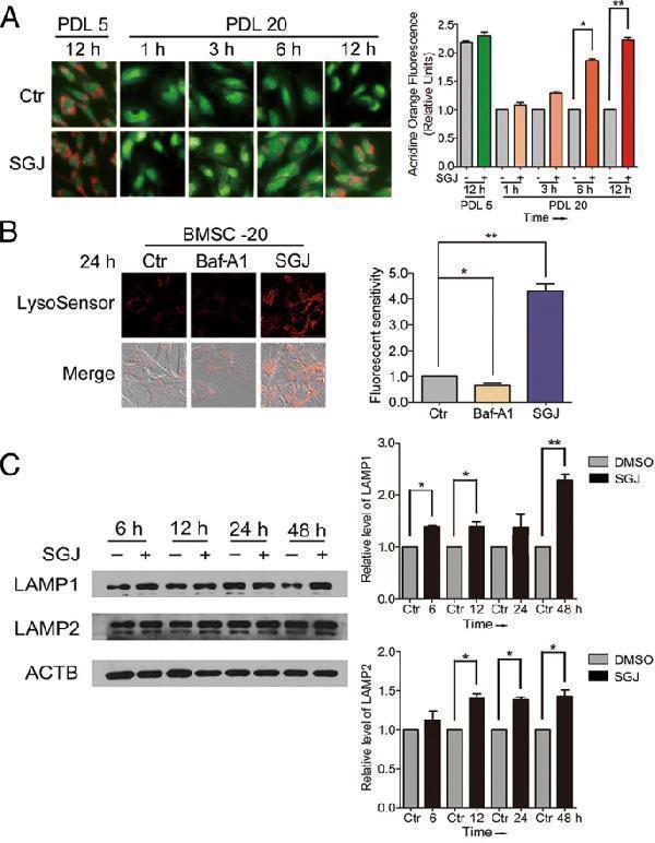
Real WB data SGJ increased the concentration of H + in lysosomes, and up-regulated LAMP1 and LAMP2 protein level. a Acridine orange staining for young (PDL 5) and senescent (PDL 20) BMSCs. Acidic vacuoles declined with age as shown in the results. Twenty-micromolar SGJ treatments for 1, 3, 6, and 12 h significantly restored the amount of acidic vacuoles (magnification × 200). b SGJ promoted lysosomal acidification. Lysosensor™ Green DND-189 was used to sense the changes of the concentration of H + in lysosomes, and quantification. BMSCs were treated with 20 nM Baf-A1 or 20 μM SGJ for 24 h. The changes of the red fluorescence reflect changes in lysosomal pH. c Western blot analysis of LAMP1 and LAMP2 protein levels with β-actin as a loading control, and quantification. BMSCs were treated with 20 μM SGJ for 6, 12, 24 and 48 h. (*, p < 0.05; **, p < 0.01, results were expressed as means ± SEM, n = 3) <b>Index in PubMed under a CC BY license. PMID: <a href='https://link.springer.com/article/10.1186/s13287-018-1081-0'>30526663</a></b>
Anti-LAMP1 Antibody Picoband®
Cat # PA1822
Real WB data Western blot analysis of LAMP1 expression in A431 cell lysate.
Anti-LAMP1/Cd107A Rabbit Monoclonal Antibody
Cat # M00780
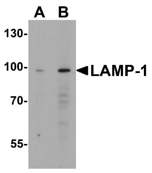
Real WB data Western blot analysis of LAMP-1 in EL4 cell lysate with LAMP-1 antibody at (A) 1 and (B) 2 μg/mL.
Anti-LAMP-1 Antibody
Cat # A00780

Three the supplier anti-LAMP1 antibodies are listed with Western blot images. The supplied evidence includes an A431 lysate blot, an EL4 lysate blot at 1 and 2 μg/mL, and a publication reporting LAMP1 blots in SGJ-treated BMSCs. These examples establish tested contexts, not broader validation.

Which to pick: For human samples, PA1822 and M00780 list human reactivity; M00780 shows an A431 lysate blot. For mouse or rat, A00780 is the only listed option with that reactivity, and its image shows an EL4 lysate blot at 1 and 2 μg/mL.

Source: BosterBio LAMP1 gene-info card — filtered to Western-blot-capable antibodies; each card shows that product's actual WB validation figure.