This website uses cookies to ensure you get the best experience on our website.


- Table of Contents
Plan LAMP3 IHC in paraffin sections around cytoplasmic staining in type II pneumocytes and non-germinal center lymphoid cells (HPA tissue IHC). This guide covers fixation, antibody titration, chromogenic detection, controls, and scoring by cell type and compartment (standard IHC practice).

Expected localisation, validated protocols, controls and antibodies — the at-a-glance facts below, then the full design guide.
| Expected localisation | Cytoplasmic staining in lung and lymphoid cells (HPA tissue IHC) | |
| Staining pattern | Type II pneumocytes: high cytoplasmic; lymphoid cells: medium (HPA tissue IHC) | |
| Antigen retrieval | Citrate pH 6.0 HIER, 95–98 °C, 20 min (rule: cytoplasmic / membrane antigen) | |
| Positive control | Lung+2 more · see all | |
| Negative control | Adipose tissue+4 more · see all |
| Fixation | Matched tissue-IHC evidence does not establish the fixation claim. Validate the specimen-specific method before use. (selected-SKU IHC image A09406) | |
| Caveat | Lymphoid staining may be weaker than type II pneumocyte staining (HPA tissue IHC) | |
| Regulation | Up-regulated in several carcinomas (UniProt) | |
| Isoform / epitope | 0 isoforms annotated; lumenal versus cytoplasmic epitope matters (UniProt) |
The catalog antibody’s IHC-P protocol is accompanied by published LAMP3 staining protocols for oral squamous cell carcinoma specimens and pediatric gastric mucosa (PMC11491047; PMC6885256).
| Sample | Paraffin-embedded human lung-cancer tissue; fixative not specified (datasheet A09406) |
| Fixation | Image fixative and duration unreported (datasheet A09406); verify before use. |
| Sectioning | 4–5 µm sections on charged slides (standard) |
| Deparaffinisation | Xylene, graded ethanol series to water (standard) |
| Antigen retrieval | Heat-induced epitope retrieval in citrate buffer, pH 6.0, 20 min at 95–98 °C (standard rule: cytoplasmic / membrane antigen) |
| Peroxidase block | 3% H2O2, 10 min, room temperature (standard) |
| Blocking | 10% normal serum of the secondary host, 30 min, room temperature (standard) |
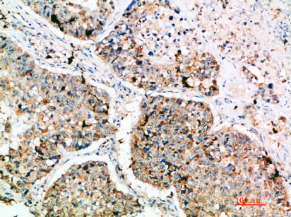
| Primary antibody | Rabbit anti-LAMP3, 1:500 recommended; image 1:200 (datasheet A09406) |
| Primary incubation | Overnight at 4 °C (standard) |
| Detection | HRP-polymer secondary, DAB chromogen 5–10 min (standard) |
| Counterstain | Hematoxylin, blue, dehydrate and mount (standard) |
| Expected result | LAMP3-positive staining in alveolar cells type II of lung (HPA tissue IHC: High). HPA tissue profile: Selective cytoplasmic expression in type II pneumocytes and lymphoid non-germinal center cells. No signal in the no-primary control. |
LAMP3 is a membrane protein found on lysosomes, endosomes, cytoplasmic vesicles and the cell surface (UniProt Q9UQV4 topology and subcellular location). In IHC, expect cytoplasmic staining strongest in lung type II pneumocytes, with moderate staining in lymph node and tonsil non-germinal center cells (HPA: High; Medium). HPA rates its tissue pattern Enhanced, citing high agreement between antibody staining and RNA expression (HPA: Enhanced reliability).
| Distinct cytoplasmic staining in alveolar type II cells. | This matches the strongest reported normal-tissue signal (HPA: Lung, alveolar type II cells, High). A granular or vesicular appearance fits LAMP3 on intracellular membranes (UniProt Q9UQV4 subcellular location); HPA reports selective cytoplasmic IHC staining, without defining one required chromogenic texture (HPA: tissue IHC profile). |
| Staining confined mainly to nuclei, with little cytoplasmic signal. | Treat a nuclear-only pattern as suspect: the supported locations are vesicle, endosome and lysosome membranes, plus the cell surface (UniProt Q9UQV4 subcellular location). Check morphology and the known-positive control before scoring it as LAMP3; localization alone cannot identify the cause of an aberrant signal (general IHC practice). |
| Strong staining in cells outside the expected positive population. | In normal lung, identify type II pneumocytes before assigning the signal to LAMP3 (HPA: Lung, alveolar type II cells, High). Unexpected staining can reflect cross-reactivity or endogenous detection activity (general IHC practice). Interpret tumor tissue separately: UniProt reports LAMP3 up-regulation in several carcinomas (UniProt Q9UQV4 tissue specificity). |
| Weak, diffuse color across many structures and empty areas. | A broad haze does not reproduce the selective cytoplasmic tissue pattern (HPA: tissue IHC profile). Consider nonspecific antibody binding, inadequate blocking, excess detection reagent or incomplete washing (general IHC practice). Judge any localized signal against a matched control section after the background is addressed. |
| No detectable signal in a lung section expected to contain type II cells. | First verify that the section contains identifiable type II pneumocytes; these cells have High LAMP3 staining in HPA tissue IHC (HPA: Lung, alveolar type II cells, High). If present, review the validated IHC conditions and detection controls before concluding that the specimen lacks LAMP3 (general IHC practice). |
| Membrane location and topology | LAMP3 has one transmembrane segment at residues 382–402, a lumenal region at 28–381 and a short cytoplasmic tail at 403–416 (UniProt Q9UQV4 topology). Its intracellular membrane locations support cytoplasmic organelle-associated IHC staining; topology alone does not establish where a particular antibody binds. |
| Protein processing and glycosylation | The signal peptide spans residues 1–27, and the annotated mature chain starts at residue 28; seven glycosylation sites are listed (UniProt Q9UQV4 processing and glycosylation). These annotations describe the antigen, but provide no evidence here for epitope accessibility or an antigen-retrieval choice. |
| Tissue and cell-type distribution | Use cell identity when comparing sections: HPA reports High staining in lung type II cells and Medium staining in lymph node and tonsil non-germinal center cells (HPA: tissue IHC). Its Not detected calls apply to the specified cells sampled, such as adipocytes in adipose tissue, rather than every cell in that organ. |
| Strength of the IHC evidence | HPA calls the tissue profile Enhanced and lists two antibodies with Enhanced IHC validation, HPA051467 and CAB025133 (HPA: tissue IHC reliability; HPA: antibody validation). This supports the reported tissue pattern, but does not validate every staining condition or establish target-specific fixation sensitivity. |
| Situation | Likely cause | Next action |
|---|---|---|
| Lung control is blank. | The section may lack identifiable type II cells, or an IHC step may have failed (HPA: High in lung type II cells; general IHC practice). | Confirm cell morphology, then review the catalog antibody’s IHC-P conditions, reagent activity and detection controls (general IHC practice). Do not use a blank, cell-poor field to call the target absent. |
| Lymphoid staining seems weaker than lung. | That difference matches the reported levels: Medium in lymph node and tonsil non-germinal center cells versus High in lung type II cells (HPA: tissue IHC). | Compare the correct cell populations and score each tissue against its expected level (HPA: tissue IHC; general IHC practice). Avoid requiring lung-level intensity in lymphoid sections. |
| Color appears mainly in nuclei. | Nuclear localization is outside the listed LAMP3 compartments (UniProt Q9UQV4 subcellular location). | Review the counterstain and chromogen localization, and compare with a known-positive control and an appropriate detection control (general IHC practice). Reassess the signal before scoring it as specific. |
| Many unrelated cells or blank areas are colored. | Nonspecific binding, endogenous detection activity or excess reagent can create misleading color (general IHC practice). | Check blocking, washing and detection controls; review antibody concentration within its validated IHC-P guidance (general IHC practice). Look for the selective cytoplasmic pattern after background is reduced (HPA: tissue IHC profile). |
| A listed negative cell type stains. | HPA reports Not detected in particular cells, including adipocytes in adipose tissue; that designation is cell-specific (HPA: tissue IHC). | Confirm the stained cell’s identity and compare a detection control (general IHC practice). Investigate persistent signal as possible cross-reactivity or endogenous activity before treating the HPA pattern as contradicted. |
| IF/ICC: what signal should appear? | HPA reports vesicles as the enhanced ICC-IF location, consistent with LAMP3 on cytoplasmic vesicle membranes (HPA: subcellular location; UniProt Q9UQV4 subcellular location). | Expect intracellular vesicular signal and assess it with appropriate IF controls (HPA: subcellular location; general IF practice). Use the separate IF/ICC guide for assay setup; this IHC-P section supplies no IF protocol. |


Comprehensive Human Protein Atlas IHC scoring per tissue (reliability: Enhanced — High consistency between antibody staining and RNA expression data.). Rows are taken directly from the HPA tissue chart — click any row's HPA link to view the source.
| Tissue | Cell type | Level | Evidence | Source |
|---|---|---|---|---|
| Lung | Alveolar cells type II | High | Protein (IHC) | HPA → |
| Lymph node | Non-germinal center cells | Medium | Protein (IHC) | HPA → |
| Tonsil | Non-germinal center cells | Medium | Protein (IHC) | HPA → |
| Tissue | Cell type | Level | Evidence | Source |
|---|---|---|---|---|
| Adipose tissue | Adipocytes | Not detected | Protein (IHC) | HPA → |
| Adrenal gland | Glandular cells | Not detected | Protein (IHC) | HPA → |
| Appendix | Glandular cells | Not detected | Protein (IHC) | HPA → |
| Bone marrow | Hematopoietic cells | Not detected | Protein (IHC) | HPA → |
| Breast | Adipocytes | Not detected | Protein (IHC) | HPA → |
Troubleshoot LAMP3 staining in paraffin sections using the catalog antibody’s tissue image, the stated retrieval setting, and expected cellular distribution.
The rendered LAMP3 antibody has an IHC image from paraffin-embedded human lung cancer (A09406 IHC image caption); IHC and IF are listed for human, mouse and rat (A09406 catalog applications and reactivity).
A09406 is the only SKU with a rendered card, and its IHC image shows paraffin-embedded human lung cancer (A09406 IHC image caption). Its catalog lists IHC and IF for human, mouse and rat, but provides no IF image (A09406 catalog applications, reactivity and image alts).
Which to pick: For tissue IHC, pick A09406 when its documented paraffin-section example is useful; the image caption does not report a fixative (A09406 IHC image caption). For IF/ICC, A09406 lists IF, while ICC validation and an IF image are unreported (A09406 catalog applications and image alts). For monkey tissue IHC, consider A09406-3: it lists monkey reactivity and IHC and is polyclonal, but has no IHC image; both SKUs list human, mouse and rat reactivity (A09406-3 catalog reactivity, applications, dilution_raw and image alts; A09406 catalog reactivity).