This website uses cookies to ensure you get the best experience on our website.


- Table of Contents
Plan chromogenic LAMTOR1 IHC in paraffin sections, from fixation through scoring. Interpret the general cytoplasmic tissue staining (HPA tissue IHC) alongside its expected lysosomal membrane localization (UniProt).

Expected localisation, validated protocols, controls and antibodies — the at-a-glance facts below, then the full design guide.
| Expected localisation | Cytoplasmic tissue staining (HPA tissue IHC); expected lysosomal membrane localization (UniProt) | |
| Staining pattern | General cytoplasmic staining, including glandular cells (HPA tissue IHC) | |
| Antigen retrieval | Citrate pH 6.0 HIER, 95–98 °C, 20 min (rule: cytoplasmic / membrane antigen) | |
| Positive control | Adrenal gland+4 more · see all | |
| Negative control | Adipose tissue+1 more · see all |
| Fixation | Keep fixation conditions consistent across paraffin sections. (standard IHC practice; not target-specific) | |
| Caveat | Cell-type variation: adipocytes have no detected staining (HPA tissue IHC) | |
| Regulation | Expression regulation is unreported (UniProt) | |
| Isoform / epitope | No isoforms annotated; the chain spans residues 2–161 (UniProt) |
The catalog antibody’s IHC-P protocol is accompanied by a published LAMTOR1 staining method for SEGA and periventricular tissue (PMC6935755).
| Sample | Tissue sections; selected-image fixative not specified (standard IHC workflow) |
| Fixation | Image fixative and duration unreported (datasheet A06290); verify before use. |
| Sectioning | 4–5 µm sections on charged slides (standard) |
| Deparaffinisation | Xylene, graded ethanol series to water (standard) |
| Antigen retrieval | Heat-induced epitope retrieval in citrate buffer, pH 6.0, 20 min at 95–98 °C (standard rule: cytoplasmic / membrane antigen) |
| Peroxidase block | 3% H2O2, 10 min, room temperature (standard) |
| Blocking | 10% normal serum of the secondary host, 30 min, room temperature (standard) |
| Primary antibody | Rabbit anti-LAMTOR1, 5 μg/mL (datasheet A06290) |
| Primary incubation | Overnight at 4 °C (standard) |
| Detection | HRP-polymer secondary, DAB chromogen 5–10 min (standard) |
| Counterstain | Hematoxylin, blue, dehydrate and mount (standard) |
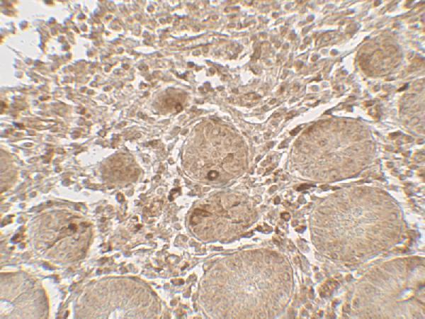
| Expected result | LAMTOR1-positive staining in glandular cells of adrenal gland (HPA tissue IHC: High). HPA tissue profile: General cytoplasmic expression. No signal in the no-primary control. |
LAMTOR1 is anchored to lysosome and late endosome membranes by N-terminal lipid modifications and has no transmembrane segment (UniProt Q6IAA8 topology). In paraffin sections, expect a generally cytoplasmic staining profile, with high staining reported in several glandular cell populations and cerebral cortex neurons (HPA: Approved tissue IHC; High in listed cells). HPA reports high consistency between antibody staining and RNA expression (HPA: Approved reliability).
| Granular cytoplasmic staining in glandular cells of colon, duodenum or appendix, with little nuclear staining. | This fits the reported high glandular-cell staining and general cytoplasmic profile (HPA: High in listed glandular cells; general cytoplasmic expression). Granularity is compatible with LAMTOR1’s endosomal and lysosomal membrane location, but chromogenic IHC alone cannot identify individual organelles (UniProt Q6IAA8 localization; standard IHC interpretation). |
| Predominantly nuclear staining, with little cytoplasmic signal in a listed positive cell population. | Treat this as a compartment mismatch requiring review: the expected tissue profile is cytoplasmic, while UniProt places LAMTOR1 on lysosome and late endosome membranes (HPA: general cytoplasmic expression; UniProt Q6IAA8 localization). Recheck staining and controls before assigning a nuclear LAMTOR1 pattern (standard IHC practice). |
| Strong staining in adipocytes or vaginal squamous epithelial cells. | These cell populations are reported as not detected, so strong staining raises concern for nonspecific antibody binding or endogenous detection activity (HPA: Not detected in adipocytes and vaginal squamous epithelial cells; standard IHC practice). A ‘not detected’ HPA result is a reference observation, not proof that every specimen must be negative (HPA: tissue IHC). |
| Uniform color across cells and extracellular areas, obscuring cell boundaries. | This does not match the reported cell-associated cytoplasmic profile (HPA: general cytoplasmic expression). Consider nonspecific background from detection reagents, inadequate blocking or incomplete washing; compare a control processed without primary antibody (standard chromogenic IHC practice). |
| No signal in colon or cerebral cortex despite visible, intact tissue. | Colon glandular cells and cerebral cortex neurons are listed as high-staining references (HPA: High in colon glandular cells and cerebral cortex neurons). A blank result warrants checking the assay and tissue quality before interpreting the specimen as LAMTOR1-negative; the HPA record does not establish target-specific fixation sensitivity (HPA: tissue IHC; standard IHC practice). |
| Membrane association and optical resolution | LAMTOR1 reaches lysosome and late endosome membranes through N-terminal myristoylation and palmitoylation and has no transmembrane segment (UniProt Q6IAA8 topology). A cytoplasmic chromogenic pattern can agree with this location, but it does not resolve membrane identity by itself (standard IHC interpretation). |
| Choice of tissue reference | High staining is reported in several glandular populations and cerebral cortex neurons; adipocytes and vaginal squamous epithelial cells are reported as not detected (HPA: tissue IHC). Compare signal within the specified cell populations, since a tissue name alone does not describe every cell in a section (standard histology practice). |
| Antibody validation scope | The listed antibody HPA002997 is Approved for IHC and Uncertain for ICC (HPA: antibody validation). The tissue profile has Approved reliability, described as high consistency between staining and RNA expression (HPA: tissue IHC reliability). Neither designation makes every unexpected staining pattern target-specific (standard IHC interpretation). |
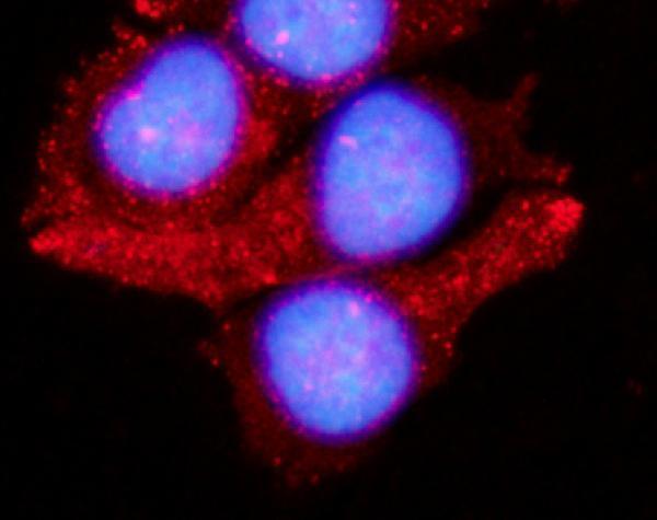
| IF/ICC Q: Should its image look identical to IHC? | No fixed match is established: HPA reports a mainly Golgi ICC-IF location as uncertain, plus vesicle and plasma membrane locations as supported (HPA: subcellular ICC-IF). UniProt places LAMTOR1 on lysosome and late endosome membranes (UniProt Q6IAA8 localization). Interpret these assay-specific observations with their stated confidence (HPA: antibody ICC Uncertain). |
| Target-specific fixation sensitivity | No target-specific fixation effect is established by the supplied UniProt topology, modifications or HPA staining categories (UniProt Q6IAA8; HPA: tissue IHC). Antigen retrieval may be evaluated as a general paraffin-IHC workflow variable, without predicting that it will restore LAMTOR1 signal (standard IHC practice). |
| Situation | Likely cause | Next action |
|---|---|---|
| Expected positive glandular cells remain unstained. | The result conflicts with reported high staining in the chosen reference population, but its cause cannot be assigned from HPA alone (HPA: High in listed glandular cells). | Confirm cell identity and section integrity, then review primary-antibody application, retrieval and detection controls using the assay’s established conditions (standard paraffin-IHC practice). |
| Signal is chiefly nuclear. | A nuclear-dominant pattern conflicts with the HPA cytoplasmic profile and UniProt endolysosomal location (HPA: general cytoplasmic expression; UniProt Q6IAA8 localization). | Inspect the no-primary control and compare staining with a listed positive cell population; avoid scoring nuclear color as expected LAMTOR1 localization without independent support (standard IHC practice). |
| Adipocytes or vaginal squamous cells stain strongly. | Strong signal differs from HPA’s not-detected observations in these cells; nonspecific binding or endogenous detection activity is possible (HPA: Not detected in listed cells; standard IHC practice). | Check a no-primary control and the detection system’s background controls, then interpret the affected cells alongside a positive reference in the same run (standard IHC practice). |
| Brown color is widespread and masks cell detail. | Diffuse background can arise from nonspecific reagent binding, endogenous detection activity or insufficient washing (standard chromogenic IHC practice). It obscures the reported cytoplasmic profile (HPA: tissue IHC). | Review blocking, washes and control sections for the chosen chromogen and detection chemistry; score LAMTOR1 only where cell-associated signal can be distinguished from background (standard IHC practice). |
| A section shows weak signal only in ovarian stroma, smooth muscle or spleen red pulp. | These cell populations are reported as low staining, so a weak result there is less informative than a blank high-staining reference (HPA: Low in listed cells; High in listed glandular cells). | Compare with a high-staining glandular or neuronal reference before judging assay performance; identify the relevant cells rather than averaging color across the whole tissue (HPA: tissue IHC; standard histology practice). |
| ICC-IF images appear Golgi-centered while paraffin IHC appears cytoplasmic. | HPA calls the Golgi ICC-IF assignment uncertain and the antibody’s ICC validation Uncertain; its IHC tissue profile is Approved (HPA: subcellular ICC-IF; antibody validation; tissue IHC). | Keep the assay-specific confidence labels when interpreting the images. For the paraffin section, judge the reported cell-associated cytoplasmic pattern against IHC controls (HPA: general cytoplasmic expression; standard IHC practice). |
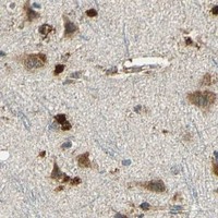
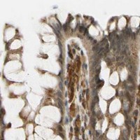
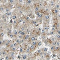
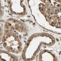


Comprehensive Human Protein Atlas IHC scoring per tissue (reliability: Approved — High consistency between antibody staining and RNA expression data.). Rows are taken directly from the HPA tissue chart — click any row's HPA link to view the source.
| Tissue | Cell type | Level | Evidence | Source |
|---|---|---|---|---|
| Adrenal gland | Glandular cells | High | Protein (IHC) | HPA → |
| Appendix | Glandular cells | High | Protein (IHC) | HPA → |
| Cerebral cortex | Neuronal cells | High | Protein (IHC) | HPA → |
| Cervix | Glandular cells | High | Protein (IHC) | HPA → |
| Colon | Glandular cells | High | Protein (IHC) | HPA → |
Troubleshoot LAMTOR1 staining in paraffin sections by checking retrieval, compartment pattern, controls, and scoring before comparing biological groups.
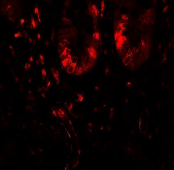
Anti-LAMTOR1 antibodies have real human small-intestine IHC and MCF-7 IF images (catalog figure tags/captions); listed reactivity covers human, mouse, rat, and monkey (catalog reactivity).
A06290 renders with human small-intestine IHC at 5 μg/mL (A06290 image caption) and lists IHC-P and IF with human, mouse, and rat reactivity (catalog applications/reactivity). A06290-1 renders with MCF-7 IF/ICC at 5 μg/mL (A06290-1 image caption) and lists IF/ICC with human, rat, and monkey reactivity (catalog applications/reactivity).
Which to pick: Choose A06290 for paraffin-section tissue IHC (catalog: IHC-P; A06290 image caption: human small intestine); the fixative is unreported (A06290 image caption). Choose A06290-1 for cell IF/ICC (catalog: IF/ICC; A06290-1 image caption: MCF-7). For cross-species planning, A06290 lists human, mouse, and rat, while A06290-1 lists human, rat, and monkey; clonality is unreported for both (catalog reactivity/clone fields).