LIF / Leukemia inhibitory factor · IHC design guide

Design Immunohistochemistry for LIF

Plan paraffin-section LIF IHC around the cytoplasmic staining reported in most tissues (HPA tissue IHC). Start with the catalog antibody’s 0.5–1 µg/mL range, and interpret staining in light of LIF secretion and the atlas’s uncertain reliability (datasheet PB9036; UniProt; HPA tissue IHC).

Evidence assembled Oct 2026 · For research use; verify linked source records and product datasheet before use
Immunohistochemistry protocol sheet for LIF (IHC for LIF): expected localisation Cytoplasmic staining in most tissues (HPA tissue IHC), antibody PB9036, validated IHC image, and IHC protocol steps
Printable LIF IHC protocol sheet — expected localisation Cytoplasmic staining in most tissues (HPA tissue IHC), antibody PB9036, controls and protocol steps. Open the full LIF IHC guide →

LIF Immunohistochemistry Experimental Design Guide

Expected localisation, validated protocols, controls and antibodies — the at-a-glance facts below, then the full design guide.

Must know before staining
Expected localisation Cytoplasmic staining in most tissues (HPA tissue IHC)
Staining pattern Cytoplasmic staining across many cell types (HPA tissue IHC)
Antigen retrieval Citrate pH 6 HIER, heat-mediated (datasheet PB9036)
Positive control ⓘ Adipose tissue+4 more · see all
Negative control ⓘ Smooth muscle
Important caveats
Reasons your staining may differ from the expected pattern.
Fixation Keep fixation consistent between sections (standard IHC practice; not target-specific)
Caveat Secreted LIF can make RNA and protein locations differ (HPA tissue IHC)
Regulation Expression regulation not annotated (UniProt)
Isoform / epitope 2 isoforms; epitope coverage is unspecified (UniProt)
Section 1

Recommended LIF IHC & IF Protocols

The catalog antibody’s IHC-P protocol is paired with four published LIF immunohistochemistry protocols covering rat uterus, mouse uterus, human oral tissue, and human liver (PMC3655613; PMC12911375; PMC12508188; PMC538256).

Recommended immunohistochemistry (IHC-P) protocol parameters
SampleParaffin-embedded human lung cancer tissues; fixative not specified (datasheet PB9036)
FixationImage fixative and duration unreported (datasheet PB9036); verify before use.
Sectioning4–5 µm sections on charged slides (standard)
DeparaffinisationXylene, graded ethanol series to water (standard)
Antigen retrievalHeat retrieval: Citrate pH 6, 20 min (datasheet PB9036)
Peroxidase block3% H2O2, 10 min, room temperature (standard)
Blocking10% goat serum (datasheet PB9036)
Primary antibodyRabbit anti-LIF, 0.5-1μg/ml (datasheet PB9036)
Primary incubationOvernight at 4 °C (datasheet PB9036)
DetectionStreptavidin-biotin complex (SABC), DAB chromogen (datasheet PB9036)
CounterstainHematoxylin, blue, dehydrate and mount (standard)
Expected resultLIF-positive staining in adipocytes of adipose tissue (HPA tissue IHC: Medium). HPA tissue profile: Cytoplasmic expression in most tissues. No signal in the no-primary control.
💡Decision noteStart with citrate pH 6 heat retrieval for the catalog antibody (datasheet PB9036); the published protocols used their stated retrieval methods with different antibodies (PMC12911375; PMC12508188).
Section 2

What Is the Expected LIF Staining Pattern?

LIF is secreted and has no transmembrane segment (UniProt P15018 topology). In tissue sections, HPA reports cytoplasmic staining in most tissues, including medium staining in adipocytes and selected glandular, epithelial, neuronal and endothelial cells (HPA tissue IHC). Treat this as a provisional pattern: HPA rates its tissue IHC profile Uncertain because secreted protein can appear away from its site of production, and retesting is pending (HPA tissue IHC reliability).

What am I looking at on my slide?
Cytoplasmic staining in adipocytes or listed glandular, epithelial, neuronal or endothelial cells.Consistent with the reported tissue pattern; HPA records medium staining in these listed cell types (HPA tissue IHC). Score cell identity and intensity together, while retaining the Uncertain reliability rating (HPA tissue IHC reliability).
Predominantly nuclear or sharply membrane-restricted staining.Neither is the reported main pattern: HPA describes cytoplasmic tissue staining and supported cytosolic ICC-IF localization (HPA tissue IHC; HPA subcellular). Recheck morphology and detection background before assigning LIF positivity; LIF has no transmembrane segment (UniProt P15018 topology).
Strong staining of smooth muscle cells while expected positive cells remain weak.This conflicts with HPA’s Not detected result for smooth muscle cells (HPA tissue IHC). Consider cross-reactivity or endogenous chromogenic detection activity as possibilities, then compare with an appropriate reagent control (general IHC practice).
Diffuse color across stroma, lumens or tissue edges without identifiable positive cells.Do not equate extracellular color with a producing cell: LIF is secreted, so protein and source may differ (UniProt P15018 localization; HPA tissue IHC reliability). Test whether the color persists in a detection-only control (general IHC practice).
No staining in a section expected to contain HPA-listed medium-positive cells.An absent result can reflect assay performance or sampling; the listed staining is an uncertain reference, not a guaranteed control outcome (HPA tissue IHC; HPA tissue IHC reliability). Confirm that the expected cell population is present before changing the staining conditions (general IHC practice).
💡Expected LIF appearanceCall a plausible positive when identifiable HPA-listed cells show medium cytoplasmic staining; predominantly nuclear color, smooth muscle staining or cell-free diffuse color warrants scrutiny (HPA tissue IHC; HPA tissue IHC reliability).
How each factor affects the staining
Secreted protein and processingLIF has a signal peptide at residues 1–22 and a mature chain at 23–202 (UniProt P15018 processing). Its secreted status complicates assignment of extracellular staining to a producing cell (UniProt P15018 localization; HPA tissue IHC reliability).
Tissue choiceAdipocytes, several listed glandular cells, respiratory epithelium, neurons and colon endothelium are medium-positive examples; smooth muscle cells are Not detected (HPA tissue IHC). Verify that the relevant cells occur in the chosen section (general IHC practice).
Antibody evidenceHPA018844 has Uncertain IHC validation; CAB025985 has no IHC status in the supplied HPA record (HPA antibodies). HPA’s tissue profile is also Uncertain, so concordant morphology should not be treated as definitive target validation (HPA tissue IHC reliability).
Isoforms and glycosylationUniProt lists 2 isoforms and 6 glycosylation sites (UniProt P15018 isoforms; glycosylation). The supplied sources do not map antibody epitopes, so they cannot predict an isoform-specific or glycosylation-dependent IHC pattern.
IF/ICC Q&A: expected compartment?HPA reports mainly cytosolic localization in ICC-IF and Supported ICC status for both listed antibodies (HPA subcellular; HPA antibodies). This informs compartment interpretation only; the primary application here is tissue IHC.
Why is my staining missing, weak or wrong?
SituationLikely causeNext action
A listed medium-positive cell population shows no color.The population may be absent from the cut, or the IHC run may have failed; HPA’s medium result remains Uncertain (HPA tissue IHC reliability).Check the section’s cell identity and run controls first. If controls fail, review retrieval, primary dilution and chromogenic detection as general IHC workflow variables (general IHC practice); no LIF-specific retrieval condition is established here.
Smooth muscle cells stain as strongly as the intended cells.HPA reports smooth muscle cells as Not detected, making this an unexpected pattern (HPA tissue IHC). Cross-reactivity or endogenous detection activity is possible (general IHC practice).Compare with a section processed without primary antibody and assess whether the signal follows tissue morphology (general IHC practice). Do not score smooth muscle color as validated LIF expression from this record.
Nuclei dominate the signal.A mainly nuclear pattern conflicts with HPA’s cytoplasmic tissue profile and supported cytosolic ICC-IF location (HPA tissue IHC; HPA subcellular).Check the counterstain and detection-only control, then reassess compartment boundaries (general IHC practice). Keep the result provisional if nuclear color remains dominant.
Broad extracellular haze obscures cell boundaries.Secreted LIF can complicate assignment of protein to a source cell (UniProt P15018 localization; HPA tissue IHC reliability); nonspecific chromogenic background is another possibility (general IHC practice).Compare primary-omission and tissue controls, then score only clearly localized cellular staining (general IHC practice). Record extracellular color separately rather than assigning it to nearby cells.
Only low-level color appears in a proposed reference tissue.HPA reports Low staining for several cell populations, including duodenal and rectal glandular cells; they provide a weaker reference than its listed medium-positive cells (HPA tissue IHC).Use an HPA-listed medium-positive cell population for a clearer comparison, while checking cell identity and the Uncertain reliability rating (HPA tissue IHC; HPA tissue IHC reliability).
ICC-IF appears cytosolic but tissue IHC is ambiguous.Supported ICC localization and Uncertain tissue IHC answer different questions (HPA subcellular; HPA antibodies; HPA tissue IHC reliability). Agreement in compartment alone cannot settle tissue specificity.Interpret the tissue slide by its cell types, compartment and IHC controls (general IHC practice); retain HPA’s Uncertain tissue rating when reporting the result (HPA tissue IHC reliability).

Sample controls for LIF IHC & IF

🧪Run bronchus first and look for staining in respiratory epithelial cells (HPA: Medium in bronchus respiratory epithelial cells). Run smooth muscle as the negative tissue (HPA: Not detected in smooth muscle cells); on the bronchus slide, use cells without discernible signal as an internal background reference, without assuming a particular cell type is LIF-negative.
Positive control tissue: Adipose tissue (Adipocytes, HPA Medium)
Negative control tissue: Smooth muscle (HPA Not detected)
ICC-IF cell lines (HPA subcellular resource): HPA ICC-IF images show LIF in A-431, U-251MG, U2OS, with annotated localisation: Cytosol (supported) (HPA subcellular).
Technical controls: Include a no-primary (secondary-only) control, a concentration-matched rabbit IgG isotype control, and a matched LIF knockout specimen if available (caption: rabbit anti-LIF primary). For chromogenic detection, quench endogenous peroxidase and check endogenous biotin background because the illustrated method uses a biotinylated secondary, streptavidin–biotin complex, and DAB (caption: PB9036 IHC).
⚠️Feasibility: No target-specific fixation window or fixation effect is reported, and the selected PB9036 paraffin-section caption does not state a fixative (caption: fixative not stated). Heat retrieval in citrate buffer at pH 6 for 20 minutes is documented, but retrieval dependency and whether frozen sections or IF are easier are unreported (caption: PB9036 IHC). In bronchus, inspect luminal mucus for trapped chromogen before scoring epithelial staining (HPA: Medium in bronchus respiratory epithelial cells).

HPA tissue IHC evidence for LIF

Comprehensive Human Protein Atlas IHC scoring per tissue (reliability: Uncertain — At least one protein variant secreted, tissue location of RNA and protein might differ and correlation is complex. Pending retesting.). Rows are taken directly from the HPA tissue chart — click any row's HPA link to view the source.

Positive expression · recommended positive controls

TissueCell typeLevelEvidenceSource
Adipose tissue Adipocytes Medium Protein (IHC) HPA →
Adrenal gland Glandular cells Medium Protein (IHC) HPA →
Appendix Glandular cells Medium Protein (IHC) HPA →
Breast Adipocytes Medium Protein (IHC) HPA →
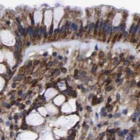
Bronchus Respiratory epithelial cells Medium Protein (IHC) HPA →

Undetected expression · recommended negative controls

TissueCell typeLevelEvidenceSource
Smooth muscle Smooth muscle cells Not detected Protein (IHC) HPA →
Section 3

Advanced LIF IHC Tips

Troubleshoot LIF staining in paraffin sections by checking retrieval, cellular pattern, detection controls, and scoring before assigning biological meaning.

What retrieval should I use when LIF staining is weak in paraffin sections?
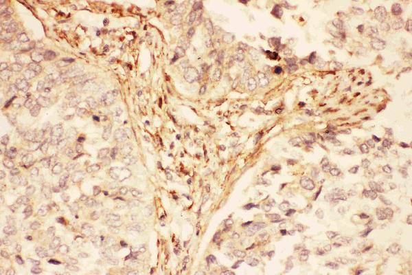
Start with heat-mediated retrieval in citrate buffer at pH 6 for 20 minutes (datasheet PB9036). The selected image used this treatment on a paraffin section of human lung cancer tissue, but its caption does not state the fixative (datasheet PB9036). Keep heating and cooling conditions consistent across cases, then compare staining with a matched positive control section processed in the same run (standard IHC practice). If signal remains weak, test another retrieval condition on adjacent sections as a fallback, while holding antibody and detection conditions constant (standard IHC practice). Assess tissue integrity and background alongside signal; stronger color alone does not establish improved LIF specificity (standard IHC practice).
Could fixation explain weak or uneven LIF staining?
Target-specific fixation sensitivity for LIF is unknown from the supplied evidence; the selected paraffin-section caption does not report a fixative (datasheet PB9036). Record the actual fixative and fixation duration for each specimen, then compare similarly handled sections before attributing different staining intensities to LIF expression (standard IHC practice). Overfixation can reduce epitope accessibility, while poor fixation can impair tissue morphology and produce uneven staining (standard IHC practice). Examine a control section from the same processing batch and inspect edges, centers, and damaged regions separately (standard IHC practice). Do not infer a LIF-specific fixation effect from cytoplasmic tissue staining or its secreted topology (HPA tissue IHC; UniProt P15018).
Where should convincing LIF staining appear within the section?
Expect predominantly cytoplasmic staining in the reported tissue pattern, and evaluate it in the context of cell identity and morphology (HPA tissue IHC). LIF is a secreted protein with a signal peptide at residues 1–22 and no transmembrane segment, so a sharp, exclusive plasma-membrane pattern needs independent validation (UniProt P15018). Supported cytosolic localisation in cell imaging makes intracellular signal plausible, while extracellular staining may require separate assessment because secreted protein can leave its producing cell (HPA subcellular; UniProt P15018). Compare stained cells with neighboring cells and a matched negative control; avoid assigning diffuse deposits or tissue-edge color to a cellular compartment (standard IHC practice).
Could LIF processing or glycosylation change what this antibody detects?
LIF has 2 annotated isoforms, a signal peptide at residues 1–22, and a mature chain spanning residues 23–202 (UniProt P15018). It also has 6 annotated glycosylation sites, which makes epitope accessibility a reasonable question, but the supplied evidence does not identify this antibody’s binding site (UniProt P15018; datasheet PB9036). Do not claim isoform selectivity or a glycosylation-dependent staining effect without direct antibody validation (standard IHC practice). If patterns conflict between specimens, compare staining under identical retrieval and detection conditions, then use an independent LIF-specific assay or antibody with a documented epitope to investigate the difference (standard IHC practice).
How can IF help check an ambiguous chromogenic LIF pattern?
Use IF as an orthogonal localisation check when a DAB pattern is hard to assign to individual cells (standard IF practice). Multiplex LIF with a validated marker for the suspected cell population, such as respiratory epithelial cells when examining bronchus, where that population has reported LIF staining (HPA tissue IHC). Choose spectrally separated fluorophores and assess unstained tissue autofluorescence before interpreting weak signal, particularly in the channel selected for LIF (standard IF practice). If assessing intracellular LIF, optimise permeabilisation for access to the cytosolic epitope; if assessing extracellular LIF, compare a condition without permeabilisation (HPA subcellular; UniProt P15018; standard IF practice). Keep IF conclusions distinct from the catalog antibody’s paraffin-section evidence (datasheet PB9036).
How do I distinguish LIF signal from DAB background?
The selected tissue image used 10% goat serum blocking, 1 µg/mL primary antibody overnight at 4°C, and biotin-based detection with DAB (datasheet PB9036). Run a no-primary control and inspect whether color persists in the same structures; persistent color points to detection background rather than primary-antibody-dependent staining (standard IHC practice). Include a peroxidase block before DAB development and consider endogenous biotin when using a biotin-based system (standard IHC practice). If background remains high, titrate primary antibody and adjust blocking or wash conditions one variable at a time (standard IHC practice). Judge improvement by clearer cellular localisation, not simply lower overall color (HPA tissue IHC; standard IHC practice).
What is a defensible way to score LIF IHC across specimens? ⚠ ANSWER MARKED FOR VERIFICATION
Define the cell population and tissue region before scoring, because reported LIF staining varies by cell type and HPA rates its tissue IHC reliability as uncertain (HPA tissue IHC). For intracellular staining, record the percentage of positive cells and an intensity-based H-score using the same thresholds and imaging settings across sections (standard IHC practice). For a sparse positive population, report positive cells per mm² alongside the counted cell population (standard IHC practice). Normalise counts to viable tissue area or the number of eligible cells, excluding folds, necrosis, and tissue edges by predefined rules (standard IHC practice). Include control-section performance and scorer agreement when comparing groups (standard IHC practice).
When should a LIF-positive pattern be treated as questionable?
Treat a convincing result as cellular staining in an identifiable population with acceptable morphology and a clean no-primary control (standard IHC practice). Cytoplasmic staining fits the reported tissue profile, while exclusive nuclear or sharply membrane-restricted staining needs independent confirmation because LIF is secreted and lacks a transmembrane segment (HPA tissue IHC; UniProt P15018). Check whether the stained cell type is plausible for that specimen; HPA reports medium staining in bronchial respiratory epithelial cells but no detection in smooth muscle cells (HPA tissue IHC). Exclude edge effects, necrotic regions, and endogenous enzyme color before scoring (standard IHC practice). Interpret apparent discordance cautiously because HPA labels its tissue IHC reliability uncertain for this secreted protein (HPA tissue IHC).
Boster reagents

Best LIF / Leukemia inhibitory factor IHC Antibodies

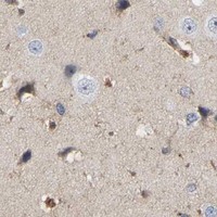
PB9036 has IHC images from human lung cancer and placenta paraffin sections (catalog PB9036 IHC captions); the payload supplies no IF/ICC images (catalog PB9036 image fields).

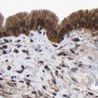
Real IHC data IHC analysis of LIF using anti-LIF antibody (PB9036). LIF was detected in paraffin-embedded section of human lung cancer tissues. Heat mediated antigen retrieval was performed in citrate buffer (pH6, epitope retrieval solution) for 20 mins. The tissue section was blocked with 10% goat serum. The tissue section was then incubated with 1μg/ml rabbit anti-LIF Antibody (PB9036) overnight at 4°C. Biotinylated goat anti-rabbit IgG was used as secondary antibody and incubated for 30 minutes at 37°C. The tissue section was developed using Strepavidin-Biotin-Complex (SABC)(Catalog # SA1022) with DAB as the chromogen.
Anti-LIF Antibody ®
Cat # PB9036

PB9036 is listed for human IHC and WB (catalog PB9036 applications and reactivity). Its IHC captions show staining in paraffin sections of human lung cancer and placenta (catalog PB9036 IHC captions).

Which to pick: Choose PB9036 for human paraffin-section IHC: its captions document citrate retrieval and chromogenic detection, but do not report the fixative (catalog PB9036 IHC captions). No SKU in this payload is listed for IF/ICC or reactivity beyond human, so it does not establish an IF/ICC or cross-species choice (catalog PB9036 applications and reactivity).

Each figure is that product's own IHC / IF validation image from its datasheet.

References

  1. UniProt Consortium. UniProt entry P15018 (LIF_HUMAN, Leukemia inhibitory factor).
  2. Human Protein Atlas. LIF tissue IHC expression (reliability: Uncertain).
  3. Human Protein Atlas. LIF subcellular location (ICC-IF): Mainly localized to the cytosol..
  4. Human Protein Atlas. LIF antibody validation summary (2 antibodies).
  5. Effects of "Bu Shen Huo Xue Decoction" on the Endometrial Morphology and Expression of Leukaemia Inhibitory Factor in the Rat Uterus during the Oestrous Cycle. Evidence-based complementary and alternative medicine : eCAM 2013 — PMC3655613.
  6. Investigation of the effects of recombinant and urinary FSH used for ovulation induction on pregnancy results in an experimental animal model in terms of endometrial receptivity markers. BMC pregnancy and childbirth 2026 — PMC12911375.
  7. Immunohistochemical expression pattern of leukemia inhibitory factor and its receptor may mark a tipping point in the progression of oral cancer. Scientific reports 2025 — PMC12508188.
  8. Expression of leukemia inhibitory factor (LIF) and its receptor gp190 in human liver and in cultured human liver myofibroblasts. Cloning of new isoforms of LIF mRNA. Comparative hepatology 2004 — PMC538256.
  9. PubMed PMID:3143918 — UniProt-cited evidence.
  10. PubMed PMID:2475312 — UniProt-cited evidence.
  11. PubMed PMID:1692837 — UniProt-cited evidence.