This website uses cookies to ensure you get the best experience on our website.


- Table of Contents
Plan LMAN1 IHC in paraffin sections around the cytoplasmic staining reported across tissues (HPA tissue IHC). Compare high-staining glandular or endocrine cells with low-staining squamous epithelia, using matched fixation and cellular intensity scoring (HPA tissue IHC; standard IHC practice).

Expected localisation, validated protocols, controls and antibodies — the at-a-glance facts below, then the full design guide.
| Expected localisation | Tissue cytoplasm (HPA tissue IHC); ER/ERGIC/Golgi membranes (UniProt) | |
| Staining pattern | Cytoplasmic staining across tissues (HPA tissue IHC) | |
| Antigen retrieval | EDTA pH 8.0 HIER, heat-mediated (datasheet A03628-1) | |
| Positive control | Adrenal gland+4 more · see all | |
| Negative control | None in HPA (detected in all 45 tissues); use no-primary + isotype controls |
| Fixation | Keep fixation consistent across sections (standard IHC practice; not target-specific); Matched tissue-IHC evidence does not establish the fixation claim. Validate the specimen-specific method before use. (selected-SKU IHC image A03628-1) | |
| Caveat | Low staining in some squamous epithelia despite broad expression (HPA tissue IHC) | |
| Regulation | Low tissue specificity (HPA tissue RNA) | |
| Isoform / epitope | No isoforms listed; cleaved signal; luminal vs cytoplasmic epitopes matter (UniProt) |
Compare the catalog antibody’s IHC-P protocol (datasheet A03628-1) with a published LMAN1 IHC method using paraffin-embedded animal organ sections (PMC13011374).
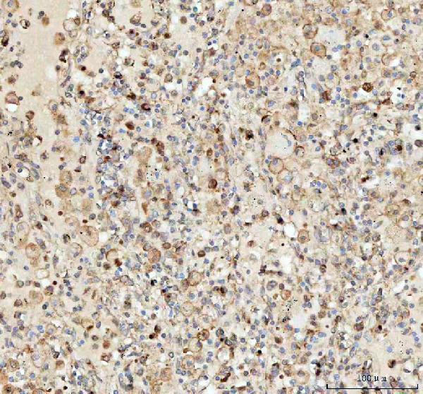
| Sample | Paraffin-embedded human lung cancer tissue; fixative not specified (datasheet A03628-1) |
| Fixation | Image fixative and duration unreported (datasheet A03628-1); verify before use. |
| Sectioning | 4–5 µm sections on charged slides (standard) |
| Deparaffinisation | Xylene, graded ethanol series to water (standard) |
| Antigen retrieval | Heat retrieval: EDTA pH 8.0 (datasheet A03628-1); 20 min, 95–100 °C (standard) |
| Peroxidase block | 3% H2O2, 10 min, room temperature (standard) |
| Blocking | 10% goat serum (datasheet A03628-1) |
| Primary antibody | Rabbit anti-LMAN1, 2-5μg/ml (datasheet A03628-1) |
| Primary incubation | Overnight at 4 °C (datasheet A03628-1) |
| Detection | HRP-conjugated secondary, DAB chromogen (datasheet A03628-1) |
| Counterstain | Hematoxylin, blue, dehydrate and mount (standard) |
| Expected result | LMAN1-positive staining in glandular cells of adrenal gland (HPA tissue IHC: High). HPA tissue profile: Cytoplasmic expression in all tissues. No signal in the no-primary control. |
LMAN1 is an ER–Golgi trafficking protein in ER–Golgi intermediate compartment, Golgi, and ER membranes, with one transmembrane segment and a lumenal domain (UniProt P49257 localization and topology). On paraffin sections, expect cytoplasmic staining across tissues, with high staining reported in selected glandular and endocrine cells (HPA tissue IHC). HPA rates the tissue pattern Supported, with medium consistency between staining and RNA expression (HPA tissue IHC).
| Cytoplasmic staining in glandular or endocrine cells, with stronger signal in an HPA high-staining tissue. | Consistent with LMAN1 IHC: HPA reports cytoplasmic expression across tissues and High staining in adrenal glandular and colon endocrine cells (HPA tissue IHC). Judge intensity against cells on the same slide; high staining in one cell group does not require equal intensity in every cell (HPA tissue IHC). |
| Signal appears predominantly nuclear, or outlines the cell surface without appreciable cytoplasmic staining. | This conflicts with the reported cytoplasmic tissue pattern and intracellular membrane locations (HPA tissue IHC; UniProt P49257 localization). Treat it as suspect and compare with an HPA high-staining tissue before assigning it to LMAN1 (general IHC practice). |
| A cell population stains intensely while expected glandular or endocrine cells remain unstained. | Consider antibody cross-reactivity or endogenous detection activity (general IHC practice). LMAN1 is broadly expressed, so staining outside one favored cell type alone is insufficient evidence of error (UniProt P49257 tissue specificity; HPA tissue IHC). Assess compartment and detection controls together. |
| Broad, diffuse chromogen obscures cell boundaries and the expected intracellular pattern. | A pattern that cannot be assigned to cells or cytoplasm is uninterpretable (general IHC practice). HPA reports cytoplasmic staining, but its Supported rating has only medium consistency with RNA expression; diffuse background should not be counted as confirmed LMAN1 signal (HPA tissue IHC). |
| Little or no signal appears in adrenal glandular or colon endocrine cells. | Those are reported High-staining examples, so absence raises concern about assay performance or specimen quality (HPA tissue IHC; general IHC practice). It does not, by itself, establish biological loss: HPA supplies no negative tissue reference here, and its tissue assessment is Supported with medium consistency (HPA tissue IHC). |
| Intracellular location and topology | LMAN1 occupies ER–Golgi intermediate compartment, Golgi, and ER membranes; residues 31–477 are lumenal and 499–510 cytoplasmic (UniProt P49257 topology). An antibody's epitope is not supplied, so topology alone cannot predict its staining intensity or retrieval response. |
| Tissue and cell context | HPA describes cytoplasmic expression in all tissues and low tissue specificity, with High staining in selected glandular and endocrine cells and Low staining in listed populations such as smooth muscle cells (HPA tissue IHC). Low staining is a poor substitute for a documented negative control. |
| Antibody evidence | HPA lists IHC as Supported for HPA002320 and CAB037163; its overall tissue assessment is Supported with medium staining–RNA consistency (HPA antibodies; HPA tissue IHC). These ratings support pattern comparison without guaranteeing specificity for every specimen. |
| IF/ICC: what localization is reported? | Mainly vesicles, with additional ER localization, are Supported in HPA ICC-IF (HPA subcellular). This answers the localization question; paraffin chromogenic IHC should be interpreted against HPA's tissue cytoplasmic pattern (HPA tissue IHC). |
| Situation | Likely cause | Next action |
|---|---|---|
| Reported High-staining cells show no chromogen. | A failed detection step or insufficient assay signal is possible (general IHC practice); the missing signal does not prove LMAN1 absence. | Check the IHC-validated antibody's documented dilution and detection conditions, then repeat with a reported High-staining tissue (general IHC practice; HPA tissue IHC). |
| Signal is mainly nuclear or confined to cell edges. | The distribution disagrees with cytoplasmic tissue staining and ER–Golgi or ER membrane localization (HPA tissue IHC; UniProt P49257 localization). | Review morphology and counterstain, and compare the compartment in a reported High-staining tissue before scoring positivity (general IHC practice; HPA tissue IHC). |
| Chromogen appears where the primary antibody was omitted. | Endogenous detection activity or reagent background may contribute signal (general IHC practice). | Address endogenous activity using controls and detection-system guidance; score only signal above the omission control (general IHC practice). |
| Most of the section has uniform haze. | Nonspecific background may hide cell-level cytoplasmic staining (general IHC practice; HPA tissue IHC). | Review blocking, antibody concentration, washes, and chromogen development; reassess cell boundaries and cytoplasm after background improves (general IHC practice). |
| Only a low-staining reference population is available. | HPA lists some Low populations but no negative tissue; a weak result there cannot establish assay failure or specificity (HPA tissue IHC). | Add a reported High-staining glandular or endocrine example for pattern comparison and retain an omission control for detection background (HPA tissue IHC; general IHC practice). |
| Staining varies between otherwise comparable paraffin sections. | Section handling or assay conditions may vary (general IHC practice). No LMAN1-specific fixation sensitivity is established by these sources. | Compare processing records and run sections together under the documented IHC conditions; avoid attributing variation specifically to LMAN1 epitope masking without evidence (general IHC practice). |


Comprehensive Human Protein Atlas IHC scoring per tissue (reliability: Supported — Medium consistency between antibody staining and RNA expression data.). Rows are taken directly from the HPA tissue chart — click any row's HPA link to view the source.
| Tissue | Cell type | Level | Evidence | Source |
|---|---|---|---|---|
| Adrenal gland | Glandular cells | High | Protein (IHC) | HPA → |
| Appendix | Endocrine cells | High | Protein (IHC) | HPA → |
| Breast | Glandular cells | High | Protein (IHC) | HPA → |
| Colon | Endocrine cells | High | Protein (IHC) | HPA → |
| Duodenum | Endocrine cells | High | Protein (IHC) | HPA → |
| Tissue | Cell type | Level | Evidence | Source |
|---|---|---|---|---|
| None in HPA: LMAN1 is detected in all 45 scored tissues. Use a no-primary (secondary-only) and an isotype control instead. | ||||
Troubleshoot LMAN1 chromogenic IHC by checking retrieval, compartment-specific staining, controls and scoring; IF considerations are covered separately below.
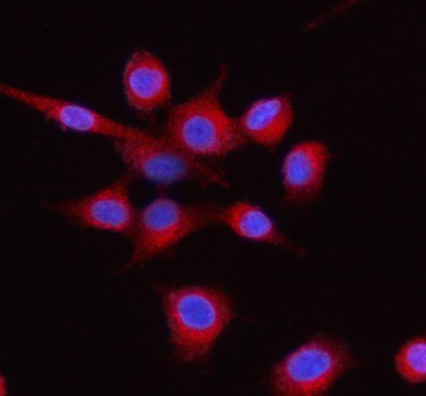
Catalog antibodies list IHC and IF/ICC for human, mouse, and rat (catalog: applications/reactivity); supplied IF images show A549 cells and human paraffin tissue (A03628-1 IF captions).
A03628-1 is listed for IHC and IF/ICC in human, mouse, and rat (catalog: applications/reactivity), with IF images of A549 cells and human paraffin placenta and ovarian cancer sections (A03628-1 IF captions). M03628-1 and M03628-3 are rabbit monoclonals listed for those applications and species (catalog: host/clone/applications/reactivity), but neither has a supplied IHC or IF image (catalog: image alts).
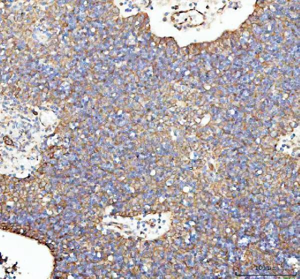
Which to pick: For tissue IHC, A03628-1 lists 2–5 μg/ml (catalog: IHC dilution); its IHC captions describe human paraffin lung cancer and spleen sections but identify a different SKU, so confirm the figure attribution before relying on those images (IHC captions). The captions report no fixative (IHC captions). For IF/ICC, A03628-1 has direct image evidence (A03628-1 IF captions); M03628-1 and M03628-3 are rabbit monoclonal alternatives listed for human, mouse, and rat, without supplied images demonstrating cross-species staining (catalog: host/clone/reactivity/applications/image alts).