Click image to see more details
-

-
-
-
-
+3
Product Info Summary
| SKU: | A03628-1 |
|---|---|
| Size: | 100 μg/vial |
| Reactive Species: | Human, Mouse, Rat |
| Host: | Rabbit |
| Application: | ELISA, IF, IHC, ICC, WB |
Customers Who Bought This Also Bought

Product info
Product Name
Anti-LMAN1 Antibody Picoband®
SKU/Catalog Number
A03628-1
Size
100 μg/vial
Form
Lyophilized
Description
Boster Bio Anti-LMAN1 Antibody Picoband® catalog # A03628-1. Tested in ELISA, IF, IHC, ICC, WB applications. This antibody reacts with Human, Mouse, Rat. The brand Picoband indicates this is a premium antibody that guarantees superior quality, high affinity, and strong signals with minimal background in Western blot applications. Only our best-performing antibodies are designated as Picoband, ensuring unmatched performance.
Storage & Handling
Store at -20˚C for one year from date of receipt. After reconstitution, at 4˚C for one month. It can also be aliquotted and stored frozen at -20˚C for six months. Avoid repeated freeze-thaw cycles.
Cite This Product
Anti-LMAN1 Antibody Picoband® (Boster Biological Technology, Pleasanton CA, USA, Catalog # A03628-1)
Host
Rabbit
Contents
Each vial contains 4 mg Trehalose, 0.9 mg NaCl and 0.2 mg Na2HPO4.
Clonality
Polyclonal
Isotype
Rabbit IgG
Immunogen
E. coli-derived human LMAN1 recombinant protein (Position: K96-M458).
Cross-reactivity
No cross-reactivity with other proteins.
Reactive Species
A03628-1 is reactive to LMAN1 in Human, Mouse, Rat
Observed Molecular Weight
58 kDa
Calculated molecular weight
57.5 kDa
Background of LMAN1
Protein ERGIC-53 also known as ER-Golgi intermediate compartment 53 kDa protein or lectin mannose-binding 1 is a protein that in humans is encoded by the LMAN1 gene. It is mapped to 18q21.32. The protein encoded by this gene is a membrane mannose-specific lectin that cycles between the endoplasmic reticulum, endoplasmic reticulum-Golgi intermediate compartment, and cis-Golgi, functioning as a cargo receptor for glycoprotein transport. The protein has an N-terminal signal sequence, a calcium-dependent and pH-sensitive carbohydrate recognition domain, a stalk region that functions in oligomerization, a transmembrane domain, and a short cytoplasmic domain required for organelle targeting. Allelic variants of this gene are associated with the autosomal recessive disorder combined factor V-factor VIII deficiency.
Antibody Validation
Boster validates all antibodies on WB, IHC, ICC, Immunofluorescence, and ELISA with known positive control and negative samples to ensure specificity and high affinity, including thorough antibody incubations.
Application & Images
Applications
A03628-1 is guaranteed for ELISA, IF, IHC, ICC, WB Boster Guarantee
Recommend Dilution
| Application | Dilution | Species |
|---|---|---|
| Western blot | 0.1-0.5μg/ml | Human, Mouse, Rat |
| Immunohistochemistry (Paraffin-embedded Section) | 2-5μg/ml | Human |
| Immunocytochemistry/Immunofluorescence | 5μg/ml | Human |
| Immunofluorescence | 5μg/ml | Human |
| ELISA | 0.1-0.5μg/ml |
Tested application
Suggested blocking solution with 5% non-fat milk or BSA; (*)Recommended protein loading: 20-40 µg per lane
Use TE buffer pH 9.0 for antigen retrieval; (*) citrate buffer pH 6.0 is an alternative.
Validation Images & Assay Conditions
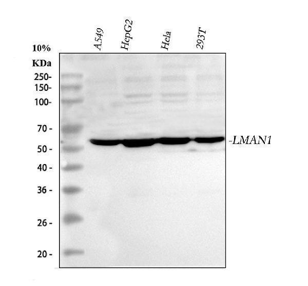
Click image to see more details
Western blot analysis of LMAN1 using anti-LMAN1 antibody (A03628-1).
Electrophoresis was performed on a 5-20% SDS-PAGE gel at 70V (Stacking gel) / 90V (Resolving gel) for 2-3 hours. The sample well of each lane was loaded with 30 ug of sample under reducing conditions.
Lane 1: human A549 whole cell lysates,
Lane 2: human HepG2 whole cell lysates,
Lane 3: human Hela whole cell lysates,
Lane 4: human 293T whole cell lysates.
After electrophoresis, proteins were transferred to a nitrocellulose membrane at 150 mA for 50-90 minutes. Blocked the membrane with 5% non-fat milk/TBS for 1.5 hour at RT. The membrane was incubated with rabbit anti-LMAN1 antigen affinity purified polyclonal antibody (Catalog # A03628-1) at 0.5 μg/mL overnight at 4°C, then washed with TBS-0.1%Tween 3 times with 5 minutes each and probed with a goat anti-rabbit IgG-HRP secondary antibody at a dilution of 1:5000 for 1.5 hour at RT. The signal is developed using an Enhanced Chemiluminescent detection (ECL) kit (Catalog # EK1002) with Tanon 5200 system. A specific band was detected for LMAN1 at approximately 58 kDa. The expected band size for LMAN1 is at 58 kDa.

Click image to see more details
Western blot analysis of LMAN1 using anti-LMAN1 antibody (A03628-1).
Electrophoresis was performed on a 5-20% SDS-PAGE gel at 70V (Stacking gel) / 90V (Resolving gel) for 2-3 hours. The sample well of each lane was loaded with 30 ug of sample under reducing conditions.
Lane 1: human A431 whole cell lysates,
Lane 2: human HepG2 whole cell lysates,
Lane 3: rat liver tissue lysates,
Lane 4: rat heart tissue lysates,
Lane 5: rat C6 whole cell lysates,
Lane 6: mouse liver tissue lysates,
Lane 7: mouse heart tissue lysates,
Lane 8: mouse heart whole cell lysates.
After electrophoresis, proteins were transferred to a nitrocellulose membrane at 150 mA for 50-90 minutes. Blocked the membrane with 5% non-fat milk/TBS for 1.5 hour at RT. The membrane was incubated with rabbit anti-LMAN1 antigen affinity purified polyclonal antibody (Catalog # A03628-1) at 0.5 μg/mL overnight at 4°C, then washed with TBS-0.1%Tween 3 times with 5 minutes each and probed with a goat anti-rabbit IgG-HRP secondary antibody at a dilution of 1:5000 for 1.5 hour at RT. The signal is developed using an Enhanced Chemiluminescent detection (ECL) kit (Catalog # EK1002) with Tanon 5200 system. A specific band was detected for LMAN1 at approximately 58 kDa. The expected band size for LMAN1 is at 58 kDa.

Click image to see more details
IHC analysis of LMAN1 using anti-LMAN1 antibody (A03768-1).
LMAN1 was detected in a paraffin-embedded section of human lung cancer tissue. Heat mediated antigen retrieval was performed in EDTA buffer (pH 8.0, epitope retrieval solution). The tissue section was blocked with 10% goat serum. The tissue section was then incubated with 2 μg/ml rabbit anti-LMAN1 Antibody (A03768-1) overnight at 4°C. Peroxidase Conjugated Goat Anti-rabbit IgG was used as secondary antibody and incubated for 30 minutes at 37°C. The tissue section was developed using HRP Conjugated Rabbit IgG Super Vision Assay Kit (Catalog # SV0002) with DAB as the chromogen.

Click image to see more details
IHC analysis of LMAN1 using anti-LMAN1 antibody (A03768-1).
LMAN1 was detected in a paraffin-embedded section of human spleen tissue. Heat mediated antigen retrieval was performed in EDTA buffer (pH 8.0, epitope retrieval solution). The tissue section was blocked with 10% goat serum. The tissue section was then incubated with 2 μg/ml rabbit anti-LMAN1 Antibody (A03768-1) overnight at 4°C. Peroxidase Conjugated Goat Anti-rabbit IgG was used as secondary antibody and incubated for 30 minutes at 37°C. The tissue section was developed using HRP Conjugated Rabbit IgG Super Vision Assay Kit (Catalog # SV0002) with DAB as the chromogen.

Click image to see more details
IF analysis of LMAN1 using anti-LMAN1 antibody (A03628-1).
LMAN1 was detected in an immunocytochemical section of A549 cells. Enzyme antigen retrieval was performed using IHC enzyme antigen retrieval reagent (AR0022) for 15 mins. The cells were blocked with 10% goat serum. And then incubated with 5 μg/mL rabbit anti-LMAN1 Antibody (A03628-1) overnight at 4°C. Cy3 Conjugated Goat Anti-Rabbit IgG (BA1032) was used as secondary antibody at 1:500 dilution and incubated for 30 minutes at 37°C. The section was counterstained with DAPI. Visualize using a fluorescence microscope and filter sets appropriate for the label used.

Click image to see more details
IF analysis of LMAN1 using anti-LMAN1 antibody (A03628-1).
LMAN1 was detected in a paraffin-embedded section of human placenta tissue. Heat mediated antigen retrieval was performed in EDTA buffer (pH 8.0, epitope retrieval solution). The tissue section was blocked with 10% goat serum. The tissue section was then incubated with 5 μg/mL rabbit anti-LMAN1 Antibody (A03628-1) overnight at 4°C. DyLight®488 Conjugated Goat Anti-Rabbit IgG (BA1127) was used as secondary antibody at 1:500 dilution and incubated for 30 minutes at 37°C. The section was counterstained with DAPI. Visualize using a fluorescence microscope and filter sets appropriate for the label used.

Click image to see more details
IF analysis of LMAN1 using anti-LMAN1 antibody (A03628-1).
LMAN1 was detected in a paraffin-embedded section of human ovarian cancer tissue. Heat mediated antigen retrieval was performed in EDTA buffer (pH 8.0, epitope retrieval solution). The tissue section was blocked with 10% goat serum. The tissue section was then incubated with 5 μg/mL rabbit anti-LMAN1 Antibody (A03628-1) overnight at 4°C. DyLight®488 Conjugated Goat Anti-Rabbit IgG (BA1127) was used as secondary antibody at 1:500 dilution and incubated for 30 minutes at 37°C. The section was counterstained with DAPI. Visualize using a fluorescence microscope and filter sets appropriate for the label used.
Specific Publications For Anti-LMAN1 Antibody Picoband® (A03628-1)
Loading publications
Recommended Resources
Here are featured tools and databases that you might find useful.
- Boster's Pathways Library
- Protein Databases
- Bioscience Research Protocol Resources
- Data Processing & Analysis Software
- Photo Editing Software
- Scientific Literature Resources
- Research Paper Management Tools
- Molecular Biology Software
- Primer Design Tools
- Bioinformatics Tools
- Phylogenetic Tree Analysis
Customer Reviews
Have you used Anti-LMAN1 Antibody Picoband®?
Share your experimental results or join a short interview to earn up to $1,000 in product credits or other rewards.
0 Reviews For Anti-LMAN1 Antibody Picoband®
Customer Q&As
Have a question?
Find answers in Q&As, reviews.
Can't find your answer?
Submit your question
1 Customer Q&As for Anti-LMAN1 Antibody Picoband®
Question
We are currently using anti-LMAN1 antibody A03628-1 for mouse tissue, and we are happy with the WB results. The species of reactivity given in the datasheet says human, mouse, rat. Is it likely that the antibody can work on dog tissues as well?
R. Johnson
Verified customer
Asked: 2013-08-23
Answer
The anti-LMAN1 antibody (A03628-1) has not been tested for cross reactivity specifically with dog tissues, though there is a good chance of cross reactivity. We have an innovator award program that if you test this antibody and show it works in dog you can get your next antibody for free. Please contact me if I can help you with anything.
Boster Scientific Support
Answered: 2013-08-23
