This website uses cookies to ensure you get the best experience on our website.


- Table of Contents
Plan chromogenic LNPEP IHC in paraffin sections with the IHC-validated antibody at 2–5 μg/mL (datasheet A05092-1). Assess cytoplasmic staining in glandular and neuronal cells, and consider the secreted form when interpreting extracellular signal (HPA tissue IHC; UniProt).

Expected localisation, validated protocols, controls and antibodies — the at-a-glance facts below, then the full design guide.
| Expected localisation | Cytoplasmic staining in most tissues; some plasma positivity (HPA tissue IHC) | |
| Staining pattern | Mostly cytoplasmic; plasma positivity in several tissues (HPA tissue IHC) | |
| Antigen retrieval | EDTA pH 8.0 HIER, heat-mediated (datasheet A05092-1) | |
| Positive control | Appendix+4 more · see all | |
| Negative control | None in HPA (detected in all 45 tissues); use no-primary + isotype controls |
| Fixation | Keep fixation consistent across sections. (standard IHC practice; not target-specific) | |
| Caveat | Secreted pregnancy serum form may add extracellular signal (UniProt) | |
| Regulation | Insulin/oxytocin shifts LNPEP to the surface (UniProt) | |
| Isoform / epitope | 3 isoforms; cytoplasmic epitopes miss soluble form (UniProt) |
The catalog antibody’s IHC protocol is paired with two published LNPEP protocols for equine paraffin sections (PMC8996865; PMC10030782).
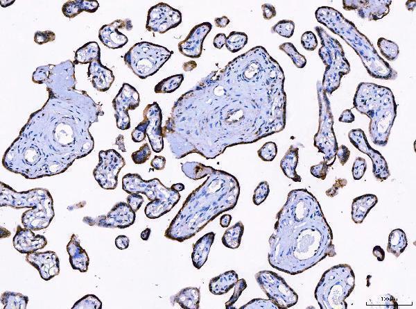
| Sample | Paraffin-embedded human placenta tissue; fixative not specified (datasheet A05092-1) |
| Fixation | Image fixative and duration unreported (datasheet A05092-1); verify before use. |
| Sectioning | 4–5 µm sections on charged slides (standard) |
| Deparaffinisation | Xylene, graded ethanol series to water (standard) |
| Antigen retrieval | Heat retrieval: EDTA pH 8.0 (datasheet A05092-1); 20 min, 95–100 °C (standard) |
| Peroxidase block | 3% H2O2, 10 min, room temperature (standard) |
| Blocking | 10% goat serum (datasheet A05092-1) |
| Primary antibody | Rabbit anti-LNPEP, 2-5 μg/ml (datasheet A05092-1) |
| Primary incubation | Overnight at 4 °C (datasheet A05092-1) |
| Detection | HRP-conjugated secondary, DAB chromogen (datasheet A05092-1) |
| Counterstain | Hematoxylin, blue, dehydrate and mount (standard) |
| Expected result | LNPEP-positive staining in glandular cells of appendix (HPA tissue IHC: High). HPA tissue profile: Cytoplasmic expression in most tissues with additional plasma positivity in several tissues. No signal in the no-primary control. |
Expect mainly cytoplasmic LNPEP staining across many tissues, with plasma positivity in several (HPA tissue IHC). LNPEP is a membrane protein whose 111–131 transmembrane segment separates a cytoplasmic N terminus from an extracellular C terminus; it also resides in intracellular vesicles and may reach the cell surface (UniProt Q9UIQ6 topology and subcellular location). HPA rates the tissue staining Approved but reports low consistency with RNA expression (HPA tissue IHC).
| Cytoplasmic signal in appendix or duodenal glandular cells. | This fits HPA's High staining in those cells and its broader cytoplasmic tissue pattern (HPA tissue IHC). Score the identified cells and their compartment alongside the chromogenic intensity; a dark field alone does not identify the stained cell (general IHC interpretation). |
| Predominantly nuclear signal, without the expected cytoplasmic pattern. | Nuclear staining is discordant with HPA's cytoplasmic tissue profile and UniProt's membrane and vesicle locations (HPA tissue IHC; UniProt Q9UIQ6 subcellular location). Treat it as suspect until controls and cell morphology support a specific signal; compartment mismatch alone cannot identify the artefact. |
| Strong signal in an unexpected cell population. | Compare cells within the same tissue: HPA reports High staining in bronchial basal cells and caudate neurons, but Low staining in thyroid glandular cells and liver cholangiocytes (HPA tissue IHC). Unexpected staining may reflect cross-reactivity or endogenous detection activity; morphology and controls must distinguish these explanations (general IHC practice). |
| Uniform brown haze over cells and surrounding tissue. | A diffuse field cannot be scored as LNPEP localisation merely because HPA describes cytoplasmic staining (HPA tissue IHC). Assess background with the primary antibody omitted, then review blocking, washing and chromogen development (general chromogenic IHC practice). |
| No signal in an appendix or duodenum section expected to stain. | HPA records High glandular-cell staining in both tissues, so an absent signal warrants a technical check (HPA tissue IHC). Confirm glandular cells are present, then check the antibody's IHC validation and the general staining workflow; HPA's low RNA–staining consistency limits any claim that every specimen must be positive (HPA tissue IHC). |
| Topology and compartment | LNPEP has a cytoplasmic 1–110 region, a 111–131 membrane span and an extracellular 132–1025 region (UniProt Q9UIQ6 topology). Epitope position would matter when interpreting surface versus intracellular access, but no epitope location is supplied for the antibody. |
| Vesicle trafficking | LNPEP resides in intracellular vesicles with GLUT4 and can translocate to the cell surface after insulin or oxytocin stimulation (UniProt Q9UIQ6 subcellular location). Cytoplasmic and membrane-associated appearances can therefore be biologically plausible; this record does not predict their proportions in a particular section. |
| Membrane and soluble forms | UniProt lists cell-membrane and secreted locations and a 155–1025 pregnancy-serum form; it reports only the membrane-bound form in brain (UniProt Q9UIQ6 subcellular location and processing). Extracellular-looking signal needs tissue context and controls before attribution to secreted LNPEP. |
| Tissue and cell context | HPA lists High staining in epididymal glandular cells and cerebral-cortex neurons, Medium staining in endometrial glandular cells, and Low staining in oral squamous epithelium (HPA tissue IHC). Use the named cell population when judging intensity; these categories do not define a universal cutoff. |
| Strength of staining evidence | HPA marks tissue staining Approved while noting low consistency with RNA expression; HPA043642 has Approved IHC status, with no Enhanced designation supplied (HPA tissue IHC; HPA antibody validation). The pattern is a useful reference, but a single staining result is insufficient to establish specificity. |
| IF/ICC evidence | HPA summarizes LNPEP as Membrane in ICC-IF but supplies no main location or cell-line images in this record (HPA subcellular ICC-IF). That summary cannot establish an IF pattern or conditions for this paraffin-section IHC assay. |
| Situation | Likely cause | Next action |
|---|---|---|
| Expected glandular cells are unstained. | The section may lack the relevant cells, or an IHC step may have failed; appendix and duodenal glandular cells are High in the HPA record (HPA tissue IHC). | Check morphology and a suitable positive section, then review the antibody's stated IHC conditions, retrieval, detection and chromogen steps (general IHC practice). No LNPEP-specific retrieval condition is supplied. |
| Nuclei dominate the staining. | That compartment conflicts with HPA's cytoplasmic profile and UniProt's membrane and vesicle locations (HPA tissue IHC; UniProt Q9UIQ6 subcellular location). | Compare the primary-omission control and inspect morphology before scoring; if nuclei remain the principal signal, report the pattern as discordant (general IHC interpretation). |
| Brown signal fills the slide evenly. | Diffuse background can arise from the detection workflow and does not reproduce HPA's cell-associated cytoplasmic profile (general IHC practice; HPA tissue IHC). | Inspect the primary-omission control, then check blocking, washing and chromogen development using the laboratory's standard chromogenic IHC workflow (general IHC practice). |
| Strong staining appears in an unexpected cell type. | Cross-reactivity or endogenous detection activity is possible; HPA's reported intensity is cell-type specific and its RNA–staining consistency is low (general IHC practice; HPA tissue IHC). | Verify cell identity on the counterstained section, compare the HPA cell-specific pattern, and review detection controls before assigning LNPEP positivity (HPA tissue IHC; general IHC practice). |
| A low-staining tissue has a weak signal. | Weak staining may be compatible with an HPA Low category: thyroid glandular cells, liver cholangiocytes and chondrocytes are listed as Low, not negative (HPA tissue IHC). | Score cell and compartment together and compare background controls; avoid using these Low entries as absolute negative controls (HPA tissue IHC; general IHC interpretation). |
| Can the IHC result predict an ICC-IF image? | The ICC-IF summary says Membrane, but this record provides no main location or cell-line images (HPA subcellular ICC-IF). | Use the separate IF/ICC guide for that application; describe the IHC slide from its observed cell and compartment pattern without claiming IF confirmation (HPA tissue IHC; HPA subcellular ICC-IF). |


Comprehensive Human Protein Atlas IHC scoring per tissue (reliability: Approved — Low consistency between antibody staining and RNA expression data.). Rows are taken directly from the HPA tissue chart — click any row's HPA link to view the source.
| Tissue | Cell type | Level | Evidence | Source |
|---|---|---|---|---|
| Appendix | Glandular cells | High | Protein (IHC) | HPA → |
| Bronchus | Basal cells | High | Protein (IHC) | HPA → |
| Caudate | Neuronal cells | High | Protein (IHC) | HPA → |
| Cerebellum | Cells in granular layer | High | Protein (IHC) | HPA → |
| Cerebral cortex | Neuronal cells | High | Protein (IHC) | HPA → |
| Tissue | Cell type | Level | Evidence | Source |
|---|---|---|---|---|
| None in HPA: LNPEP is detected in all 45 scored tissues. Use a no-primary (secondary-only) and an isotype control instead. | ||||
Troubleshoot LNPEP staining in paraffin sections by checking retrieval, compartment, cell identity and controls before comparing staining intensity.
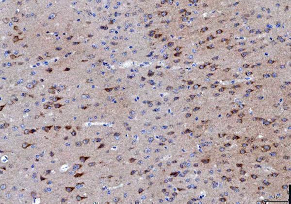
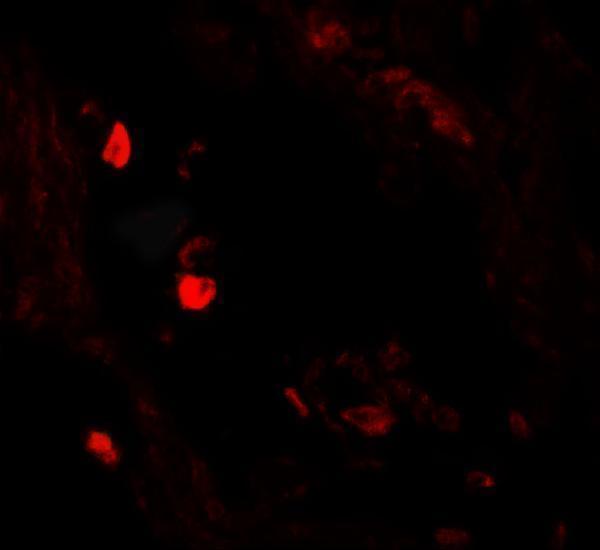
Validated LNPEP antibodies have IHC data from human placenta and mouse and rat brain paraffin sections, plus IF data from human lung tissue (catalog image captions).
A05092-1 is listed for IHC in human, mouse and rat samples, with paraffin-section images from human placenta and mouse and rat brain (catalog applications, reactivity and image captions). A05092 is listed for human IF, with an image from human lung tissue (catalog applications, reactivity and image caption).
Which to pick: Choose A05092-1 for tissue IHC, including cross-species work, because IHC is listed for human, mouse and rat and its images show paraffin sections from all three species (catalog applications, reactivity and image captions). Its captions do not report the fixative, so the paraffin images do not establish fixation (A05092-1 image captions). Choose A05092 for human IF; ICC is not listed, and clone information is unreported for both SKUs (catalog applications, reactivity and clone fields). The selected A05092-1 tissue-IHC caption documents paraffin sections, but does not specify the fixative (selected-SKU IHC image A05092-1).