LRP8 / Low-density lipoprotein receptor-related protein 8 · IHC design guide

Design Immunohistochemistry for LRP8

Plan LRP8 paraffin IHC with a testis control showing high staining in pachytene spermatocytes (HPA tissue IHC). Interpret staining with its membrane location and potentially secreted fragment in mind (UniProt).

Evidence assembled Oct 2026 · For research use; verify linked source records and product datasheet before use
Immunohistochemistry protocol sheet for LRP8 (IHC for LRP8): expected localisation Cell membrane; a fragment may be secreted (UniProt), antibody A03444-2, validated IHC image, and IHC protocol steps
Printable LRP8 IHC protocol sheet — expected localisation Cell membrane; a fragment may be secreted (UniProt), antibody A03444-2, controls and protocol steps. Open the full LRP8 IHC guide →

LRP8 Immunohistochemistry Experimental Design Guide

Expected localisation, validated protocols, controls and antibodies — the at-a-glance facts below, then the full design guide.

Must know before staining
Expected localisation Cell membrane; a fragment may be secreted (UniProt)
Staining pattern Secreted protein profile; high in pachytene spermatocytes (HPA tissue IHC)
Antigen retrieval EDTA pH 8.0 HIER, heat-mediated (datasheet A03444-2)
Positive control ⓘ Testis
Negative control ⓘ Adipose tissue+4 more · see all
Important caveats
Reasons your staining may differ from the expected pattern.
Fixation Keep tissue fixation consistent across samples. (standard IHC practice; not target-specific)
Caveat Shedding may shift staining away from intact cells (UniProt)
Regulation No defined expression inducer in record (UniProt)
Isoform / epitope 5 isoforms; epitope position matters for shed forms (UniProt)
Section 1

Recommended LRP8 IHC & IF Protocols

The catalog antibody’s IHC-P protocol is presented alongside 4 published LRP8 IHC protocols (datasheet A03444-2; PMC12339728; PMC11954824; PMC12964802; PMC12412626).

Recommended immunohistochemistry (IHC-P) protocol parameters
SampleParaffin-embedded human lung cancer tissue; fixative not specified (datasheet A03444-2)
FixationImage fixative and duration unreported (datasheet A03444-2); verify before use.
Sectioning4–5 µm sections on charged slides (standard)
DeparaffinisationXylene, graded ethanol series to water (standard)
Antigen retrievalHeat retrieval: EDTA pH 8.0 (datasheet A03444-2); 20 min, 95–100 °C (standard)
Peroxidase block3% H2O2, 10 min, room temperature (standard)
Blocking10% goat serum (datasheet A03444-2)
Primary antibodyRabbit anti-LRP8, 0.5-1μg/ml (datasheet A03444-2)
Primary incubationOvernight at 4 °C (datasheet A03444-2)
DetectionStreptavidin-biotin complex (SABC), DAB chromogen (datasheet A03444-2)
CounterstainHematoxylin, blue, dehydrate and mount (standard)
Expected resultLRP8-positive staining in pachytene spermatocytes of testis (HPA tissue IHC: High). HPA tissue profile: secreted protein expression. No signal in the no-primary control.
💡Decision noteStart with heat-mediated EDTA retrieval at pH 8.0 for the catalog antibody (datasheet A03444-2); a published tissue protocol used sodium citrate retrieval (PMC12412626).
Section 2

What Is the Expected LRP8 Staining Pattern?

LRP8 is a cell-surface receptor with an extracellular domain and cytoplasmic tail, so membrane-associated staining is biologically plausible (UniProt Q14114 topology). HPA reports High staining in testicular pachytene spermatocytes and rates its tissue IHC evidence Enhanced (HPA tissue IHC). Some isoforms release a secreted receptor fragment, so extracellular signal may also be plausible; compartment alone cannot establish specificity (UniProt Q14114 processing).

What am I looking at on my slide?
Pachytene spermatocytes stain strongly, with signal associated with cell outlines.This matches the High cell-level result in testis (HPA tissue IHC). Membrane association fits the receptor topology (UniProt Q14114 topology); HPA does not specify a required outline pattern.
Nuclei are the dominant stained compartment.Treat this as suspect: HPA observed nuclear off-target binding and disregarded it (HPA tissue IHC). LRP8 topology instead supports a membrane-associated receptor, with a possible secreted fragment (UniProt Q14114 topology and processing).
Cells recorded as unstained in the HPA profile show strong signal.For example, staining of adipocytes in adipose tissue or granular-layer cells in cerebellum conflicts with those cell-level observations (HPA tissue IHC). Check antibody cross-reactivity and endogenous detection activity (general IHC practice); these observations do not prove whole tissues lack LRP8.
Color is spread broadly across the section without a discernible cell pattern.This does not reproduce HPA's defined pachytene-spermatocyte result (HPA tissue IHC). A secreted fragment is possible (UniProt Q14114 processing), but broad signal alone cannot distinguish it from nonspecific background; inspect detection controls (general IHC practice).
Pachytene spermatocytes have no detectable stain.The result conflicts with HPA's High testis observation (HPA tissue IHC). Check section identity, antibody and detection performance, and whether the positive tissue worked in the same run (general IHC practice) before interpreting absence as biology.
💡Expected LRP8 appearanceCall a result consistent when testicular pachytene spermatocytes show High staining (HPA tissue IHC), with membrane association biologically plausible (UniProt Q14114 topology); dominant nuclear staining is suspect (HPA tissue IHC).
How each factor affects the staining
Receptor topologyThe extracellular region spans residues 42–826, the membrane segment 827–847, and the cytoplasmic tail 848–963 (UniProt Q14114 topology). These support a membrane-associated expectation; the antibody epitope is unspecified here, so they cannot predict its precise staining pattern.
Processing and secretionIsoforms containing a furin-type cleavage site can release a receptor fragment (UniProt Q14114 processing). HPA also labels the tissue profile secreted protein expression and cautions that RNA and protein locations may differ (HPA tissue IHC). Interpret extracellular signal alongside controls.
Isoforms and epitope coverageUniProt lists 5 isoforms (UniProt Q14114 isoforms). Because no antibody epitope is supplied, the record cannot establish which isoforms the stain detects or whether a secreted fragment retains that epitope; avoid assigning a stained structure to a specific isoform.
Tissue contextUniProt describes expression mainly in brain and placenta (UniProt Q14114 tissue specificity), while HPA records High staining in testicular pachytene spermatocytes and no detection in specified cerebellar granular-layer cells (HPA tissue IHC). Compare the named cells and assays, rather than treating either statement as a universal tissue rule.
Antibody evidenceHPA073031 has Enhanced IHC validation, meaning its pattern was supported by independent antibodies or orthogonal data (HPA antibody validation). That strengthens the reported tissue pattern but does not make every stained compartment specific, especially given HPA's nuclear off-target observation (HPA tissue IHC).
IF/ICC?HPA summarizes LRP8 as membrane or secreted but provides no ICC-IF images or ICC validation for HPA073031 (HPA subcellular; HPA antibody validation). Use the separate IF/ICC guide for that application; this IHC pattern does not establish an IF/ICC result.
Why is my staining missing, weak or wrong?
SituationLikely causeNext action
Pachytene spermatocytes are negative.The slide conflicts with HPA's High staining report; the present evidence does not identify a target-specific fixation or retrieval failure (HPA tissue IHC).Confirm tissue and cell identification, then compare a known-positive section processed in the same IHC run and verify reagent performance (general IHC practice).
Nuclear staining dominates.HPA explicitly reports nuclear off-target binding (HPA tissue IHC).Do not score nuclear signal as LRP8 positivity; assess the expected cells and review antibody and detection controls (HPA tissue IHC; general IHC practice).
A named HPA-negative cell population stains strongly.Cross-reactivity or endogenous detection activity is possible (general IHC practice); HPA reports no detection in the specified cell population (HPA tissue IHC).Confirm the cell type, examine an appropriate detection control, and compare staining with the pachytene-spermatocyte positive pattern (HPA tissue IHC; general IHC practice).
Color is diffuse across tissue and blank areas.Nonspecific background is possible (general IHC practice). LRP8 shedding alone cannot explain staining in blank areas (UniProt Q14114 processing).Inspect background and detection controls and compare cellular versus extracellular signal; avoid calling diffuse color specific without a reproducible cell pattern (general IHC practice).
Signal appears extracellular instead of on cell outlines.Some isoforms can release a receptor fragment (UniProt Q14114 processing), but its detection depends on an antibody epitope that is unspecified here.Record the compartment separately from cellular staining and assess controls; do not assign extracellular color to the shed fragment solely from appearance (UniProt Q14114 processing; general IHC practice).
Different tissues give apparently conflicting results.UniProt's broad tissue-expression statement and HPA's cell-level IHC observations describe different evidence (UniProt Q14114 tissue specificity; HPA tissue IHC).Compare the specific cells and sections tested. Use HPA's pachytene-spermatocyte result as the stated IHC positive reference, without extrapolating a negative cell result to an entire organ (HPA tissue IHC).

Sample controls for LRP8 IHC & IF

🧪Run testis first and look for staining in pachytene spermatocytes (HPA: High in pachytene spermatocytes). Run adipose tissue as the negative tissue, focusing on adipocytes (HPA: Not detected in adipocytes); on the testis slide, compare other cells with the stained spermatocytes as a background check, without assuming those cells are LRP8-negative (HPA: High is reported specifically in pachytene spermatocytes).
Positive control tissue: Testis (Pachytene spermatocytes, HPA High)
Negative control tissue: Adipose tissue (HPA Not detected)
ICC-IF cell lines (HPA subcellular resource): HPA carries no ICC-IF cell line for LRP8; derive a cell-line control from the positive tissue's cell type (Pachytene spermatocytes) and confirm it by RNA or western blot first.
Technical controls: Include a no-primary, secondary-only control, a matched rabbit IgG isotype control for the rabbit catalog antibody, and LRP8 knockout tissue as a biological negative if available (selected-SKU caption: rabbit primary; standard IHC practice). For the caption’s biotin-based DAB detection, check endogenous peroxidase and biotin background on the testis section (selected-SKU caption: biotinylated secondary and DAB; standard IHC practice).
⚠️Feasibility: A target-specific fixation window or fixation effect is unreported, and the selected-SKU paraffin-section caption does not state a fixative (selected-SKU caption: fixative unreported). The reported IHC method uses heat retrieval in EDTA at pH 8.0; this supports a starting condition but does not establish that retrieval is required (selected-SKU caption). There is no supplied evidence that frozen sections or IF are easier; with biotin-based detection in testis, assess endogenous biotin background before scoring staining (HPA subcellular: no ICC-IF image cell lines; selected-SKU caption: biotin-based detection; standard IHC practice).

HPA tissue IHC evidence for LRP8

Comprehensive Human Protein Atlas IHC scoring per tissue (reliability: Enhanced — At least one protein variant secreted, tissue location of RNA and protein might differ and correlation is complex. Nuclear off target binding observed and disregarded.). Rows are taken directly from the HPA tissue chart — click any row's HPA link to view the source.

Positive expression · recommended positive controls

TissueCell typeLevelEvidenceSource
Testis Pachytene spermatocytes High Protein (IHC) HPA →

Undetected expression · recommended negative controls

TissueCell typeLevelEvidenceSource
Adipose tissue Adipocytes Not detected Protein (IHC) HPA →
Adrenal gland Glandular cells Not detected Protein (IHC) HPA →
Appendix Glandular cells Not detected Protein (IHC) HPA →
Bone marrow Hematopoietic cells Not detected Protein (IHC) HPA →
Breast Adipocytes Not detected Protein (IHC) HPA →
Section 3

Advanced LRP8 IHC Tips

Troubleshoot LRP8 staining in paraffin sections by checking retrieval, cellular compartment, controls, and scoring before assigning biological meaning.

What retrieval should I start with if LRP8 staining is weak?
Start with heat-mediated retrieval in EDTA at pH 8.0 for paraffin sections (datasheet A03444-2). The selected tissue-IHC image used that retrieval before incubation with 1 μg/mL antibody overnight at 4°C, so keep those conditions together for an initial comparison (datasheet A03444-2). If staining remains weak, adjust heating duration on adjacent sections while holding antibody concentration and detection constant; excessive heating can damage morphology or raise background (standard IHC practice). Compare the change in a positive control, such as pachytene spermatocytes, against a negative-control section, and judge both staining and tissue preservation (HPA: High in pachytene spermatocytes; standard IHC practice).
Could fixation explain weak or patchy LRP8 staining?
Target-specific fixation sensitivity is unknown because the selected paraffin-section caption does not state a fixative (datasheet A03444-2). Record the actual fixative, fixation duration, processing history, and section age before comparing cases; those variables can alter antigen accessibility in routine IHC (standard IHC practice). Run affected and well-stained sections together using EDTA at pH 8.0 and the same antibody concentration and detection sequence, then check whether the difference follows a processing batch (datasheet A03444-2; standard IHC practice). Do not assign a particular fixation effect to LRP8 from its membrane topology, glycosylation sites, or HPA tissue pattern; those records do not test fixation (UniProt Q14114 topology and glycosylation; HPA tissue IHC).
Where should I expect LRP8 staining in a tissue section?
Prioritise membrane-associated staining in cells with plausible expression: LRP8 spans the membrane at residues 827–847, with an extracellular region at 42–826 and a cytoplasmic tail at 848–963 (UniProt Q14114 topology). Diffuse extracellular signal may also be biologically plausible because isoforms containing a furin-type cleavage site can release a receptor fragment (UniProt Q14114 processing). Interpret cytoplasmic puncta cautiously because LRP8 may function as an endocytic receptor, but morphology alone cannot establish that mechanism (UniProt Q14114 function; standard IHC practice). Treat isolated nuclear staining as suspect: HPA reported nuclear off-target binding and disregarded it (HPA tissue IHC).
How could the antibody epitope change my interpretation of LRP8 staining?
Confirm the catalog antibody’s immunogen or mapped epitope before deciding which LRP8 species the chromogenic signal represents; this payload does not provide an epitope for A03444-2 (datasheet A03444-2). LRP8 has 5 listed isoforms, and isoforms with the furin-type cleavage-site exon can generate a secreted fragment (UniProt Q14114 isoforms and processing). An extracellular epitope within residues 42–826 could detect membrane receptor and retained extracellular fragment, whereas a cytoplasmic epitope within 848–963 would favor the receptor tail (UniProt Q14114 topology and processing). Compare cellular and extracellular compartments separately, and avoid calling a staining difference isoform-specific without epitope information and an independent control (standard IHC practice).
How should I plan an IF follow-up to the chromogenic LRP8 result?
Treat IF as a separate validation experiment: the selected A03444-2 evidence is paraffin-section chromogenic IHC, and no ICC/IF images are supplied here (datasheet A03444-2; HPA subcellular). Multiplex LRP8 with a validated marker for the expected cell type, and assess colocalisation at single-cell resolution rather than assigning a diffuse IHC signal to nearby cells (standard IF practice). Choose fluorophores after inspecting unstained tissue autofluorescence, reserving a spectrally distinct channel for weak LRP8 signal and including single-color controls (standard IF practice). Match permeabilisation to the mapped epitope: the extracellular domain is 42–826, while the cytoplasmic tail is 848–963; test access accordingly (UniProt Q14114 topology; standard IF practice).
How can I reduce diffuse or misleading LRP8 DAB staining?
First distinguish diffuse extracellular signal from background, since cleavage-site-containing LRP8 isoforms can release a receptor fragment (UniProt Q14114 processing). The selected image used 10% goat serum blocking, 1 μg/mL primary antibody overnight at 4°C, and a biotinylated secondary with DAB development (datasheet A03444-2). Include no-primary and secondary-only controls, assess endogenous peroxidase and endogenous biotin where relevant, and shorten development if the chromogen obscures cellular boundaries (standard IHC practice). Compare any change against a positive control and inspect section edges and damaged regions, where nonspecific staining can mimic a broad LRP8 distribution (HPA: High in pachytene spermatocytes; standard IHC practice).
What is a defensible way to quantify LRP8 IHC across samples? ⚠ ANSWER MARKED FOR VERIFICATION
Define the cell population and compartment before scoring; record membrane-associated cellular staining separately from extracellular deposition because LRP8 is a membrane receptor with potentially secreted fragments (UniProt Q14114 topology and processing). For cellular signal, report the percentage of positive cells and an H-score from 0–300 using intensity categories 0–3 across the same regions (standard IHC practice). For extracellular signal, report positive area or density per mm², excluding folds, necrosis, and section edges by a prespecified rule (standard IHC practice). Normalise cellular counts to eligible nucleated cells and area measurements to analysed tissue area, while keeping retrieval, illumination, and DAB development consistent across slides (standard IHC practice).
When is an apparent LRP8-positive cell likely to be an artefact?
A convincing call combines the expected compartment, a plausible cell population, and signal above matched control sections; LRP8 is membrane-associated and can yield a secreted fragment (UniProt Q14114 localisation and processing; standard IHC practice). Pachytene spermatocytes offer a documented positive reference, whereas adipocytes in adipose tissue were reported as not detected in the HPA tissue set (HPA tissue IHC). Be cautious with nuclear-only signal because HPA observed and disregarded nuclear off-target binding, and scrutinise edge staining, necrosis, and endogenous enzyme deposits against controls (HPA tissue IHC; standard IHC practice). The selected lung-cancer section establishes a staining example for A03444-2, but its caption does not establish the fixative or prove that every stained structure contains intact surface receptor (datasheet A03444-2; UniProt Q14114 processing).
Boster reagents

Best LRP8 / Low-density lipoprotein receptor-related protein 8 IHC Antibodies

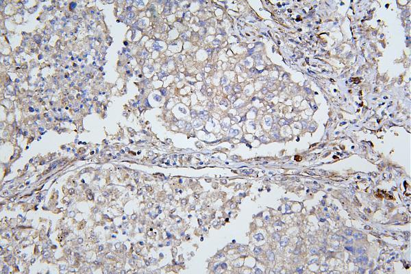
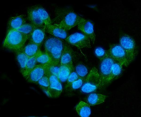
A03444-2 has pictured IHC in paraffin sections of human lung, mammary and rectal cancer tissue (catalog IHC captions), and pictured IF in HepG2 cells (catalog IF caption).

Real IHC data IHC analysis of ApoER2/LRP8 using anti-ApoER2/LRP8 antibody (A03444-2). ApoER2/LRP8 was detected in paraffin-embedded section of human lung cancer tissue. Heat mediated antigen retrieval was performed in EDTA buffer (pH8.0, epitope retrieval solution). The tissue section was blocked with 10% goat serum. The tissue section was then incubated with 1μg/ml rabbit anti-ApoER2/LRP8 Antibody (A03444-2) overnight at 4°C. Biotinylated goat anti-rabbit IgG was used as secondary antibody and incubated for 30 minutes at 37°C. The tissue section was developed using Strepavidin-Biotin-Complex (SABC) (Catalog # SA1022) with DAB as the chromogen.
Anti-ApoER2/LRP8 Antibody ®
Cat # A03444-2

A03444-2 is listed for IHC in human, mouse and rat (catalog applications and reactivity), with pictured paraffin-section IHC in human lung, mammary and rectal cancer tissue (A03444-2 IHC captions). The same SKU is listed for IF/ICC and has pictured IF in HepG2 cells (catalog applications; A03444-2 IF caption).

Which to pick: For tissue IHC, choose A03444-2: its human lung cancer paraffin-section caption documents EDTA retrieval at pH 8.0 and 1 μg/ml primary antibody (A03444-2 IHC caption); the fixative is unreported (A03444-2 IHC caption). For IF/ICC, the same SKU has a HepG2-cell fluorescence image using 4 μg/ml primary antibody (A03444-2 IF caption). For human, mouse or rat samples, A03444-2 is the listed rabbit-host option with IHC reactivity in all three species (catalog host, applications and reactivity); its pictured tissue IHC is human (A03444-2 IHC captions).

Each figure is that product's own IHC / IF validation image from its datasheet.

References

  1. UniProt Consortium. UniProt entry Q14114 (LRP8_HUMAN, Low-density lipoprotein receptor-related protein 8).
  2. Human Protein Atlas. LRP8 tissue IHC expression (reliability: Enhanced).
  3. Human Protein Atlas. LRP8 subcellular location (ICC-IF): Membrane, Secreted.
  4. Human Protein Atlas. LRP8 antibody validation summary (1 antibodies).
  5. Reelin-LRP8 signaling mediates brain dissemination of breast cancer cells via abluminal migration. EMBO molecular medicine 2025 — PMC12339728.
  6. Enhanced LRP8 expression induced by Helicobacter pylori drives gastric cancer progression by facilitating β-Catenin nuclear translocation. Journal of advanced research 2025 — PMC11954824.
  7. LRP8 Promotes colorectal cancer progression by suppressing ferroptosis through the SLC3A2/GPX4 signalling axis. European journal of medical research 2026 — PMC12964802.
  8. LRP8 Regulates Lipid Metabolism to Stimulate Malignant Progression and Cisplatin Resistance in Bladder Cancer. The Kaohsiung journal of medical sciences 2025 — PMC12412626.
  9. PubMed PMID:8626535 — UniProt-cited evidence.
  10. PubMed PMID:9079678 — UniProt-cited evidence.
  11. PubMed PMID:11152697 — UniProt-cited evidence.