This website uses cookies to ensure you get the best experience on our website.


- Table of Contents
Plan LRRK2 paraffin IHC with the catalog antibody at 1:25 (datasheet M00221-3). Use strongly stained kidney tubules as a reference and assess cytoplasmic staining in the relevant cell types (HPA tissue IHC).

Expected localisation, validated protocols, controls and antibodies — the at-a-glance facts below, then the full design guide.
| Expected localisation | Cytoplasmic staining in several cell types (HPA tissue IHC) | |
| Staining pattern | High in renal tubules; medium in alveolar cells (HPA tissue IHC) | |
| Antigen retrieval | Citrate pH 6 HIER, heat-mediated (datasheet M00221-3) | |
| Positive control | Kidney+1 more · see all | |
| Negative control | Adipose tissue+4 more · see all |
| Fixation | Formaldehyde-fixed paraffin sections (selected-SKU IHC image M00221-3) | |
| Caveat | Brain expression may yield low neuronal staining (UniProt; HPA tissue IHC) | |
| Regulation | Lysosomal stress recruits LRRK2 (UniProt) | |
| Isoform / epitope | No annotated isoforms or transmembrane segment (UniProt) |
The catalog antibody’s IHC-P protocol uses citrate pH 6 retrieval (datasheet: M00221-3). The published IHC protocols below cover rodent brain and tissue sections (PMC4076169; PMC5289148; PMC7812009).
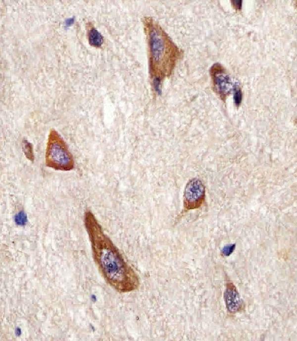
| Sample | Formaldehyde-fixed, paraffin-embedded Human brain tissue (datasheet M00221-3) |
| Fixation | Image formalin-fixed; duration unreported (datasheet M00221-3); verify before use. |
| Sectioning | 4–5 µm sections on charged slides (standard) |
| Deparaffinisation | Xylene, graded ethanol series to water (standard) |
| Antigen retrieval | Heat retrieval: Citrate pH 6 (datasheet M00221-3); 20 min, 95–100 °C (standard) |
| Peroxidase block | 3% H2O2, 10 min, room temperature (standard) |
| Blocking | 10% normal serum of the secondary host, 30 min, room temperature (standard) |
| Primary antibody | Rabbit anti-LRRK2, 1:25 (datasheet M00221-3) |
| Primary incubation | 1 hours at 37°C (datasheet M00221-3) |
| Detection | HRP-polymer secondary, DAB chromogen 5–10 min (standard) |
| Counterstain | Hematoxylin, blue, dehydrate and mount (standard) |
| Expected result | LRRK2-positive staining in cells in tubules of kidney (HPA tissue IHC: High). HPA tissue profile: Cytoplasmic expression in several cell types, including pneumocytes and cells in renal tubules. No signal in the no-primary control. |
LRRK2 is expected mainly in cytoplasmic and vesicle-associated compartments; it has no transmembrane segment (UniProt Q5S007 topology and subcellular location). In paraffin-section IHC, look first at renal tubular cells, reported High, and lung alveolar cells, reported Medium (HPA tissue IHC). HPA calls its tissue IHC reliability Approved, with medium consistency between antibody staining and RNA expression (HPA tissue IHC reliability).
| Distinct cytoplasmic staining in renal tubular cells, with weaker staining in lung alveolar cells. | This matches the reported High tubular and Medium alveolar staining levels (HPA tissue IHC). Compare cells within each section: these levels describe HPA observations, not intensity thresholds for every run (HPA tissue IHC; general IHC practice). |
| Strong nuclear-only staining or a uniform nuclear outline dominates the positive cells. | Recheck compartment assignment and controls: UniProt places LRRK2 chiefly in cytoplasmic and vesicle-associated locations (UniProt Q5S007 subcellular location). HPA ICC-IF lists nucleoplasm and nuclear membrane only as uncertain additional locations, so nuclear-only IHC does not establish the expected pattern (HPA subcellular ICC-IF). |
| Prominent staining appears in cells reported as undetected, such as adipocytes in adipose tissue. | Consider antibody cross-reactivity or endogenous chromogenic detection activity (HPA tissue IHC: adipocytes Not detected; general IHC practice). Check cell identity and a matched negative control before assigning that signal to LRRK2 (general IHC practice). |
| Colour covers the section diffusely, obscuring cell borders and cytoplasmic detail. | The pattern cannot be scored against HPA's cell-specific IHC observations (HPA tissue IHC; general IHC practice). Inadequate blocking, insufficient washing, or excess detection reagent can produce diffuse background; inspect the no-primary control to locate the source (general IHC practice). |
| Renal tubular cells show no discernible signal in an otherwise readable section. | HPA reports High staining in renal tubular cells, making them a useful reference for a missed signal (HPA tissue IHC). Check the IHC-validated antibody, detection reagents, and retrieval conditions with controls before interpreting an unstained specimen as LRRK2-negative (general IHC practice). |
| Tissue and cell selection | Use renal tubular cells as the stronger reference and lung alveolar cells as a second observed site (HPA tissue IHC: High; Medium). HPA reports several low-level sites, so a faint result there needs careful comparison with controls (HPA tissue IHC; general IHC practice). |
| Subcellular interpretation | LRRK2 is associated with vesicles, Golgi, endosomes and lysosomes, among other cytoplasmic sites (UniProt Q5S007 subcellular location). Its lack of a transmembrane segment does not make crisp cell-surface staining the expected IHC result (UniProt Q5S007 topology; general IHC practice). |
| Antibody validation | HPA lists HPA014293 and CAB037160 as IHC Approved; HPA014293 is also ICC Supported (HPA antibodies). These labels support the reported uses but do not independently verify an unexpected cell or compartment in a new specimen (HPA antibodies; general IHC practice). |
| IF/ICC comparison | On its separate IF/ICC guide, the relevant comparison is vesicular localisation: HPA marks vesicles Supported, while basal body and listed nuclear locations are Uncertain (HPA subcellular ICC-IF). Treat those uncertain locations as prompts for validation, not IHC acceptance criteria (HPA subcellular ICC-IF; general IHC practice). |
| Antigen retrieval and detection | Target-specific fixation effects are not established by the supplied assay evidence. Verify with a matched IHC source before attributing a result to fixation. |
| Situation | Likely cause | Next action |
|---|---|---|
| Renal tubules are negative while the section and counterstain look intact. | A failed primary, retrieval, or detection step is possible; HPA reports High tubular staining (general IHC practice; HPA tissue IHC). | Run a known-positive kidney section alongside the specimen; verify the catalog antibody's IHC instructions and the detection controls before scoring absence (general IHC practice). |
| Lung alveolar cells stain weakly, with a clear kidney positive control. | This may fit the reported Medium alveolar versus High tubular levels (HPA tissue IHC). | Compare the signal with nearby background and the no-primary control; avoid calling weak alveolar staining a failure solely because it is less intense than kidney (HPA tissue IHC; general IHC practice). |
| A tissue reported as Not detected shows broad staining in many cell types. | Nonspecific antibody binding or endogenous chromogenic activity may account for the mismatch (HPA tissue IHC; general IHC practice). | Check the exact cell type against HPA, examine a no-primary control, and use the appropriate endogenous-enzyme block for the chosen chromogen system (HPA tissue IHC; general IHC practice). |
| Signal is mainly nuclear or outlines the plasma membrane. | That distribution conflicts with the expected cytoplasmic and vesicle-associated pattern; HPA's additional nuclear ICC-IF locations remain Uncertain (UniProt Q5S007 subcellular location; HPA subcellular ICC-IF). | Review morphology and control sections, then confirm the pattern with another independently validated antibody if available (general IHC practice). |
| Diffuse colour hides individual positive cells. | Background from blocking, washing, primary concentration, or detection can prevent cell-specific interpretation (general IHC practice). | Compare with a no-primary control; adjust one general IHC variable at a time using the catalog antibody's IHC instructions (general IHC practice). |
| Cortical glia are negative despite reported LRRK2 expression in brain. | The records refer to different cells: HPA reports cerebral-cortex glial cells Not detected, while UniProt reports cortical pyramidal neurons expressing LRRK2 (HPA tissue IHC; UniProt Q5S007 tissue specificity). | Identify neurons and glia separately before scoring; use the observed kidney tubular pattern as a positive IHC reference (HPA tissue IHC; UniProt Q5S007 tissue specificity). |


Comprehensive Human Protein Atlas IHC scoring per tissue (reliability: Approved — Medium consistency between antibody staining and RNA expression data.). Rows are taken directly from the HPA tissue chart — click any row's HPA link to view the source.
| Tissue | Cell type | Level | Evidence | Source |
|---|---|---|---|---|
| Kidney | Cells in tubules | High | Protein (IHC) | HPA → |
| Lung | Alveolar cells | Medium | Protein (IHC) | HPA → |
| Tissue | Cell type | Level | Evidence | Source |
|---|---|---|---|---|
| Adipose tissue | Adipocytes | Not detected | Protein (IHC) | HPA → |
| Adrenal gland | Glandular cells | Not detected | Protein (IHC) | HPA → |
| Bone marrow | Hematopoietic cells | Not detected | Protein (IHC) | HPA → |
| Breast | Adipocytes | Not detected | Protein (IHC) | HPA → |
| Cerebral cortex | Glial cells | Not detected | Protein (IHC) | HPA → |
Troubleshoot LRRK2 staining in paraffin sections by checking retrieval, cellular pattern and controls before comparing chromogenic signal across samples.
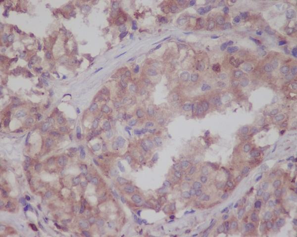
The catalog includes IHC images from human brain and thyroid paraffin sections; IF/ICC applications are listed for human, mouse, and rat, but no IF image is supplied (catalog applications and reactivity; IHC image captions).
M00221-3 has an IHC image from formaldehyde-fixed human brain paraffin sections (M00221-3 IHC image caption). M00221-2 has an IHC image from human thyroid paraffin sections; its fixative is unreported, and IF/ICC is listed without an IF image (M00221-2 IHC image caption; catalog applications).
Which to pick: For tissue IHC with a documented processing example, choose rabbit polyclonal M00221-3: its human brain caption reports formaldehyde fixation, citrate retrieval at pH 6, and primary antibody at 1:25 (catalog clonality; M00221-3 IHC image caption). For IF/ICC or work across human, mouse, and rat, choose rabbit monoclonal M00221-2 based on its listed applications and reactivity; its own IHC image documents human thyroid paraffin sections, with the fixative unreported (catalog applications, reactivity, and clonality; M00221-2 IHC image caption).