LYN / Tyrosine-protein kinase Lyn · IHC design guide

Design Immunohistochemistry for LYN

Plan chromogenic LYN IHC in paraffin sections around the observed membranous and cytoplasmic tissue pattern (HPA tissue IHC). Use high-staining germinal centers or splenic red pulp to assess staining while scoring defined cell populations (HPA tissue IHC).

Evidence assembled Oct 2026 · For research use; verify linked source records and product datasheet before use
Immunohistochemistry protocol sheet for LYN (IHC for LYN): expected localisation Membranous and cytoplasmic staining in selected tissues (HPA tissue IHC), antibody A01424-2, validated IHC image, and IHC protocol steps
Printable LYN IHC protocol sheet — expected localisation Membranous and cytoplasmic staining in selected tissues (HPA tissue IHC), antibody A01424-2, controls and protocol steps. Open the full LYN IHC guide →

LYN Immunohistochemistry Experimental Design Guide

Expected localisation, validated protocols, controls and antibodies — the at-a-glance facts below, then the full design guide.

Must know before staining
Expected localisation Membranous and cytoplasmic staining in selected tissues (HPA tissue IHC)
Staining pattern Germinal centers and splenic red pulp show membranous and cytoplasmic staining (HPA tissue IHC)
Antigen retrieval Citrate pH 6 HIER, heat-mediated (datasheet A01424-2)
Positive control ⓘ Lymph node+4 more · see all
Negative control ⓘ Adipose tissue+4 more · see all
Important caveats
Reasons your staining may differ from the expected pattern.
Fixation Keep fixation conditions consistent across paraffin sections. (standard IHC practice; not target-specific)
Caveat Strong lymphoid staining can confound whole-section scoring (HPA tissue IHC)
Regulation Lymphoid-enriched expression (HPA tissue IHC)
Isoform / epitope 2 isoforms; no transmembrane region; check epitope coverage (UniProt)
Section 1

Recommended LYN IHC & IF Protocols

The catalog antibody’s IHC-P protocol (datasheet: A01424-2) is presented alongside three published LYN IHC protocols (PMC12174511; PMC6400685; PMC5342753).

Recommended immunohistochemistry (IHC-P) protocol parameters
SampleParaffin-embedded mouse intestine tissue; fixative not specified (datasheet A01424-2)
FixationImage fixative and duration unreported (datasheet A01424-2); verify before use.
Sectioning4–5 µm sections on charged slides (standard)
DeparaffinisationXylene, graded ethanol series to water (standard)
Antigen retrievalHeat retrieval: Citrate pH 6, 20 min (datasheet A01424-2)
Peroxidase block3% H2O2, 10 min, room temperature (standard)
Blocking10% goat serum (datasheet A01424-2)
Primary antibodyRabbit anti-LYN, 0.5-1μg/ml (datasheet A01424-2)
Primary incubationOvernight at 4 °C (datasheet A01424-2)
DetectionStreptavidin-biotin complex (SABC), DAB chromogen (datasheet A01424-2)
CounterstainHematoxylin, blue, dehydrate and mount (standard)
Expected resultLYN-positive staining in germinal center cells of lymph node (HPA tissue IHC: High). HPA tissue profile: Distinct membranous and cytoplasmic expression in selected tissues at variable levels, most abundant in lymphoid tissues. No signal in the no-primary control.
💡Decision noteStart with heat-mediated citrate retrieval at pH 6 (datasheet: A01424-2); the colorectal study also used citrate at pH 6 (PMC12174511).
Section 2

What Is the Expected LYN Staining Pattern?

LYN is a non-receptor kinase with no transmembrane segment (UniProt P07948 topology). In paraffin-section IHC, expect mainly membranous and cytoplasmic staining, strongest in selected lymphoid and immune-cell populations (HPA: Enhanced tissue IHC reliability; high in lymph-node germinal-center cells, placental Hofbauer cells, splenic red-pulp cells, and tonsillar germinal-center cells). UniProt also records Golgi, perinuclear, and nuclear localization; those locations need context when interpreting a slide (UniProt P07948 subcellular location).

What am I looking at on my slide?
Distinct cell outlines with cytoplasmic staining in germinal-center cells.This fits the reported tissue pattern: lymph-node and tonsillar germinal-center cells stain at a high level, and HPA describes selected tissues as membranous and cytoplasmic (HPA: tissue IHC). Score the named cells and their compartments together; intensity elsewhere need not match because reported tissue levels vary (HPA: tissue IHC).
Predominantly nuclear staining, with little membrane or cytoplasmic signal.Treat this as discordant with the usual tissue IHC pattern and check controls before calling it LYN (HPA: tissue IHC). Nuclear localization is possible, particularly when CRM1-mediated export or kinase activity is inhibited, so nuclear signal alone does not prove an artefact (UniProt P07948 subcellular location).
Strong signal in cells reported as undetected, such as adipocytes or cardiomyocytes.HPA reports no detection in those named cell types, so unexpected staining warrants a specificity check rather than an automatic positive call (HPA: tissue IHC). Compare the same run with known-positive cells and a detection-only control; cross-reactivity or endogenous chromogenic activity are general IHC possibilities (standard IHC practice).
Color spread across stroma, empty spaces, and many unrelated cells.A diffuse deposit lacks the cell-associated membranous and cytoplasmic distribution described for LYN (HPA: tissue IHC). Review detection-only and blocking controls for nonspecific deposition or endogenous activity before assigning cellular staining (standard IHC practice).
No signal in lymph-node or tonsillar germinal-center cells.These cells are reported as high-staining references, so an absent signal makes the run or antibody performance uncertain (HPA: tissue IHC). Check that the positive reference and detection controls worked; the supplied sources do not establish a LYN-specific fixation sensitivity (HPA: tissue IHC; UniProt P07948).
💡Expected LYN appearanceCall a positive result when distinct membranous and cytoplasmic signal is evident in high-staining germinal-center cells or Hofbauer cells (HPA: tissue IHC); widespread, cell-independent color is a false-positive warning (standard IHC practice).
How each factor affects the staining
Which cells provide the clearest tissue reference?HPA reports high staining in lymph-node and tonsillar germinal-center cells, placental Hofbauer cells, and splenic red-pulp cells; marrow hematopoietic cells are medium (HPA: tissue IHC). Use the stated cell population, since a whole-tissue average can hide a restricted positive pattern (standard IHC practice).
How much weight does HPA validation carry?HPA rates tissue IHC reliability Enhanced and lists HPA001231 and CAB004492 as IHC Enhanced (HPA: tissue IHC; HPA: antibodies). This supports the reported pattern, while a new assay still needs its own positive and detection controls (standard IHC practice).
Does localization require a transmembrane signal?LYN has no transmembrane segment, yet UniProt places it at the cell membrane, including lipid rafts, and in cytoplasm, Golgi, and nucleus (UniProt P07948 topology; subcellular location). Interpret an outlined cell together with its cytoplasmic signal, as observed in tissue IHC (HPA: tissue IHC).
Can isoform or modification differences change the call?UniProt lists two isoforms and multiple modified residues (UniProt P07948 isoforms; modified residues). The supplied records give no antibody epitope or isoform coverage, so they cannot predict which form a particular IHC stain detects; use the observed HPA cell pattern as the reference (HPA: tissue IHC).
IF/ICC: what localization should be expected?HPA ICC-IF reports mainly plasma-membrane localization, with Golgi and vesicular signal also observed (HPA: subcellular ICC-IF). This is a separate application: its cell-image localization can inform interpretation, but its reported pattern does not set an IHC staining intensity threshold (HPA: subcellular ICC-IF; tissue IHC).
Why is my staining missing, weak or wrong?
SituationLikely causeNext action
Known-positive germinal centers are blank.A failed staining or detection step is possible; these cells are reported high in lymph node and tonsil (HPA: tissue IHC).Check the positive reference, reagent application, retrieval record, and detection control for the run (standard IHC practice). The supplied sources do not identify a LYN-specific retrieval condition or fixation effect (HPA: tissue IHC; UniProt P07948).
Only nuclear staining appears.The pattern diverges from HPA tissue IHC, although regulated nuclear accumulation is recorded by UniProt (HPA: tissue IHC; UniProt P07948 subcellular location).Compare with a known-positive tissue and detection-only control; record nuclear staining separately rather than scoring it as the expected membranous and cytoplasmic pattern (standard IHC practice; HPA: tissue IHC).
A broadly positive adipocyte or cardiomyocyte population obscures the result.Those cell types are reported as undetected by HPA; nonspecific binding or detection activity may explain unexpected color (HPA: tissue IHC; standard IHC practice).Inspect cell boundaries, compare a detection-only control, and reassess the antibody signal in a high-staining reference population before assigning LYN positivity (standard IHC practice; HPA: tissue IHC).
Background color covers the section.Cell-independent color is inconsistent with HPA's distinct membranous and cytoplasmic pattern (HPA: tissue IHC); blocking, wash, or chromogenic detection issues are general possibilities (standard IHC practice).Review blocking and wash steps, then compare a detection-only control with the stained section (standard IHC practice). Interpret only cell-associated signal after the background source is resolved (HPA: tissue IHC).
Signal is weaker than expected in marrow.HPA rates marrow hematopoietic cells medium, while several named lymphoid or immune-cell populations are high (HPA: tissue IHC).Check the identified cell population and compare the run with a high-staining lymph-node or tonsil reference before calling a medium signal a technical failure (HPA: tissue IHC; standard IHC practice).
A punctate pattern is mistaken for the main IHC result.Golgi and vesicular localization is reported in ICC-IF, whereas HPA's tissue IHC summary emphasizes distinct membranous and cytoplasmic expression (HPA: subcellular ICC-IF; tissue IHC).Record puncta as an additional observation and judge IHC positivity against the expected cell types and membranous and cytoplasmic pattern; use controls if puncta occur across unrelated cells (HPA: tissue IHC; standard IHC practice).

Sample controls for LYN IHC & IF

🧪Run lymph node first and score germinal center cells, where LYN staining is High (HPA: High in lymph node germinal center cells). Use adipose tissue adipocytes as the negative comparator (HPA: Not detected in adipocytes); on the lymph node slide, cells without specific staining should lack brown DAB deposit, but cells outside germinal centers should not be presumed LYN-negative (standard IHC interpretation; UniProt P07948 tissue specificity).
Positive control tissue: Lymph node (Germinal center cells, HPA High)
Negative control tissue: Adipose tissue (HPA Not detected)
ICC-IF cell lines (HPA subcellular resource): HPA ICC-IF images show LYN in A-431, U-251MG, U2OS, Sperm, with annotated localisation: Plasma membrane (supported) (HPA subcellular).
Technical controls: Include a no-primary control, a secondary-only control, and an isotype control matched to the rabbit primary antibody’s immunoglobulin class (selected-SKU IHC caption: rabbit anti-Lyn; standard IHC practice). A LYN knockout specimen can test biological specificity; block endogenous peroxidase and check endogenous biotin background in lymph node when using the caption’s SABC/DAB detection method (standard IHC practice; selected-SKU IHC caption: SABC/DAB).
⚠️Feasibility: A target-specific fixation window or fixation effect is unreported, and the selected A01424-2 paraffin-section caption does not state the fixative (selected-SKU IHC caption). Heat retrieval in citrate buffer at pH 6 for 20 minutes is a documented IHC-P starting condition, but retrieval dependency has not been established (selected-SKU IHC caption). Whether frozen sections or IF/ICC are easier is unreported; endogenous peroxidase or biotin can create misleading lymph node signal with the reported SABC/DAB method (standard IHC practice; selected-SKU IHC caption: SABC/DAB).

HPA tissue IHC evidence for LYN

Comprehensive Human Protein Atlas IHC scoring per tissue (reliability: Enhanced — High consistency between antibody staining and RNA expression data.). Rows are taken directly from the HPA tissue chart — click any row's HPA link to view the source.

Positive expression · recommended positive controls

TissueCell typeLevelEvidenceSource
Lymph node Germinal center cells High Protein (IHC) HPA →
Placenta Hofbauer cells High Protein (IHC) HPA →
Spleen Cells in red pulp High Protein (IHC) HPA →
Tonsil Germinal center cells High Protein (IHC) HPA →
Appendix Lymphoid tissue Medium Protein (IHC) HPA →

Undetected expression · recommended negative controls

TissueCell typeLevelEvidenceSource
Adipose tissue Adipocytes Not detected Protein (IHC) HPA →
Adrenal gland Glandular cells Not detected Protein (IHC) HPA →
Breast Adipocytes Not detected Protein (IHC) HPA →
Endometrium Cells in endometrial stroma Not detected Protein (IHC) HPA →
Epididymis Glandular cells Not detected Protein (IHC) HPA →
Section 3

Advanced LYN IHC Tips

Troubleshoot LYN staining in paraffin sections by checking retrieval, cell identity, subcellular pattern, and detection controls before interpreting signal.

What retrieval should I try first when LYN staining is weak in paraffin sections?
Start with heat-mediated retrieval in citrate buffer at pH 6 (datasheet A01424-2). The catalog antibody’s mouse-intestine IHC image used 20 minutes of retrieval, followed by 1 μg/mL primary antibody overnight at 4°C (datasheet A01424-2). Keep heating, cooling, and section handling consistent across the test and control slides so changes in staining can be attributed to retrieval (standard IHC practice). Compare staining in the expected cells with the no-primary control, and inspect tissue morphology for damage from excessive heating (standard IHC practice). A weak result alone does not establish that the retrieval buffer is unsuitable (standard IHC practice).
Can I infer the best fixative for LYN from the available tissue staining?
The selected mouse-intestine caption describes a paraffin section but does not state its fixative (datasheet A01424-2). Target-specific fixation sensitivity is therefore unknown from the supplied evidence; the reported HPA staining pattern does not establish a fixation method (datasheet A01424-2; HPA: tissue IHC profile). For an IHC comparison, document the actual fixative and fixation duration for each specimen, and process matched controls together (standard IHC practice). If signal differs between batches, compare sections with similar tissue preservation before changing antibody concentration or retrieval (standard IHC practice). Record morphology and background alongside staining intensity because poorly preserved sections can mislead scoring (standard IHC practice).
Should I reject LYN staining that appears outside the plasma membrane?
Expect membrane-associated and cytoplasmic staining in selected tissues, with the strongest HPA tissue signal concentrated in lymphoid tissues (HPA: tissue IHC profile). LYN has no transmembrane segment, and its reported locations include membrane, cytoplasm, perinuclear region, Golgi apparatus, and nucleus (UniProt P07948 topology and subcellular annotation). Its supported main subcellular location is the plasma membrane, with additional Golgi localisation (HPA: subcellular profile). Score membrane, cytoplasmic, and nuclear patterns separately instead of combining them into one intensity value (standard IHC practice). Treat an unexpected compartment as a finding to verify against cell identity, morphology, and controls before assigning biological meaning (standard IHC practice).
How can I tell whether the antibody detects both LYN isoforms in tissue?
LYN has 2 annotated isoforms and contains SH3, SH2, and kinase domains (UniProt P07948 isoforms and domains). The supplied caption establishes staining with A01424-2 in a paraffin mouse-intestine section but does not map the antibody epitope or establish isoform coverage (datasheet A01424-2). Check the antibody’s documented immunogen or epitope against each isoform sequence before describing tissue staining as isoform-specific (standard IHC practice). Phosphorylated residues are annotated for LYN, but the supplied IHC evidence does not establish whether modification changes antibody recognition (UniProt P07948 modified residues; datasheet A01424-2). Report the result as LYN immunoreactivity unless isoform specificity has been independently verified (standard IHC practice).
How should I plan IF to check which cells carry the LYN signal?
Use IF as a separate validation experiment alongside the chromogenic tissue readout, and pair LYN with a marker for the expected cell population, such as a B-cell marker in germinal centers (HPA: High in lymph-node germinal center cells; standard IF practice). Choose spectrally separated fluorophores and check an unstained section for tissue autofluorescence before assigning a weak LYN channel (standard IF practice). LYN lacks a transmembrane segment and is reported at both membrane and intracellular sites, so select permeabilisation according to the antibody epitope’s accessibility (UniProt P07948 topology and subcellular annotation; standard IF practice). Compare gentle and stronger permeabilisation only if the epitope’s orientation is uncertain, while monitoring loss of membrane detail (standard IF practice). The paraffin IHC caption does not establish IF fixation or permeabilisation conditions (datasheet A01424-2).
What should I check when DAB staining is widespread or diffuse?
The selected IHC image used a biotinylated secondary antibody, an avidin-biotin detection complex, and DAB (datasheet A01424-2). If staining is diffuse, run no-primary and detection-only controls to locate background from secondary binding or the detection system (standard IHC practice). Include a peroxidase block before DAB development and compare background with and without the biotin-based detection reagents where appropriate (standard IHC practice). The caption reports 10% goat-serum blocking and 1 μg/mL primary antibody overnight at 4°C; use these as documented starting conditions for this antibody (datasheet A01424-2). Judge improvements by retained staining in expected cells, not by a lighter slide alone (HPA: tissue IHC profile; standard IHC practice).
How should I score LYN across tissues with different cell mixtures? ⚠ ANSWER MARKED FOR VERIFICATION
Define the cell population and compartment before scoring, because HPA reports variable membranous and cytoplasmic expression across selected tissues (HPA: tissue IHC profile). For a defined population, record the percentage of positive cells and staining intensity; an H-score can combine intensity grades 0–3 with percentages into a 0–300 score (standard IHC practice). If the question concerns infiltrating positive cells, report their density per mm² of viable tissue instead (standard IHC practice). Normalise each result to the relevant cell population or viable tissue area, and apply the same threshold and compartment rule to every section (standard IHC practice). Document whether lymphoid, glandular, and stromal cells were scored separately (HPA: tissue IHC profile; standard IHC practice).
How do I distinguish plausible LYN staining from an IHC artefact?
First locate the stained cells: high signal in lymph-node germinal center cells and placental Hofbauer cells is consistent with the reported tissue profile (HPA: High in germinal center cells and Hofbauer cells). Membranous or cytoplasmic staining also fits the reported tissue pattern, while nuclear LYN is annotated and needs separate verification rather than automatic rejection (HPA: tissue IHC profile; UniProt P07948 subcellular annotation). Inspect section edges and necrotic areas for disproportionate staining, and compare them with well-preserved regions (standard IHC practice). Check no-primary and peroxidase-control sections when DAB signal follows tissue damage or endogenous enzyme activity (standard IHC practice). Interpret any apparent change only after cell composition and staining controls support it (standard IHC practice).
Boster reagents

Best LYN / Tyrosine-protein kinase Lyn IHC Antibodies

Two antibodies have IHC images from paraffin sections: A01424-2 in mouse intestine and A01424Y508 in human brain (catalog image captions). Both list human, mouse and rat reactivity (catalog applications/reactivity).

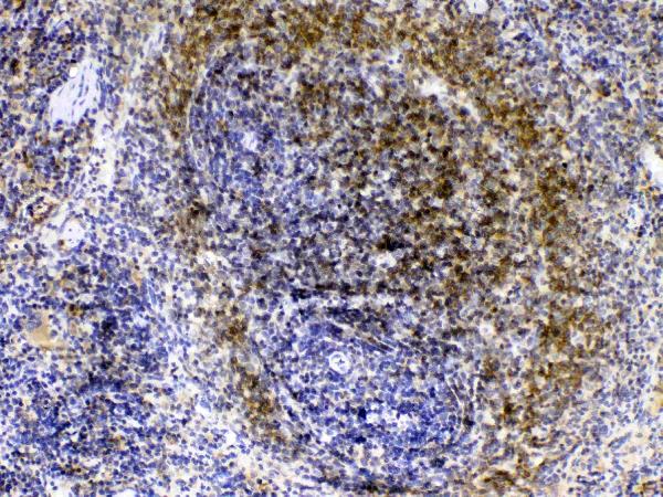
Real IHC data IHC analysis of Lyn using anti-Lyn antibody (A01424-2). Lyn was detected in paraffin-embedded section of mouse intestine tissue . Heat mediated antigen retrieval was performed in citrate buffer (pH6, epitope retrieval solution) for 20 mins. The tissue section was blocked with 10% goat serum. The tissue section was then incubated with 1μg/ml rabbit anti-Lyn Antibody (A01424-2) overnight at 4°C. Biotinylated goat anti-rabbit IgG was used as secondary antibody and incubated for 30 minutes at 37°C. The tissue section was developed using Strepavidin-Biotin-Complex (SABC)(Catalog # SA1022) with DAB as the chromogen.
Anti-Lyn Antibody ®
Cat # A01424-2
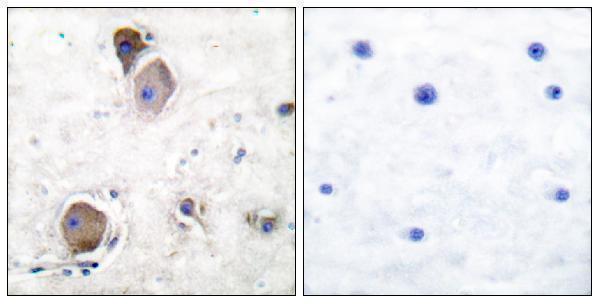
Real IHC data Immunohistochemistry analysis of paraffin-embedded human brain, using Lyn (Phospho-Tyr507) Antibody. The picture on the right is blocked with the phospho peptide.
Anti-Phospho-Lyn (Y508) Antibody
Cat # A01424Y508

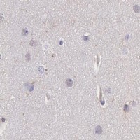
A01424-2 has IHC images from mouse intestine, rat and mouse spleen, and human tonsil paraffin sections (catalog image captions). A01424Y508 has a phospho-Lyn IHC image from a human brain paraffin section with a phospho-peptide blocking control (catalog image caption).

Which to pick: For tissue IHC, choose A01424-2 for Lyn staining across the depicted tissues, or A01424Y508 when phospho-Lyn staining is the goal (catalog titles and image captions). For IF/ICC, P01424-1 lists both applications and human/mouse reactivity; A01424Y508 lists IF and human/mouse/rat reactivity (catalog applications/reactivity). For cross-species IHC, both rendered SKUs list human, mouse and rat reactivity, though their images show different tissues; both captions specify paraffin sections and leave the fixative unreported (catalog applications/reactivity and image captions).

Each figure is that product's own IHC / IF validation image from its datasheet.

References

  1. UniProt Consortium. UniProt entry P07948 (LYN_HUMAN, Tyrosine-protein kinase Lyn).
  2. Human Protein Atlas. LYN tissue IHC expression (reliability: Enhanced).
  3. Human Protein Atlas. LYN subcellular location (ICC-IF): Mainly localized to the plasma membrane. In addition localized to the Golgi apparatus and vesicles..
  4. Human Protein Atlas. LYN antibody validation summary (2 antibodies).
  5. Role of Lyn Immunohistochemical Staining in Progression of Colorectal Carcinoma. Asian Pacific journal of cancer prevention : APJCP 2025 — PMC12174511.
  6. Lyn Kinase Promotes the Proliferation of Malignant Melanoma Cells through Inhibition of Apoptosis and Autophagy via the PI3K/Akt Signaling Pathway. Journal of Cancer 2019 — PMC6400685.
  7. Tyrosine kinase LYN is an oncotarget in human cervical cancer: A quantitative proteomic based study. Oncotarget 2016 — PMC5342753.
  8. Minocycline binds and inhibits LYN activity to prevent STAT3-meditated metastasis of colorectal cancer. International journal of biological sciences 2022 — PMC8990469.
  9. PubMed PMID:3561390 — UniProt-cited evidence.
  10. PubMed PMID:8125304 — UniProt-cited evidence.
  11. PubMed PMID:15489334 — UniProt-cited evidence.