This website uses cookies to ensure you get the best experience on our website.


- Table of Contents
Plan LZTR1 IHC on paraffin sections around granular cytoplasmic staining in gastrointestinal tissue (HPA tissue IHC). Start antibody A06363 optimization at 5 μg/mL (datasheet), and interpret staining in light of its very low consistency with RNA expression (HPA tissue IHC).

Expected localisation, validated protocols, controls and antibodies — the at-a-glance facts below, then the full design guide.
| Expected localisation | Granular cytoplasm in gastrointestinal tissue (HPA tissue IHC) | |
| Staining pattern | Granular cytoplasmic staining in GI glandular cells (HPA tissue IHC) | |
| Antigen retrieval | Citrate pH 6.0 HIER, 95–98 °C, 20 min (rule: cytoplasmic / membrane antigen) | |
| Positive control | Appendix+4 more · see all | |
| Negative control | Adipose tissue+4 more · see all |
| Fixation | Keep fixation consistent across sections (standard IHC practice; not target-specific) | |
| Caveat | Antibody staining and RNA show very low consistency (HPA tissue IHC) | |
| Regulation | No expression regulator annotated (UniProt) | |
| Isoform / epitope | No isoforms annotated; no transmembrane segment (UniProt) |
The catalog antibody’s IHC-P protocol is accompanied by published LZTR1 staining methods for human liver tissue (PMC11237970) and mouse and human tissues (PMC12572372).
| Sample | Tissue sections; selected-image fixative not specified (standard IHC workflow) |
| Fixation | Image fixative and duration unreported (datasheet A06363); verify before use. |
| Sectioning | 4–5 µm sections on charged slides (standard) |
| Deparaffinisation | Xylene, graded ethanol series to water (standard) |
| Antigen retrieval | Heat-induced epitope retrieval in citrate buffer, pH 6.0, 20 min at 95–98 °C (standard rule: cytoplasmic / membrane antigen) |
| Peroxidase block | 3% H2O2, 10 min, room temperature (standard) |
| Blocking | 10% normal serum of the secondary host, 30 min, room temperature (standard) |
| Primary antibody | Rabbit anti-LZTR1, 5 μg/mL (datasheet A06363) |
| Primary incubation | Overnight at 4 °C (standard) |
| Detection | HRP-polymer secondary, DAB chromogen 5–10 min (standard) |
| Counterstain | Hematoxylin, blue, dehydrate and mount (standard) |
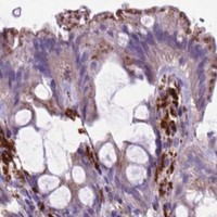
| Expected result | LZTR1-positive staining in glandular cells of appendix (HPA tissue IHC: Medium). HPA tissue profile: Granular cytoplasmic expression in gastrointestinal tract. No signal in the no-primary control. |
In paraffin-section IHC, expect granular cytoplasmic LZTR1 staining, particularly in gastrointestinal glandular cells (HPA tissue IHC). UniProt places LZTR1 in the Golgi apparatus, recycling endosome and endomembrane system, with no transmembrane segment (UniProt Q8N653). HPA rates its tissue IHC profile Approved but reports very low consistency between antibody staining and RNA expression; use the cell-level pattern as an observed reference, not proof of specificity (HPA tissue IHC).
| Granular cytoplasmic staining in appendix or duodenal glandular cells. | This matches the reported gastrointestinal pattern; both cell populations have Medium staining (HPA tissue IHC). Score the cellular pattern alongside intensity, because HPA reports very low staining–RNA consistency even though external characterization supports its antibody staining (HPA tissue IHC). |
| Predominantly nuclear staining in a paraffin section, without the expected granular cytoplasm. | Recheck interpretation and controls: this differs from the tissue IHC profile (HPA tissue IHC). Nucleoplasm is an approved main location in ICC-IF, so nuclear signal alone cannot be declared biologically impossible across applications (HPA subcellular ICC-IF). |
| Strong staining restricted to a cell population listed as Not detected, such as adipocytes. | Treat this as a possible false positive and check morphology, background and detection controls (HPA tissue IHC; general IHC practice). The HPA designation concerns the listed cell type, not every cell in that tissue (HPA tissue IHC). |
| Diffuse staining across tissue compartments, including areas without a clear cellular pattern. | This does not resemble the reported granular cytoplasmic profile (HPA tissue IHC). Review blocking, washing and chromogen development as general IHC checks; diffuse color alone cannot establish LZTR1 expression (general IHC practice). |
| No staining in appendix or duodenal glandular cells. | These are useful comparison cells because HPA reports Medium staining there (HPA tissue IHC). First inspect section quality, retrieval, antibody and detection controls; a blank section by itself does not establish absent LZTR1 (general IHC practice). |
| Tissue and cell selection | HPA reports Medium staining in several distinct populations, including appendix and duodenal glandular cells, breast myoepithelial cells and colon endothelial cells (HPA tissue IHC). Identify the scored cell type before comparing sections; whole-tissue labels are too broad for that comparison (general IHC practice). |
| IHC evidence strength | The tissue profile is Approved, while antibody staining has very low consistency with RNA expression; external characterization supports staining (HPA tissue IHC). HPA lists IHC as Approved for HPA068772 and HPA071248, without an IHC Enhanced designation in the supplied record (HPA antibodies). |
| Subcellular readout across applications | The IHC profile is granular cytoplasmic (HPA tissue IHC), whereas ICC-IF reports approved nucleoplasm, cytosol and centrosome locations (HPA subcellular ICC-IF). IF/ICC Q: Should its nuclear signal be the IHC scoring criterion? A: Use the paraffin-section IHC pattern for this guide; interpret ICC-IF on its own guide page (HPA tissue IHC; HPA subcellular ICC-IF). |
| Topology and epitope information | UniProt reports no transmembrane segment, signal peptide or propeptide, and describes LZTR1 as an endomembrane, recycling-endosome and Golgi protein (UniProt Q8N653). The supplied record gives no antibody epitope position, so it cannot predict whether a particular retrieval condition will expose that epitope. |
| Antigen retrieval and detection | Check retrieval and detection against the chosen IHC-validated antibody's documented conditions when optimizing paraffin sections (general IHC practice). Neither the supplied HPA nor UniProt record establishes LZTR1-specific fixation sensitivity, an optimal retrieval setting or an IHC dilution. |
| Situation | Likely cause | Next action |
|---|---|---|
| Expected positive cells are blank. | The result could reflect a missed staining step, unsuitable antibody conditions or insufficient detection (general IHC practice); HPA reports Medium appendix and duodenal glandular staining (HPA tissue IHC). | Verify section integrity and the staining run's controls, then review the antibody's IHC conditions and retrieval against its own documentation (general IHC practice). |
| Color is diffuse across cells and extracellular areas. | Background from blocking, washing or chromogen development can obscure a cellular pattern (general IHC practice). HPA instead describes granular cytoplasmic gastrointestinal staining (HPA tissue IHC). | Inspect a detection control and adjust the relevant blocking, washing or development step before scoring LZTR1 (general IHC practice). |
| Strong signal appears in adipocytes or respiratory epithelial cells. | Those specific populations are Not detected in the HPA tissue profile; cross-reactivity or endogenous detection activity is possible (HPA tissue IHC; general IHC practice). | Confirm cell identity, compare a documented positive cell population and run an appropriate detection control (general IHC practice). |
| Only nuclear staining is visible in IHC. | The result differs from HPA's granular cytoplasmic tissue profile, although ICC-IF separately reports approved nucleoplasmic localization (HPA tissue IHC; HPA subcellular ICC-IF). | Check controls and cellular morphology, then record the compartment explicitly; do not substitute the ICC-IF location for the paraffin-section IHC reference (general IHC practice; HPA tissue IHC). |
| A low-staining tissue gives a weak, patchy result. | HPA lists, for example, thyroid glandular cells and hepatocytes as Low; weak staining there offers a less clear comparison than Medium cells (HPA tissue IHC). | Compare with appendix or duodenal glandular cells, both reported Medium, in a controlled staining run (HPA tissue IHC; general IHC practice). |
| Results vary between antibodies or sections. | HPA reports very low consistency between antibody staining and RNA expression despite an Approved tissue profile (HPA tissue IHC); run conditions can also vary (general IHC practice). | Record antibody identity, retrieval and detection conditions, compare the same named cell populations, and avoid treating intensity alone as confirmation of specificity (general IHC practice; HPA tissue IHC). |
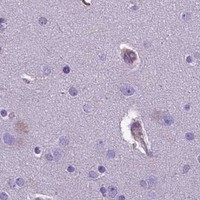


![HPA ICC-IF BJ [Human fibroblast] — LZTR1 (HPA067852)](https://images.proteinatlas.org/67852/1866_G5_33_blue_red_green.jpg)
![HPA ICC-IF BJ [Human fibroblast] — LZTR1 (HPA067852)](https://images.proteinatlas.org/67852/1866_G5_35_blue_red_green.jpg)
Comprehensive Human Protein Atlas IHC scoring per tissue (reliability: Approved — Very low consistency between antibody staining and RNA expression data. External characterization data supports antibody staining.). Rows are taken directly from the HPA tissue chart — click any row's HPA link to view the source.
| Tissue | Cell type | Level | Evidence | Source |
|---|---|---|---|---|
| Appendix | Glandular cells | Medium | Protein (IHC) | HPA → |
| Breast | Myoepithelial cells | Medium | Protein (IHC) | HPA → |
| Colon | Endothelial cells | Medium | Protein (IHC) | HPA → |
| Duodenum | Glandular cells | Medium | Protein (IHC) | HPA → |
| Epididymis | Glandular cells | Medium | Protein (IHC) | HPA → |
| Tissue | Cell type | Level | Evidence | Source |
|---|---|---|---|---|
| Adipose tissue | Adipocytes | Not detected | Protein (IHC) | HPA → |
| Adrenal gland | Glandular cells | Not detected | Protein (IHC) | HPA → |
| Bronchus | Respiratory epithelial cells | Not detected | Protein (IHC) | HPA → |
| Caudate | Glial cells | Not detected | Protein (IHC) | HPA → |
| Cerebellum | Cells in granular layer | Not detected | Protein (IHC) | HPA → |
Troubleshoot LZTR1 staining in paraffin sections by checking retrieval, compartment, controls, and cell-specific scoring before interpreting signal.
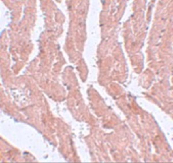
A06363 has real LZTR1 IHC data from mouse heart tissue (catalog image caption). The catalog lists human, mouse, and rat reactivity (catalog reactivity); no IF image is supplied (catalog images).
A06363 lists IHC-P and human, mouse, and rat reactivity (catalog applications and reactivity). Its own image shows LZTR1 IHC in mouse heart tissue at 5 μg/mL (catalog image caption).
Which to pick: Choose A06363 for paraffin-section tissue IHC (catalog: IHC-P); its own image shows mouse heart staining at 5 μg/mL (catalog image caption), but the fixative is unreported (catalog image caption). No SKU in this payload has IF/ICC validation or an IF dilution (catalog applications and IF fields). A06363 lists human, mouse, and rat reactivity (catalog reactivity), while its pictured IHC data cover mouse tissue only (catalog image caption).