This website uses cookies to ensure you get the best experience on our website.
- Table of Contents
Facts about Transcription factor MafA.
Binds the insulin enhancer C1/RIPE3b element (PubMed:15665000). Binds to consensus TRE-type MARE 5'-TGCTGACTCAGCA-3' DNA sequence (By similarity).
Rat | |
---|---|
Gene Name: | Mafa |
Uniprot: | D3ZNT6 |
Entrez: | 366949 |
Belongs to: |
---|
bZIP family |
hMafA; Pancreatic beta-cell-specific transcriptional activator; RIPE3b1; transcription factor MafA; Transcription factor RIPE3b1; v-maf musculoaponeurotic fibrosarcoma oncogene homolog A (avian); V-maf musculoaponeurotic fibrosarcoma oncogene homolog A
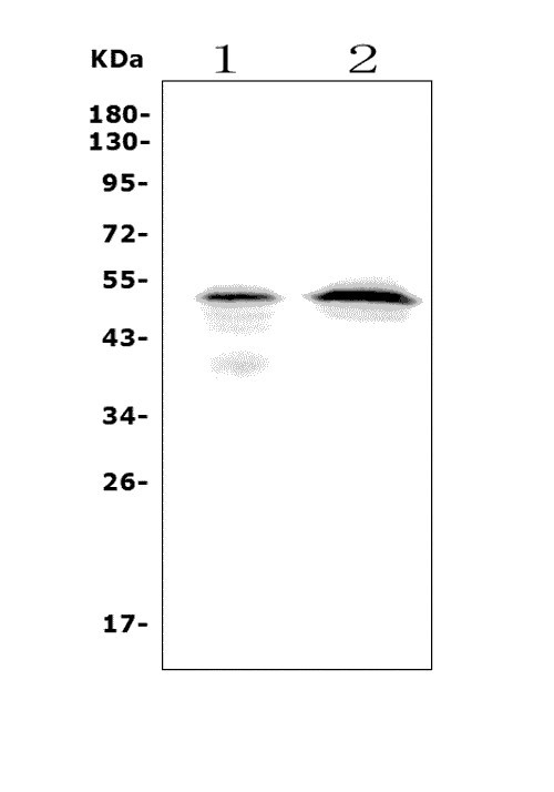
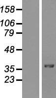
Mass (kDA):
37.78 kDA
Rat | |
---|---|
Location: | 7q34 |
Sequence: | 7; |
When using the MAFA marker, the antibodies must be diluted at 1:200 to get a desired signal. For best results, mix the MAFA with the antibody in a sterile container. This process removes any background signal. After a few minutes, the antibody should be ready for use. The MAFA is also known as the mafa marker. You can see this marker in a variety of studies.
If you are looking for MAFA ELISA Kits, look no further than Boster Bio. Founded in 1993 by a celebrated histologist, the company specializes in manufacturing high-specificity and sensitivity antibodies. Over 12,000 Boster Bio antibodies have been validated for use in WB, ELISA, and Flow Cytometry assays. All of their reagents are backed by Boster's quality guarantee.
The company also develops ELISA kits and research antibodies for detection of biomarkers in various areas, such as neurosciences, developmental biology, and inflammation. Its MAFA ELISA Kits have sensitivity levels up to the picogram range. These kits are available in a variety of formats. The company also offers custom services and BeNeLux deliveries. These kits provide high sensitivity and specificity and are backed by tebu-bio's technical support.
Boster Bio's IHC-optimized polyclonals for immunohistochemistry are made from recombinant proteins produced in Escherichia coli and eukaryotic cell lines. These antibodies recognize multiple epitopes on a single antigen with high specificity but modest affinity. They are characterized by high sensitivity, but are often limited by poor stability and batch-to-bath variability.
For a wide range of applications, Boster Bio has optimized its products for IHC. These antibodies have sensitivity at the picogram level and are backed by extensive validation for IHC and ELISA. Customers can request validation images and procedures. Supervision(tm) saves up to 30 min of IHC and improves background. The company's Picoband(tm) platform is powered by insights into the most effective immunogen design and a dedicated technical support team.
Boster Bio's IHC-optimized polyclonals for immunohistochemistry (IHC) are designed to detect the protein of interest in complex tissue samples. Their unique formulations prevent cross-reactivity, which can be useful for the detection of small molecules. As a result, researchers can be confident of finding high-quality antibodies for IHC. Ultimately, they can help them improve the results of their research.
Unlike monoclonal antibodies, polyclonal antibodies are more stable in the presence of a variety of protein types. They recognize multiple epitopes and are less likely to cross-react with other proteins. They also reduce background staining, a common issue with monoclonal antibodies. And, polyclonal antibodies are more versatile than monoclonal antibodies.
Monoclonal antibodies are less sensitive and have a low binding affinity. They must be rigorously washed to remove cross-reactive antigens. Polyclonal antibodies are made from host antigens. To obtain these antibodies, scientists immunize host animals with the antigens and extract the antibodies from their sera or eggs. Monoclonal antibodies are also produced in the same way, but they are not as highly specific as their monoclonal counterparts.
The ELISA kit for the MAFA marker is a solid-phase, 1.5-hour assay. It is intended for research purposes only. Because human C-peptide has a half-life three to four times that of insulin, it is a useful marker of insulin production from beta cells. This kit is designed to detect low levels of the marker in serum and plasma. Its sensitivity ranges from 0.1 to 10 micrograms.
SpectraMax readers were used to prepare ELISA Boosters. The Sword ELISA Booster assays detected inflammatory cytokines at low levels of IL-1ss and TNF-a. Both assays used a Molecular Devices SpectraMax(r) reader to measure the levels of the two cytokines. A Sword ELISA Booster contains 3.2 mL of 5X Sword Development solution in 12.8 mL deionized water.
The MAFA ELISA kit uses a competitive enzyme immunoassay technique. The MAFA antigen is bound to a polyclonal antibody called MAFA-HRP conjugate. The assay sample is incubated for one hour. The sample is then decanted and washed five times. The wells are then incubated with the HRP enzyme, which binds the MAFA-HRP conjugate and forms a blue color complex. After the reaction is complete, the color intensity of each sample is recorded.
During beta cell development, MafA and NEUROG3 play an important role. Recently, ectopic expression of these genes has been used to reprogram other cell types into insulin-producing cells. For example, NEUROG3 and PDX1 have been used to reprogram pancreatic exocrine cells into insulin-producing cells. Similarly, ectopic expression of MAFA has been used to reprogram pluripotent stem cells into insulin-producing cells.
To identify the mafA gene, we first performed PCR. Using the N primer (5'-GGATCCATCGCGCGCTGCGTGT-3'), we isolated the coding region of the gene (pGEM(T)). Then, the cloned gene fragment was digested with Bamh2 restriction enzyme. Finally, we subcloned this fragment into a lentivirus-based expression vector obtained from BiOSETTIA.
PMID: 15665000 by Zhao L., et al. The islet beta cell-enriched MafA activator is a key regulator of insulin gene transcription.
PMID: 18042454 by Rocques N., et al. GSK-3-mediated phosphorylation enhances Maf-transforming activity.