This website uses cookies to ensure you get the best experience on our website.


- Table of Contents
Plan MAP2K1 paraffin IHC around general cytoplasmic staining (HPA tissue IHC). Lung macrophages show medium staining, while breast adipocytes are undetected; interpret these references in light of the low consistency between staining and RNA expression (HPA tissue IHC).

Expected localisation, validated protocols, controls and antibodies — the at-a-glance facts below, then the full design guide.
| Expected localisation | General cytoplasmic tissue staining (HPA tissue IHC) | |
| Staining pattern | Cytoplasmic staining across multiple cell types (HPA tissue IHC) | |
| Antigen retrieval | EDTA pH 8.0 HIER, heat-mediated (datasheet A00292) | |
| Positive control | Adrenal gland+4 more · see all | |
| Negative control | Breast+4 more · see all |
| Fixation | Keep fixation consistent across paraffin sections (standard IHC practice; not target-specific) | |
| Caveat | Staining has low consistency with RNA expression (HPA tissue IHC) | |
| Regulation | Low tissue specificity (HPA tissue RNA) | |
| Isoform / epitope | 2 isoforms; full-length chain, epitope coverage unknown (UniProt) |
The catalog antibody’s IHC-P protocol is accompanied by published MAP2K1 staining protocols for hypopharyngeal squamous cell carcinoma and liver sections (PMC11984367; PMC7641625).
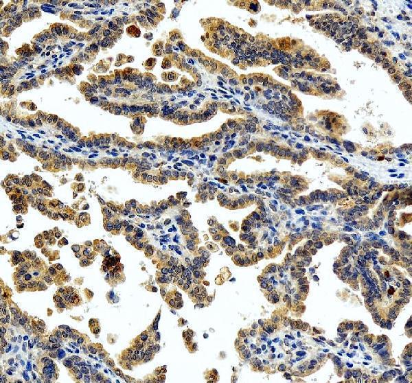
| Sample | Paraffin-embedded human ovarian cancer tissue; fixative not specified (datasheet A00292) |
| Fixation | Image fixative and duration unreported (datasheet A00292); verify before use. |
| Sectioning | 4–5 µm sections on charged slides (standard) |
| Deparaffinisation | Xylene, graded ethanol series to water (standard) |
| Antigen retrieval | Heat retrieval: EDTA pH 8.0 (datasheet A00292); 20 min, 95–100 °C (standard) |
| Peroxidase block | 3% H2O2, 10 min, room temperature (standard) |
| Blocking | 10% goat serum (datasheet A00292) |
| Primary antibody | Rabbit anti-MAP2K1, 2-5 μg/ml (datasheet A00292) |
| Primary incubation | Overnight at 4 °C (datasheet A00292) |
| Detection | HRP-conjugated secondary, DAB chromogen (datasheet A00292) |
| Counterstain | Hematoxylin, blue, dehydrate and mount (standard) |
| Expected result | MAP2K1-positive staining in glandular cells of adrenal gland (HPA tissue IHC: Medium). HPA tissue profile: General cytoplasmic expression. No signal in the no-primary control. |
MAP2K1 staining in paraffin sections is expected mainly in the cytoplasm: HPA reports general cytoplasmic expression, including medium staining in duodenal glandular cells and lung macrophages (HPA tissue IHC). UniProt also lists nuclear, membrane and mitotic structures, and reports no transmembrane segment (UniProt Q02750). Interpret tissue comparisons cautiously: HPA rates its IHC data Approved but reports low consistency between antibody staining and RNA expression (HPA tissue IHC).
| Cytoplasmic staining in duodenal glandular cells or lung macrophages. | This fits the reported IHC pattern and medium staining level in those specific cell populations (HPA tissue IHC). Judge the cells on the slide; the HPA entries do not imply that every cell in either tissue should stain. |
| Strong staining confined to nuclei, with little cytoplasmic signal. | Recheck the pattern against HPA's general cytoplasmic IHC profile (HPA tissue IHC). UniProt also lists nuclear localization (UniProt Q02750), so nuclear signal alone does not prove an artefact; compare it with a positive tissue and controls. |
| Signal mainly in a cell population reported as undetected. | For example, HPA reports no detection in bronchial respiratory epithelial cells (HPA tissue IHC). Consider antibody cross-reactivity or endogenous chromogenic activity, then examine a no-primary control. An HPA negative entry is a comparison point, not a universal absence claim. |
| Diffuse colour across tissue, stroma and blank areas. | An indiscriminate deposit does not match HPA's cell-resolved cytoplasmic pattern (HPA tissue IHC). In general IHC practice, assess background from detection reagents, blocking and washing with a no-primary control before scoring MAP2K1-positive cells. |
| No staining in a selected HPA medium-staining cell population. | Absence in duodenal glandular cells or lung macrophages conflicts with the cited HPA observations (HPA tissue IHC), but does not establish biological loss. First check tissue preservation, antibody and detection performance, and whether those cells are present in the section. |
| Tissue and cell selection | HPA reports medium staining in several glandular and neuronal populations and lung macrophages, but no detection in selected epithelial and other cells (HPA tissue IHC). Select and score the named cell population rather than treating an entire organ as uniformly positive. |
| Compartment and topology | General cytoplasmic IHC staining is the tissue reference (HPA tissue IHC). UniProt also records nucleus, membrane and mitotic structures, with no transmembrane segment (UniProt Q02750); these annotations do not predict that every location will resolve in chromogenic sections. |
| Antibody interpretation | Three listed antibodies have Approved IHC status; HPA reports low consistency between staining and RNA expression for this tissue profile (HPA antibody validation; HPA tissue IHC). Treat an unexpected distribution as a result to validate, not a definitive expression map. |
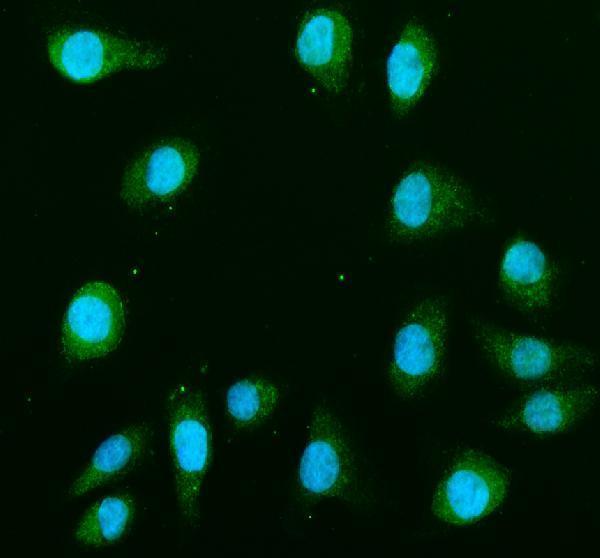
| IF/ICC Q: Should the section resemble an ICC image? | A: HPA supports plasma membrane and cytosol localization, plus basal body localization, in ICC-IF (HPA subcellular). Use that as compartment context; assess paraffin-section staining against HPA tissue IHC. IF/ICC methods belong in the separate guide. |
| Situation | Likely cause | Next action |
|---|---|---|
| A selected positive population has no visible chromogen. | The chosen cells may be absent, or the staining run may have failed; HPA reports medium staining in duodenal glandular cells and lung macrophages (HPA tissue IHC). | Verify cell identity on the counterstained section and inspect a same-run positive control. As general IHC practice, check antibody, retrieval and detection steps against the catalog antibody's IHC-P instructions. |
| Colour appears throughout the slide, including regions without target cells. | Noncellular background can obscure HPA's general cytoplasmic pattern (HPA tissue IHC). In general IHC practice, detection reagent background or insufficient washing are possible causes. | Review a no-primary control and the detection blank; adjust blocking, washing or chromogen development using standard IHC controls before interpreting faint cellular staining. |
| Macrophages show unusually intense chromogen or a no-primary control stains. | Endogenous detection activity is a possible source of chromogenic signal in general IHC practice. HPA reports medium MAP2K1 staining in lung macrophages (HPA tissue IHC), so cell identity alone cannot resolve the signal's origin. | Compare the no-primary control on the same tissue and check the detection system's endogenous-activity blocking step. Score MAP2K1 only where specific cellular staining exceeds that control. |
| Nuclear signal dominates while cytoplasmic staining is weak. | This departs from the general tissue IHC profile (HPA tissue IHC), although nuclear localization is listed by UniProt (UniProt Q02750). | Compare a known-positive section and no-primary control under the same scoring criteria. Report the compartment observed; avoid calling nuclear staining either specific or artefactual from location alone. |
| Cells reported as undetected show clear staining. | HPA reports no detection in bronchial respiratory epithelial cells and breast adipocytes (HPA tissue IHC). Unexpected signal could reflect background or antibody cross-reactivity; the HPA profile has low RNA–staining consistency (HPA tissue IHC). | Confirm the cell type and examine no-primary and positive controls. If the signal persists, document the discrepancy and seek independent antibody or orthogonal evidence before assigning MAP2K1 expression. |
| ICC-IF membrane signal seems inconsistent with cytoplasmic tissue IHC. | HPA supports plasma membrane and cytosol localization in ICC-IF, while its tissue IHC profile is generally cytoplasmic (HPA subcellular; HPA tissue IHC). The observations come from different applications. | Score the paraffin section using its visible cellular pattern and tissue controls. Use the separate IF/ICC guide for fluorescence interpretation; do not transfer an ICC-IF appearance into an IHC-P protocol expectation. |


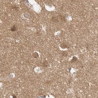
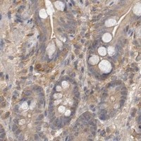
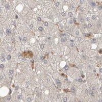
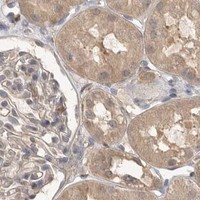


Comprehensive Human Protein Atlas IHC scoring per tissue (reliability: Approved — Low consistency between antibody staining and RNA expression data.). Rows are taken directly from the HPA tissue chart — click any row's HPA link to view the source.
| Tissue | Cell type | Level | Evidence | Source |
|---|---|---|---|---|
| Adrenal gland | Glandular cells | Medium | Protein (IHC) | HPA → |
| Appendix | Glandular cells | Medium | Protein (IHC) | HPA → |
| Caudate | Neuronal cells | Medium | Protein (IHC) | HPA → |
| Cerebral cortex | Neuronal cells | Medium | Protein (IHC) | HPA → |
| Duodenum | Glandular cells | Medium | Protein (IHC) | HPA → |
| Tissue | Cell type | Level | Evidence | Source |
|---|---|---|---|---|
| Breast | Adipocytes | Not detected | Protein (IHC) | HPA → |
| Bronchus | Respiratory epithelial cells | Not detected | Protein (IHC) | HPA → |
| Cervix | Squamous epithelial cells | Not detected | Protein (IHC) | HPA → |
| Esophagus | Squamous epithelial cells | Not detected | Protein (IHC) | HPA → |
| Liver | Cholangiocytes | Not detected | Protein (IHC) | HPA → |
Troubleshoot MAP2K1 staining in paraffin sections by checking retrieval, compartment, cell identity and controls before comparing chromogenic signal across samples.
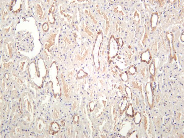
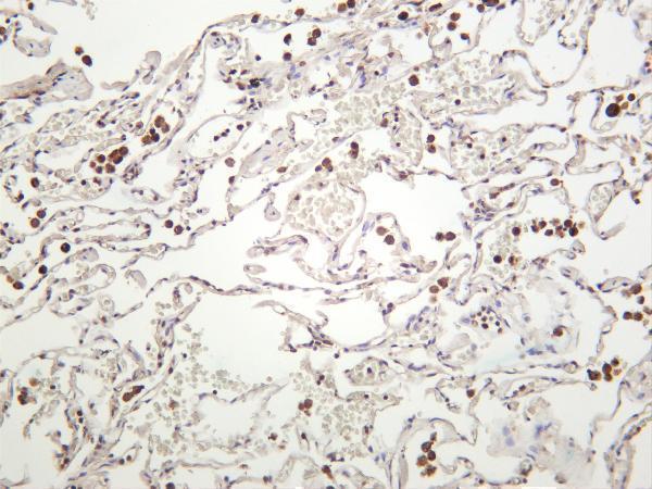
The catalog includes IHC images of human ovarian cancer, kidney and lung, plus IF images; listed reactivity extends to Human, Mouse and Rat (catalog image captions and reactivity).
A00292 has IHC data from a human ovarian cancer paraffin section and IF data from TPC1 and U2OS cells; M00292-5 has IHC images of human kidney and lung (catalog image captions). M00292-1 has IHC data from a human kidney paraffin section and IF data from HeLa cells; M00292-2 has an IF figure whose caption does not identify the specimen (catalog image captions).
Which to pick: For tissue IHC, A00292 provides the most detailed starting conditions: its own paraffin-section caption specifies EDTA retrieval at pH 8.0, 2 μg/ml primary antibody and DAB detection; the fixative is unreported (A00292 IHC image caption). For IF/ICC, M00292-2 is listed for both applications and has an IF figure at 1:50, though its caption does not identify the specimen (M00292-2 catalog applications and IF image caption). For cross-species planning, monoclonal M00292-5 lists Human, Mouse and Rat reactivity, while its IHC images show human kidney and lung only; section preparation and fixative are unreported (M00292-5 catalog description, reactivity and IHC image captions).