This website uses cookies to ensure you get the best experience on our website.


- Table of Contents
Plan paraffin-section MAP4K1 IHC with lymphoid tissue controls and a cytoplasmic staining expectation (HPA tissue IHC). The guide covers fixation consistency, antigen retrieval, antibody dilution, chromogenic detection, and interpretation of possible off-target staining (HPA tissue IHC).

Expected localisation, validated protocols, controls and antibodies — the at-a-glance facts below, then the full design guide.
| Expected localisation | Cytoplasmic staining in lymphoid tissue (HPA tissue IHC) | |
| Staining pattern | Cytoplasmic staining in germinal center cells (HPA tissue IHC) | |
| Antigen retrieval | EDTA pH 8.0 HIER, heat-mediated (datasheet A07909-3) | |
| Positive control | Bone marrow+4 more · see all | |
| Negative control | Adipose tissue+4 more · see all |
| Fixation | Keep fixation conditions consistent across sections (standard IHC practice; not target-specific) | |
| Caveat | Presumed off-target staining has been observed (HPA tissue IHC) | |
| Regulation | Primarily expressed in hematopoietic organs (UniProt) | |
| Isoform / epitope | Two isoforms; confirm antibody epitope coverage (UniProt) |
The catalog antibody protocol uses EDTA pH 8.0 retrieval (datasheet A07909-3); the three published MAP4K1 IHC protocols below provide sample-specific alternatives (PMC10514360; PMC11472321; PMC6491656).
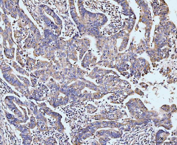
| Sample | Paraffin-embedded human colonic adenocarcinoma tissue; fixative not specified (datasheet A07909-3) |
| Fixation | Image fixative and duration unreported (datasheet A07909-3); verify before use. |
| Sectioning | 4–5 µm sections on charged slides (standard) |
| Deparaffinisation | Xylene, graded ethanol series to water (standard) |
| Antigen retrieval | Heat retrieval: EDTA pH 8.0 (datasheet A07909-3); 20 min, 95–100 °C (standard) |
| Peroxidase block | 3% H2O2, 10 min, room temperature (standard) |
| Blocking | 10% goat serum (datasheet A07909-3) |
| Primary antibody | Rabbit anti-MAP4K1, 2-5 μg/ml (datasheet A07909-3) |
| Primary incubation | Overnight at 4 °C (datasheet A07909-3) |
| Detection | Streptavidin-biotin complex (SABC), DAB chromogen (datasheet A07909-3) |
| Counterstain | Hematoxylin, blue, dehydrate and mount (standard) |
| Expected result | MAP4K1-positive staining in hematopoietic cells of bone marrow (HPA tissue IHC: High). HPA tissue profile: Cytoplasmic expression in lymphoid tissues. No signal in the no-primary control. |
MAP4K1 staining in paraffin-section IHC is expected mainly in the cytoplasm of lymphoid and hematopoietic cells (HPA: tissue IHC profile). Bone marrow hematopoietic cells and lymph node and tonsil germinal center cells show high staining (HPA: tissue IHC). MAP4K1 has no transmembrane segment (UniProt Q92918: topology). HPA rates the tissue profile Enhanced, with medium consistency between antibody staining and RNA data and presumed off-target staining disregarded (HPA: reliability description).
| Cytoplasmic staining in bone marrow hematopoietic cells or lymph node and tonsil germinal center cells. | This matches the expected positive pattern; HPA reports high staining in those cell populations (HPA: tissue IHC). Judge the labeled cells and their compartment against the section’s morphology, rather than treating every cell in a positive tissue as positive (general IHC interpretation). |
| Cytoplasmic staining in appendix germinal center cells or spleen white-pulp cells. | A moderate signal can fit the reference pattern: both populations are reported at medium staining, below the high-staining populations listed above (HPA: tissue IHC). Compare comparable cell populations when judging intensity (general IHC interpretation). |
| Predominantly nuclear staining, with little staining in the expected cytoplasmic compartment. | This conflicts with the reported cytoplasmic tissue-IHC profile (HPA: tissue IHC). Treat it as a possible artifact or nonspecific result and inspect morphology and controls before assigning it to MAP4K1 (general IHC interpretation). |
| Strong staining in adipocytes or respiratory epithelial cells while expected lymphoid cells are unstained. | HPA reports no detection in adipose-tissue adipocytes or bronchial respiratory epithelial cells (HPA: tissue IHC). Consider off-target antibody binding or endogenous chromogenic activity; HPA’s reliability note acknowledges presumed off-target staining (HPA: reliability description; general IHC practice). |
| Diffuse color across tissue, or no signal in a bone marrow or tonsil positive control. | Diffuse color without a recognizable cell pattern is difficult to score as specific staining (general IHC interpretation). A blank expected positive control prevents a reliable negative call on test sections; bone marrow and tonsil contain high-staining populations (HPA: tissue IHC). |
| Tissue and cell population | Expression is concentrated in hematopoietic organs (UniProt Q92918: tissue specificity). HPA reports high staining in bone marrow hematopoietic cells and lymph node and tonsil germinal center cells, and medium staining in appendix germinal centers and spleen white pulp (HPA: tissue IHC). |
| Antibody validation and specificity | Both listed antibodies, HPA007419 and CAB007754, have Enhanced IHC status (HPA: antibodies). The overall tissue profile has medium agreement with RNA data, and presumed off-target binding was disregarded; a labeled cell outside the reference pattern still needs scrutiny (HPA: reliability description). |
| Compartment and protein architecture | The tissue-IHC profile is cytoplasmic (HPA: tissue IHC). UniProt lists no transmembrane segment or signal peptide for MAP4K1 (UniProt Q92918: topology, processing). These features do not establish a target-specific antigen-retrieval or fixation effect (UniProt Q92918: record scope). |
| Isoforms and epitope coverage | UniProt lists two MAP4K1 isoforms (UniProt Q92918: isoforms). The supplied evidence gives no epitope location or isoform coverage for the catalog antibody, so an isoform-specific staining difference cannot be predicted from this record (UniProt Q92918: record scope). |
| IF/ICC Q&A | Q: Should membrane staining in IF/ICC change the paraffin-IHC expectation? A: HPA reports an approved plasma-membrane location in ICC-IF, while its tissue-IHC profile is cytoplasmic (HPA: subcellular; HPA: tissue IHC). Interpret each assay against its own reported pattern; the supplied sources do not explain the difference. |
| Situation | Likely cause | Next action |
|---|---|---|
| No chromogenic signal in a test section or expected positive control. | The run has not demonstrated detection in a population reported as high staining (HPA: bone marrow, lymph node and tonsil tissue IHC). Tissue content or a general staining-workflow failure may be responsible (general IHC practice). | Confirm that the positive-control section contains the expected cells; check antibody application, detection reagents and control performance before calling the test tissue negative (general IHC practice). |
| Signal appears mainly nuclear. | Nuclear dominance differs from the cytoplasmic tissue-IHC profile (HPA: tissue IHC). Counterstain overlap or nonspecific staining can complicate compartment calls (general IHC interpretation). | Review the counterstain and cell boundaries at higher magnification; compare the expected cytoplasmic positive-control cells and appropriate negative controls (general IHC practice). |
| Strong color appears in adipocytes or bronchial respiratory epithelium. | Those populations are reported as not detected (HPA: tissue IHC). Off-target binding is plausible given HPA’s reliability note; endogenous chromogenic activity is another general IHC possibility (HPA: reliability description; general IHC practice). | Check no-primary and detection controls, then compare staining with the expected hematopoietic or germinal-center pattern before scoring it as MAP4K1 (general IHC practice; HPA: tissue IHC). |
| Color is diffuse across the section, with poor cell-level definition. | Background from the staining or detection workflow can obscure compartment and cell-type assessment (general IHC practice). The HPA reference pattern requires recognizable cytoplasmic staining in lymphoid cells (HPA: tissue IHC). | Inspect control sections and review blocking, washing, antibody concentration and chromogen development as general IHC workflow variables; reassess only where cell boundaries remain clear (general IHC practice). |
| Spleen or appendix staining looks weaker than tonsil or lymph node. | HPA rates spleen white-pulp cells and appendix germinal-center cells medium, versus high in tonsil and lymph node germinal-center cells (HPA: tissue IHC). | Score the relevant cell populations against their tissue-specific reference levels; use a high-staining reference population to confirm the run if the result remains uncertain (HPA: tissue IHC; general IHC practice). |
| The catalog antibody stains a region outside the expected tissue pattern. | Enhanced IHC status supports the reference pattern, but does not make every observed signal specific; HPA reports medium staining–RNA consistency and disregarded presumed off-target binding (HPA: antibodies; HPA: reliability description). | Document the affected cell type and compartment, compare the tissue-IHC reference and controls, and withhold a MAP4K1-positive call if the discordance persists (HPA: tissue IHC; general IHC interpretation). |


Comprehensive Human Protein Atlas IHC scoring per tissue (reliability: Enhanced — Medium consistency between antibody staining and RNA expression data. Presumed off target binding observed and disregarded.). Rows are taken directly from the HPA tissue chart — click any row's HPA link to view the source.
| Tissue | Cell type | Level | Evidence | Source |
|---|---|---|---|---|
| Bone marrow | Hematopoietic cells | High | Protein (IHC) | HPA → |
| Lymph node | Germinal center cells | High | Protein (IHC) | HPA → |
| Tonsil | Germinal center cells | High | Protein (IHC) | HPA → |
| Appendix | Germinal center cells | Medium | Protein (IHC) | HPA → |
| Spleen | Cells in white pulp | Medium | Protein (IHC) | HPA → |
| Tissue | Cell type | Level | Evidence | Source |
|---|---|---|---|---|
| Adipose tissue | Adipocytes | Not detected | Protein (IHC) | HPA → |
| Adrenal gland | Glandular cells | Not detected | Protein (IHC) | HPA → |
| Breast | Adipocytes | Not detected | Protein (IHC) | HPA → |
| Bronchus | Respiratory epithelial cells | Not detected | Protein (IHC) | HPA → |
| Caudate | Glial cells | Not detected | Protein (IHC) | HPA → |
Troubleshoot MAP4K1 staining in paraffin sections using the catalog antibody’s tissue protocol and cell-specific controls.
Three human-reactive antibodies have paraffin-section IHC images (catalog captions); A30542 also lists mouse reactivity and IF/ICC applications (catalog applications/reactivity).
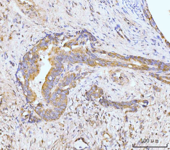
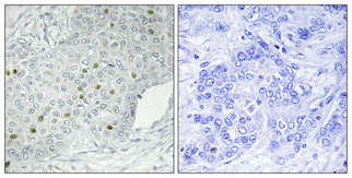
A07909-3 has IHC images of human colonic adenocarcinoma and gall bladder adenosquamous carcinoma paraffin sections; A30542 has an IHC image of human breast cancer paraffin tissue (catalog image captions). M07909 has IHC images of human epityphlon and tonsil paraffin sections (catalog image captions).
Which to pick: For human paraffin-section IHC, choose A07909-3 for the documented EDTA pH 8 retrieval and 2 μg/ml overnight staining with DAB detection, or rabbit polyclonal M07909 for its documented automated 1:500 IHC protocol (A07909-3 and M07909 image captions; M07909 catalog dilution_raw). For IF/ICC or a human- and mouse-reactive option, choose rabbit polyclonal A30542, which lists those applications and species; its IHC image shows human breast cancer paraffin tissue, while no IF image is supplied (A30542 catalog applications/reactivity/dilution_raw and image captions). The fixative is unreported for all three IHC examples (catalog image captions).