MAVS / Mitochondrial antiviral-signaling protein · IHC design guide

Design Immunohistochemistry for MAVS

Plan chromogenic MAVS IHC in paraffin sections using granular cytoplasmic staining as a tissue benchmark (HPA tissue IHC). Compare staining by cell type and verify epitope coverage across six isoforms (HPA tissue IHC; UniProt).

Evidence assembled Oct 2026 · For research use; verify linked source records and product datasheet before use
Immunohistochemistry protocol sheet for MAVS (IHC for MAVS): expected localisation Granular cytoplasm in most tissues (HPA tissue IHC), antibody A00169-1, validated IHC image, and IHC protocol steps
Printable MAVS IHC protocol sheet — expected localisation Granular cytoplasm in most tissues (HPA tissue IHC), antibody A00169-1, controls and protocol steps. Open the full MAVS IHC guide →

MAVS Immunohistochemistry Experimental Design Guide

Expected localisation, validated protocols, controls and antibodies — the at-a-glance facts below, then the full design guide.

Must know before staining
Expected localisation Granular cytoplasm in most tissues (HPA tissue IHC)
Staining pattern Most tissues show granular cytoplasmic staining (HPA tissue IHC)
Antigen retrieval Citrate pH 6 HIER, heat-mediated (datasheet A00169-1)
Positive control ⓘ Adrenal gland+4 more · see all
Negative control ⓘ None in HPA (detected in all 44 tissues); use no-primary + isotype controls
Important caveats
Reasons your staining may differ from the expected pattern.
Fixation Keep fixation consistent across sections (standard IHC practice; not target-specific)
Caveat Hematopoietic-cell staining may complicate scoring (HPA tissue IHC)
Regulation Expression changes are not specified (UniProt)
Isoform / epitope Six isoforms; verify antibody epitope coverage (UniProt)
Section 1

Recommended MAVS IHC & IF Protocols

The catalog antibody protocol is accompanied by one published MAVS IHC protocol using paraffin-embedded tissue microarrays (PMC10468804).

Recommended immunohistochemistry (IHC-P) protocol parameters
SampleParaffin-embedded human mammary cancer tissue; fixative not specified (datasheet A00169-1)
FixationImage fixative and duration unreported (datasheet A00169-1); verify before use.
Sectioning4–5 µm sections on charged slides (standard)
DeparaffinisationXylene, graded ethanol series to water (standard)
Antigen retrievalHeat retrieval: Citrate pH 6, 20 min (datasheet A00169-1)
Peroxidase block3% H2O2, 10 min, room temperature (standard)
Blocking10% goat serum (datasheet A00169-1)
Primary antibodyRabbit anti-MAVS, 0.5-1μg/ml (datasheet A00169-1)
Primary incubationOvernight at 4 °C (datasheet A00169-1)
DetectionStreptavidin-biotin complex (SABC), DAB chromogen (datasheet A00169-1)
CounterstainHematoxylin, blue, dehydrate and mount (standard)
Expected resultMAVS-positive staining in glandular cells of adrenal gland (HPA tissue IHC: High). HPA tissue profile: Granular cytoplasmic expression in most tissues. No signal in the no-primary control.
💡Decision noteStart with heat-mediated citrate retrieval at pH 6 (datasheet A00169-1); the published protocol used 95–98°C for 20 min (PMC10468804).
Section 2

What Is the Expected MAVS Staining Pattern?

MAVS should appear as granular cytoplasmic staining in many tissues (HPA: tissue IHC profile, Enhanced reliability). Its outer mitochondrial membrane anchor leaves most of the protein facing the cytoplasm; UniProt also places MAVS on peroxisomes (UniProt Q7Z434 topology and subcellular location). High staining is documented in bronchial respiratory epithelium, breast glandular cells, bone marrow hematopoietic cells and cerebral cortex glia (HPA: tissue IHC).

What am I looking at on my slide?
Granular cytoplasmic staining in bronchial respiratory epithelial cells, with tissue structure still clear (HPA: High in these cells).This matches the reported IHC profile and is consistent with membrane-associated MAVS (HPA: granular cytoplasm in most tissues; UniProt Q7Z434: outer mitochondrial membrane). Chromogenic IHC alone cannot assign each granule to a mitochondrion or peroxisome.
Predominantly nuclear staining, or a crisp cell-surface outline instead of cytoplasmic granules.Neither compartment matches the reported MAVS locations (UniProt Q7Z434: mitochondrion outer membrane and peroxisome; HPA: granular cytoplasm). Treat the pattern as suspect and compare it with a known-positive section and detection controls before scoring it.
Strong staining in chondrocytes while documented high-staining cells on the same run are weak (HPA: Low in soft-tissue chondrocytes).The reversed pattern raises concern for cross-reactivity or endogenous detection activity. Low HPA staining does not mean zero expression, so judge the cell distribution and controls together rather than declaring every stained chondrocyte false.
Uniform color over cells, extracellular material and empty spaces, with little granular detail.This is diffuse background, not the cellular pattern reported for MAVS (HPA: granular cytoplasmic expression). In general chromogenic IHC practice, insufficient blocking or excess detection reagent can obscure specific staining; use the no-primary control to locate background.
No cellular signal in an adequately represented bronchial section (HPA: High in respiratory epithelial cells).First suspect a failed or insensitive IHC run, while checking that respiratory epithelium is present. HPA reports high staining there, but tissue expression does not guarantee every antibody and processing condition will produce a visible result.
💡Expected MAVS appearanceCall a section positive when expected cells show discernible granular cytoplasmic color, especially HPA high-staining cell populations; predominantly nuclear, cell-surface or structure-free diffuse color is suspect (HPA: tissue IHC profile; UniProt Q7Z434: membrane location).
How each factor affects the staining
Compartment and resolutionMAVS has a transmembrane segment at residues 514–534, with residues 2–513 cytoplasmic (UniProt Q7Z434 topology). Expect cytoplasmic granules in routine IHC (HPA: tissue IHC); their appearance does not distinguish mitochondrial from peroxisomal MAVS.
Antibody and epitopeHPA rates IHC with HPA049850 and HPA053524 Enhanced, and CAB009187 Supported (HPA: antibody validation). MAVS has six isoforms (UniProt Q7Z434); without a supplied antibody epitope, isoform coverage and any retrieval requirement are undetermined.
Reference tissue selectionUse a documented high-staining population, such as appendix glandular cells or bone marrow hematopoietic cells, to assess the run (HPA: High in each). Chondrocytes are reported Low and are a weak reference; HPA supplies no verified negative tissue.
IF/ICC Q: What localisation should fluorescence show?A: HPA reports enhanced mitochondrial localisation in ICC-IF images from A-431, U-251MG and U2OS (HPA: subcellular). That supports a mitochondrial comparison, while this page's positive call remains the observed granular cytoplasmic IHC pattern (HPA: tissue IHC).
Why is my staining missing, weak or wrong?
SituationLikely causeNext action
Known-positive respiratory epithelium has no stain (HPA: High in bronchus).The section may lack the expected cells, or an IHC reagent or detection step may have failed.Confirm tissue identity and morphology, then check a documented high-staining control in the same run; review the antibody's validated IHC instructions before adjusting general workflow settings.
Signal is faint across documented high-staining tissues (HPA: tissue IHC).The run may be insensitive; HPA staining levels do not establish MAVS-specific retrieval or fixation sensitivity.Check primary-antibody dilution, retrieval and detection against the antibody's IHC instructions, changing one general workflow variable at a time and retaining a high-staining control.
Color is strongest in nuclei rather than cytoplasmic granules.The compartment conflicts with MAVS localisation (UniProt Q7Z434: outer mitochondrial membrane and peroxisome; HPA: granular cytoplasm).Review the no-primary control, stain development and antibody specificity; withhold a MAVS-positive call until the expected cytoplasmic pattern is visible.
Bone marrow shows widespread color without distinct stained cells (HPA: High in hematopoietic cells).Background or endogenous chromogenic detection activity may conceal the cellular pattern (general IHC practice).Compare a no-primary section and the assay's endogenous-enzyme control; address control-positive color in the detection workflow before interpreting hematopoietic staining.
Soft-tissue chondrocytes stain more strongly than the chosen high-staining control (HPA: Low in chondrocytes).Cross-reactivity or background is possible; HPA's Low category is not a negative-control designation.Check the control section and no-primary section, then compare the cell-specific distribution with HPA rather than assigning specificity from intensity alone.
A punctate IF/ICC image seems inconsistent with broader chromogenic granules.HPA reports mitochondrial ICC-IF localisation and granular tissue IHC, which are read at different spatial resolution (HPA: subcellular and tissue IHC).Interpret each against its own HPA reference pattern; reserve mitochondrial assignment for the IF/ICC evidence and score the IHC section by cell type and cytoplasmic distribution.

Sample controls for MAVS IHC & IF

🧪Run bronchus first: respiratory epithelial cells should stain (HPA: High in bronchus respiratory epithelial cells). HPA detects MAVS in all 44 scored tissues, so use no-primary and isotype controls for negative comparisons; any unstained cells on the positive slide should show counterstain only, but cannot be assumed to be biological negatives (HPA: no negative tissue rows).
Positive control tissue: Adrenal gland (Glandular cells, HPA High)
Negative control tissue: None in HPA: MAVS is detected in all 44 scored tissues. Use a no-primary (secondary-only) and an isotype control instead.
ICC-IF cell lines (HPA subcellular resource): HPA ICC-IF images show MAVS in A-431, U-251MG, U2OS, with annotated localisation: Mitochondria (enhanced) (HPA subcellular).
Technical controls: Include a secondary-only slide, a concentration-matched nonimmune rabbit IgG isotype control, and identically processed MAVS-knockout material as a biological negative (caption: rabbit anti-MAVS antibody; standard IHC controls). For DAB detection, quench endogenous peroxidase and check bronchial background; if using the caption’s streptavidin–biotin method, control for endogenous biotin (caption: SABC/DAB).
⚠️Feasibility: A target-specific fixation window or fixation effect is unreported, and the selected A00169-1 tissue-IHC caption does not state its fixative (caption: fixative not stated). The paraffin-section example uses citrate retrieval at pH 6 for 20 minutes; retrieval dependence beyond that example is unreported (caption: citrate retrieval). No matched evidence establishes that frozen sections or IF are easier; HPA shows mitochondrial ICC-IF localization in A-431, U-251MG and U2OS, while endogenous peroxidase can complicate chromogenic scoring in bronchial inflammatory cells (HPA: subcellular ICC-IF; standard IHC practice).

HPA tissue IHC evidence for MAVS

Comprehensive Human Protein Atlas IHC scoring per tissue (reliability: Enhanced — High consistency between antibody staining and RNA expression data.). Rows are taken directly from the HPA tissue chart — click any row's HPA link to view the source.

Positive expression · recommended positive controls

TissueCell typeLevelEvidenceSource
Adrenal gland Glandular cells High Protein (IHC) HPA →
Appendix Glandular cells High Protein (IHC) HPA →
Bone marrow Hematopoietic cells High Protein (IHC) HPA →
Breast Glandular cells High Protein (IHC) HPA →
Bronchus Respiratory epithelial cells High Protein (IHC) HPA →

Undetected expression · recommended negative controls

TissueCell typeLevelEvidenceSource
None in HPA: MAVS is detected in all 44 scored tissues. Use a no-primary (secondary-only) and an isotype control instead.
Section 3

Advanced MAVS IHC Tips

Troubleshooting guidance for chromogenic MAVS IHC in paraffin sections, with one Q&A on IF multiplexing.

How should I retrieve MAVS when staining paraffin sections?
Start with heat-mediated citrate buffer at pH 6 for 20 minutes (datasheet A00169-1). The catalog image used that retrieval before overnight incubation with 1 µg/mL antibody at 4°C (datasheet A00169-1). If staining is weak, compare a modestly adjusted heating time on adjacent sections while keeping detection and exposure to chromogen constant; inspect morphology as well as signal (standard IHC practice). Include a reference section in each run, because granular cytoplasmic staining is the reported tissue pattern and the expected pattern helps distinguish improved retrieval from diffuse background (HPA tissue IHC).
Could fixation explain weak or uneven MAVS staining?
Target-specific sensitivity to fixation is unknown from the supplied evidence, and the selected paraffin-section caption does not state a fixative (datasheet A00169-1). Record the actual fixative, fixation duration, and processing history for each specimen, then compare matched sections with the same citrate pH 6 retrieval and detection conditions (standard IHC practice; datasheet A00169-1). Variable fixation can alter antigen access and tissue morphology in IHC generally, so assess both before changing antibody concentration (standard IHC practice). Keep a reference section in the run; its expected granular cytoplasmic pattern provides a practical comparison for uneven staining (HPA tissue IHC).
What MAVS staining pattern should I expect in tissue?
Expect predominantly granular cytoplasmic staining in tissue sections (HPA tissue IHC). MAVS is associated with the mitochondrial outer membrane and peroxisomes, with residues 2–513 on the cytoplasmic side and a membrane-spanning segment at 514–534 (UniProt Q7Z434 topology and subcellular location). In chromogenic IHC, assess whether granules lie within intact cell cytoplasm rather than treating every brown deposit as organelle-resolved evidence (standard IHC interpretation). Compare the pattern across adjacent cells and a reference section; prominent nuclear-only staining or deposits outside cells should prompt review of counterstain, tissue integrity, and detection background (HPA tissue IHC; standard IHC practice).
How do I troubleshoot staining when the antibody epitope is unknown?
MAVS has 6 reported isoforms, a CARD domain at residues 10–77, and a transmembrane segment at 514–534 (UniProt Q7Z434). The supplied catalog caption does not identify the antibody epitope, so staining alone cannot establish which isoforms it detects (datasheet A00169-1; standard IHC interpretation). Check the antibody’s documented immunogen or epitope before interpreting differences between specimens, and compare matched sections under identical retrieval conditions (standard IHC practice). If signal changes after retrieval adjustment, confirm that the expected granular cytoplasmic pattern remains and avoid assigning the change to a particular isoform or modified residue without direct evidence (HPA tissue IHC; UniProt Q7Z434).
How should I design MAVS multiplex IF alongside this IHC guide?
For IF, pair MAVS with a marker for the expected cell population in the specimen, and include a mitochondrial marker when assessing organelle overlap (HPA tissue IHC; HPA subcellular). Select spectrally separated fluorophores and place the weaker signal in a channel with less tissue autofluorescence, using single-stain and unstained controls to check bleed-through and background (standard IF practice). MAVS residues 2–513 face the cytoplasm, so test gentle permeabilisation that gives antibody access while preserving mitochondrial structure; the antibody epitope is unspecified here (UniProt Q7Z434 topology; datasheet A00169-1). Interpret overlap cautiously because MAVS also has a reported peroxisomal location (UniProt Q7Z434 subcellular location).
What should I check when MAVS IHC shows diffuse brown staining?
Compare the affected section with a no-primary control and a reference section before increasing antibody concentration (standard IHC practice). The selected image used 10% goat-serum blocking, 1 µg/mL primary antibody overnight at 4°C, biotinylated secondary detection, and DAB (datasheet A00169-1). Check blocking and washes, include an endogenous peroxidase block in the chromogenic workflow, and consider endogenous biotin when using biotin-based detection (standard IHC practice). True tissue staining is reported as granular cytoplasmic, so diffuse extracellular deposits or uniformly brown nuclei warrant review of detection background, counterstain, and section integrity (HPA tissue IHC; standard IHC interpretation).
How should I score MAVS staining across specimens? ⚠ ANSWER MARKED FOR VERIFICATION
Define the cell population and analysis area before scoring, then exclude damaged tissue and staining artefacts consistently (standard IHC practice). For cellular expression, report the percentage of positive cells and an H-score based on the proportion at each intensity; use positive-cell density per mm² when cell abundance itself is the endpoint (standard IHC quantification). Normalise intensity-based comparisons to the number of evaluable cells in the same compartment, and density measurements to the measured tissue area (standard IHC quantification). Keep retrieval, DAB development, imaging, and scoring thresholds constant, using granular cytoplasmic staining as the target pattern (HPA tissue IHC; standard IHC practice).
How can I distinguish a true MAVS signal from artefact?
A credible positive signal is granular and cytoplasmic in intact cells, consistent with the reported tissue pattern and MAVS mitochondrial localisation (HPA tissue IHC; HPA subcellular). Check whether the stained cells match the population being evaluated; high staining is reported in breast glandular cells and bone-marrow hematopoietic cells, among others, so cell identity matters (HPA tissue IHC). Treat nuclear-only signal, section-edge accumulation, and necrotic deposits as possible artefacts, and compare them with a no-primary control and preserved adjacent tissue (standard IHC interpretation). If brown signal persists without primary antibody, investigate endogenous peroxidase or biotin-based detection background before assigning it to MAVS (standard IHC practice).
Boster reagents

Best MAVS / Mitochondrial antiviral-signaling protein IHC Antibodies

The catalog includes anti-MAVS antibodies with IHC images from human tissue and IF images from human cells (catalog image captions); mouse and rat reactivity is listed for one antibody (catalog: A00169-2).

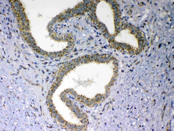
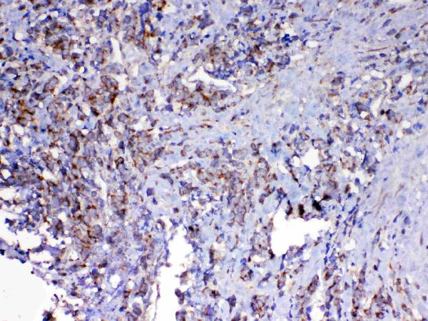
Real IHC data IHC analysis of MAVS using anti-MAVS antibody (A00169-1). MAVS was detected in paraffin-embedded section of human mammary cancer tissue. Heat mediated antigen retrieval was performed in citrate buffer (pH6, epitope retrieval solution) for 20 mins. The tissue section was blocked with 10% goat serum. The tissue section was then incubated with 1μg/ml rabbit anti-MAVS Antibody (A00169-1) overnight at 4°C. Biotinylated goat anti-rabbit IgG was used as secondary antibody and incubated for 30 minutes at 37°C. The tissue section was developed using Strepavidin-Biotin-Complex (SABC)(Catalog # SA1022) with DAB as the chromogen.
Anti-MAVS Antibody ®
Cat # A00169-1
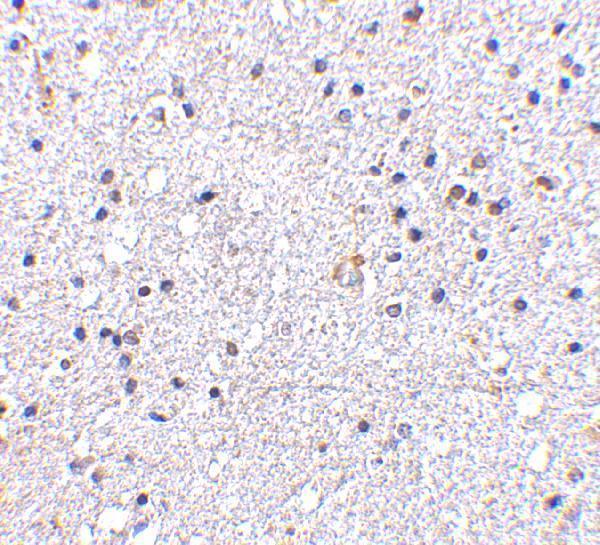
Real IHC data Immunohistochemistry of VISA in human brain tissue with VISA antibody at 5 μg/mL.
Anti-VISA MAVS Antibody
Cat # A00169-2
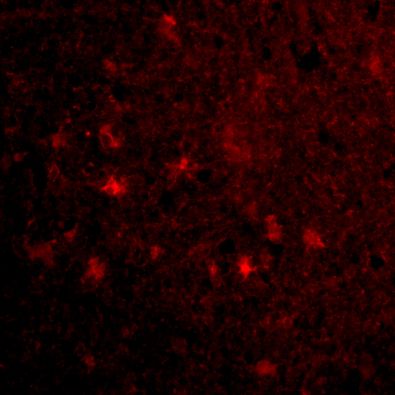
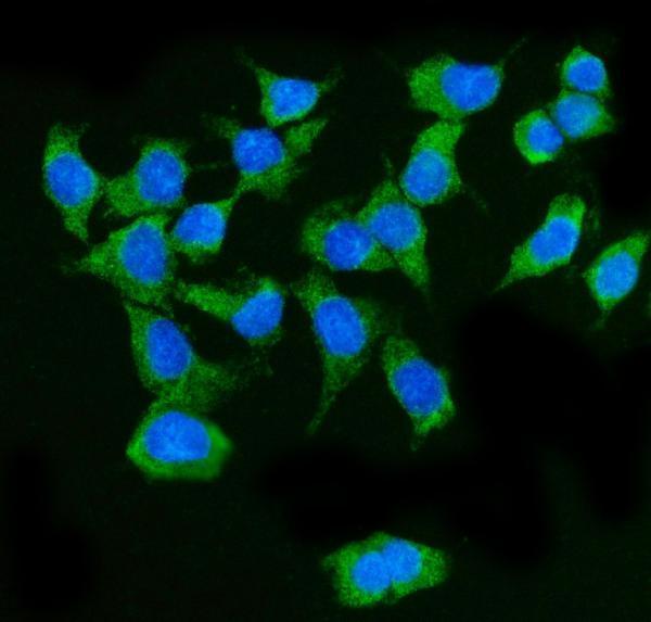
Real IF data IF analysis of MAVS using anti-MAVS antibody (A00169-3). MAVS was detected in an immunocytochemical section of Caco-2 cells. Enzyme antigen retrieval was performed using IHC enzyme antigen retrieval reagent (AR0022) for 15 mins. The cells were blocked with 10% goat serum. And then incubated with 5 μg/mL rabbit anti-MAVS Antibody (A00169-3) overnight at 4°C. DyLight®488 Conjugated Goat Anti-Rabbit IgG (BA1127) was used as secondary antibody at 1:100 dilution and incubated for 30 minutes at 37°C. The section was counterstained with DAPI. Visualize using a fluorescence microscope and filter sets appropriate for the label used.
Anti-MAVS Antibody ®
Cat # A00169-3

A00169-1 shows IHC in paraffin-embedded human mammary cancer tissue, while A00169-2 shows IHC in human brain tissue and IF in human brain cells (catalog image captions). A00169-3 shows IF/ICC in Caco-2 cells and lists human reactivity (catalog image caption; application and reactivity lists).

Which to pick: For human tissue IHC, choose A00169-1 when working with paraffin sections; its image documents citrate pH 6 retrieval, but does not report the fixative (A00169-1 IHC image caption). For human IF/ICC, choose A00169-3, a polyclonal antibody with an IF image in Caco-2 cells (A00169-3 dilution notes; IF image caption). For mouse or rat samples, consider A00169-2 because those species are listed as reactive, while its pictured IHC result is from human brain tissue and its fixative is unreported (A00169-2 reactivity list; IHC image caption).

Each figure is that product's own IHC / IF validation image from its datasheet.

References

  1. UniProt Consortium. UniProt entry Q7Z434 (MAVS_HUMAN, Mitochondrial antiviral-signaling protein).
  2. Human Protein Atlas. MAVS tissue IHC expression (reliability: Enhanced).
  3. Human Protein Atlas. MAVS subcellular location (ICC-IF): Localized to the mitochondria..
  4. Human Protein Atlas. MAVS antibody validation summary (3 antibodies).
  5. An Analysis Regarding the Prognostic Significance of MAVS and Its Underlying Biological Mechanism in Ovarian Cancer. Frontiers in cell and developmental biology 2021 — PMC8551630.
  6. MAVS/CMTM6 axis couples mitochondrial homeostasis to immunogenic senescence via CCL3-driven T-cell recruitment in renal carcinoma. Journal for immunotherapy of cancer 2025 — PMC12766799.
  7. Overexpression of the mitochondrial anti-viral signaling protein, MAVS, in cancers is associated with cell survival and inflammation. Molecular therapy. Nucleic acids 2023 — PMC10468804.
  8. Novel insights into stress-induced susceptibility to influenza: corticosterone impacts interferon-β responses by Mfn2-mediated ubiquitin degradation of MAVS. Signal transduction and targeted therapy 2020 — PMC7499204.
  9. PubMed PMID:16125763 — UniProt-cited evidence.
  10. PubMed PMID:16153868 — UniProt-cited evidence.
  11. PubMed PMID:16177806 — UniProt-cited evidence.