This website uses cookies to ensure you get the best experience on our website.


- Table of Contents
Plan MBNL1 paraffin-section IHC around the nuclear staining seen in most tissues (HPA tissue IHC). Use HPA high-staining cells as positive controls and start the catalog antibody at 2–5 µg/mL (HPA tissue IHC; datasheet A02309-1).

Expected localisation, validated protocols, controls and antibodies — the at-a-glance facts below, then the full design guide.
| Expected localisation | Nuclear tissue staining; cytoplasm also annotated (HPA tissue IHC; UniProt) | |
| Staining pattern | Nuclear staining in most tissues (HPA tissue IHC) | |
| Antigen retrieval | EDTA pH 8.0 HIER, heat-mediated (datasheet A02309-1) | |
| Positive control | Cerebellum+4 more · see all | |
| Negative control | Adipose tissue+2 more · see all |
| Fixation | Matched tissue-IHC evidence does not establish the fixation claim. Validate the specimen-specific method before use. (selected-SKU IHC image A02309-1) | |
| Caveat | Skeletal myocytes lack detectable IHC staining despite reported muscle expression (HPA tissue IHC; UniProt) | |
| Regulation | Higher during myoblast differentiation (UniProt) | |
| Isoform / epitope | Seven isoforms; antibody epitope coverage is unknown (UniProt) |
Compare the catalog antibody’s IHC-P protocol (datasheet: A02309-1) with published MBNL1 staining protocols for transplanted tPVAT (PMC8698607), human brain and glioblastoma (PMC7642170), and stenotic intestine (PMC13297613).
| Sample | Paraffin-embedded human thyroid cancer tissue; fixative not specified (datasheet A02309-1) |
| Fixation | Image fixative and duration unreported (datasheet A02309-1); verify before use. |
| Sectioning | 4–5 µm sections on charged slides (standard) |
| Deparaffinisation | Xylene, graded ethanol series to water (standard) |
| Antigen retrieval | Heat retrieval: EDTA pH 8.0 (datasheet A02309-1); 20 min, 95–100 °C (standard) |
| Peroxidase block | 3% H2O2, 10 min, room temperature (standard) |
| Blocking | 10% goat serum (datasheet A02309-1) |
| Primary antibody | Rabbit anti-MBNL1, 2-5μg/ml (datasheet A02309-1) |
| Primary incubation | Overnight at 4 °C (datasheet A02309-1) |
| Detection | HRP-conjugated secondary, DAB chromogen (datasheet A02309-1) |
| Counterstain | Hematoxylin, blue, dehydrate and mount (standard) |
| Expected result | MBNL1-positive staining in cells in molecular layer of cerebellum (HPA tissue IHC: High). HPA tissue profile: Nuclear expression in most tissues. No signal in the no-primary control. |
MBNL1 staining in paraffin sections is expected mainly in nuclei across many tissues (HPA tissue IHC: nuclear expression in most tissues). Strong examples include cerebellar molecular-layer cells, nasopharyngeal respiratory epithelium, skin arrector pili muscle cells and testicular spermatogonia (HPA tissue IHC: High). Cytoplasmic signal can also be biologically plausible (UniProt Q9NR56: nucleus and cytoplasm). MBNL1 has no transmembrane segment, so a membrane-restricted pattern is unexpected (UniProt Q9NR56 topology). HPA rates its tissue staining reliability Enhanced, with medium staining–RNA consistency (HPA tissue IHC).
| Distinct nuclear chromogen in the expected cells, with stronger staining in a documented High example (HPA tissue IHC: High). | This fits the reported nuclear pattern (HPA tissue IHC: nuclear expression in most tissues). Score the relevant cell population and compartment; compare intensity with controls processed in the same run (general IHC practice). |
| Staining is confined to cell borders or an extracellular structure, with little nuclear signal. | A membrane-restricted pattern conflicts with MBNL1’s lack of a transmembrane segment (UniProt Q9NR56 topology). Review morphology, background and antibody controls before interpreting it as target staining (general IHC practice). Cytoplasmic signal alone is less decisive because MBNL1 also localizes there (UniProt Q9NR56). |
| Prominent chromogen appears in an unexpected cell population, or persists when primary antibody is omitted. | Consider antibody cross-reactivity or endogenous chromogenic detection activity (general IHC practice). Check the omitted-primary control and tissue morphology before assigning MBNL1 expression; HPA documents cell-specific differences within tissues (HPA tissue IHC). |
| Diffuse color covers nuclei, cytoplasm and tissue spaces without clear cell boundaries. | This does not provide a reliable compartment call (general IHC practice). Review background controls, blocking, washing and chromogen development (general IHC practice); the reference pattern is chiefly nuclear (HPA tissue IHC). |
| No signal appears in a section containing a documented High cell population (HPA tissue IHC: High). | First treat the run as potentially uninformative: confirm that the relevant cells are present and that positive controls and detection worked (general IHC practice). Absence alone cannot distinguish low abundance from an assay failure (general IHC practice). |
| Tissue and cell selection | Use the named cell population when judging a section: HPA reports High staining in cerebellar molecular-layer cells and nasopharyngeal respiratory epithelium, but Not detected in adipocytes and spleen red-pulp cells (HPA tissue IHC). A tissue name alone is an imprecise control designation (general IHC practice). |
| Tissue-source disagreement | UniProt describes high protein expression in cardiac and skeletal muscle, while HPA reports Low staining in cardiomyocytes and Not detected in skeletal myocytes (UniProt Q9NR56: tissue specificity; HPA tissue IHC). Treat those HPA cells cautiously as positive IHC controls; the records do not establish why they differ. |
| Antibody validation | HPA lists CAB016398 as IHC Enhanced and HPA035098 as IHC Supported (HPA antibodies). These are validation labels for the listed antibodies, not a guarantee that every section or detection run will reproduce a given intensity (HPA antibodies; general IHC practice). |
| Protein form and topology | UniProt lists seven MBNL1 isoforms, one annotated 1–388 chain, no signal peptide or propeptide, and no transmembrane segment (UniProt Q9NR56). Epitope coverage across isoforms is unspecified here; do not infer an isoform-specific stain from the observed compartment alone. |
| IF/ICC Q&A: Where should signal appear? | Mainly in nucleoplasm, with additional cytosol reported in ICC-IF (HPA subcellular: nucleoplasm Enhanced; cytosol Supported). UniProt also reports stress-granule localization upon stress (UniProt Q9NR56). These cell-image observations inform localization; they do not define a paraffin-section staining intensity. |
| Situation | Likely cause | Next action |
|---|---|---|
| Known High cells are present, but the section and run control are blank (HPA tissue IHC: High). | A shared staining or detection failure is plausible (general IHC practice). | Check the detection reagents and run control, then review the IHC-validated antibody’s stated IHC-P conditions before repeating the run (general IHC practice). |
| The run control stains, but the chosen test population is blank. | The selected cells may have little detectable staining: cardiomyocytes are Low and skeletal myocytes Not detected in HPA IHC (HPA tissue IHC). | Verify cell identity and compare with a documented High population on a suitable control section (HPA tissue IHC; general IHC practice). Do not call the entire assay failed from this population alone. |
| Brown color persists in the omitted-primary control. | Endogenous detection activity or another primary-independent background source may contribute (general chromogenic IHC practice). | Review the detection system’s background-blocking step and compare matched controls before scoring the test section (general IHC practice). |
| Staining is widespread and obscures nuclear boundaries. | Excess background or chromogen development can prevent compartment assessment (general IHC practice). | Review washing, blocking and development against the run controls; score MBNL1 only where cell boundaries and nuclear localization remain interpretable (general IHC practice; HPA tissue IHC: nuclear pattern). |
| Only cell borders stain while expected nuclei remain clear. | A border-only pattern does not match a protein without a transmembrane segment or HPA’s chiefly nuclear pattern (UniProt Q9NR56 topology; HPA tissue IHC). | Inspect morphology and background controls, then reassess antibody specificity before reporting the border signal as MBNL1 (general IHC practice). |
| Cytoplasmic staining accompanies nuclear staining. | Cytoplasmic localization is documented, although the HPA tissue IHC profile is chiefly nuclear (UniProt Q9NR56; HPA tissue IHC). | Record nuclear and cytoplasmic staining separately, compare with controls and avoid labeling cytoplasmic signal an artefact solely because it is extranuclear (general IHC practice; UniProt Q9NR56). |
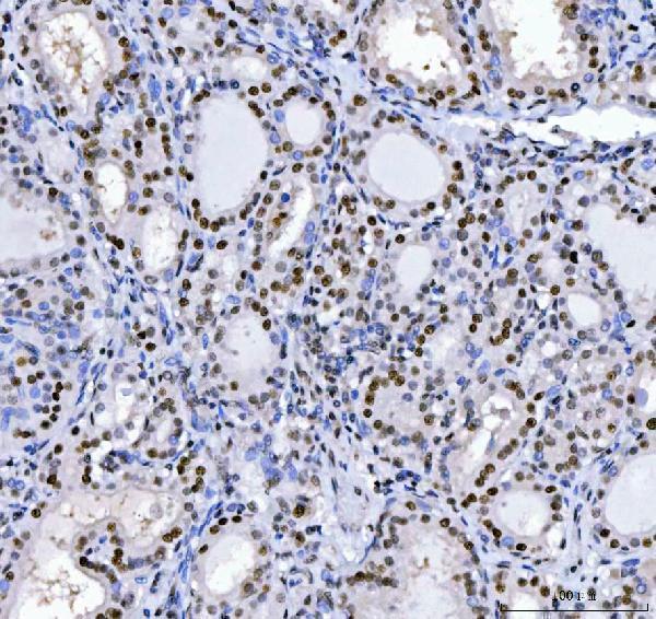
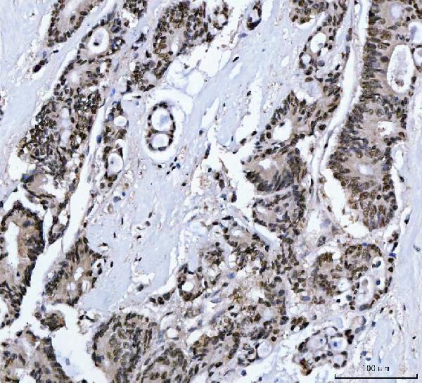
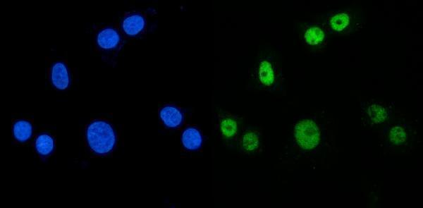


Comprehensive Human Protein Atlas IHC scoring per tissue (reliability: Enhanced — Medium consistency between antibody staining and RNA expression data.). Rows are taken directly from the HPA tissue chart — click any row's HPA link to view the source.
| Tissue | Cell type | Level | Evidence | Source |
|---|---|---|---|---|
| Cerebellum | Cells in molecular layer | High | Protein (IHC) | HPA → |
| Nasopharynx | Respiratory epithelial cells | High | Protein (IHC) | HPA → |
| Skin | Arrector pili muscle cells | High | Protein (IHC) | HPA → |
| Testis | Spermatogonia cells | High | Protein (IHC) | HPA → |
| Adrenal gland | Glandular cells | Medium | Protein (IHC) | HPA → |
Troubleshoot MBNL1 staining in paraffin sections by checking retrieval, compartment, controls and cell-specific scoring before interpreting changes in signal.
A02309-1 has IHC images from human paraffin sections and IF images from A549 cells and human colon cancer sections; the catalog lists Human, Monkey, Mouse and Rat reactivity (catalog image captions; catalog reactivity).
A02309-1 has IHC images from paraffin sections of human thyroid, colorectal, laryngeal and liver cancers (catalog IHC image captions). A02309-1 also has IF images from A549 cells and paraffin sections of human colon cancer (catalog IF image captions).
Which to pick: For chromogenic tissue IHC, choose A02309-1: its human paraffin section images use EDTA retrieval at pH 8.0, 2 μg/ml primary antibody and DAB detection; the fixative is unreported (catalog IHC image captions). For IF/ICC, A02309-1 has images from A549 cells and human colon cancer sections, with 5 μg/ml primary antibody (catalog IF image captions). For cross-species planning, the catalog lists Human, Monkey, Mouse and Rat reactivity for A02309-1, while the supplied IHC and IF images show human samples (catalog reactivity; catalog image captions).